. 2003 Mar 28;278(13):11086-93.
doi: 10.1074/jbc.M212157200. Epub 2003 Jan 21.

Muscarinic receptor activation protects cells from apoptotic effects of DNA damage, oxidative stress, and mitochondrial inhibition

Affiliations

Muscarinic receptor activation protects cells from apoptotic effects of DNA damage, oxidative stress, and mitochondrial inhibition

Patrizia De Sarno et al. J Biol Chem. .

Abstract

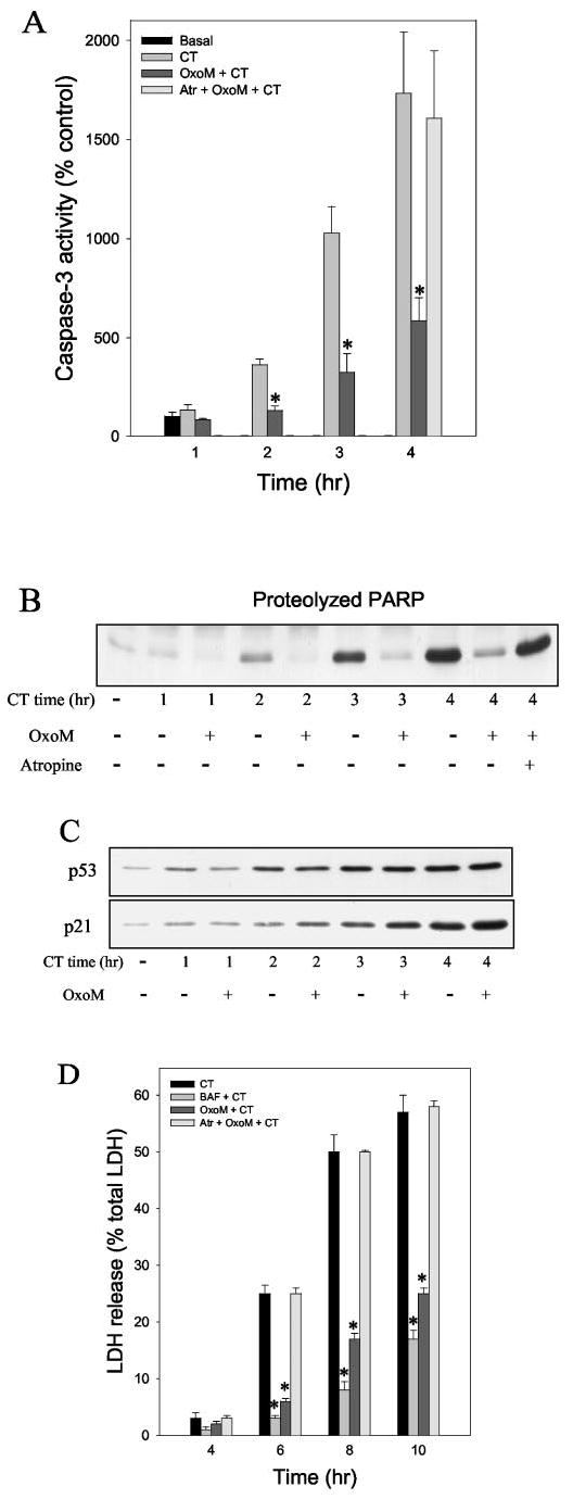
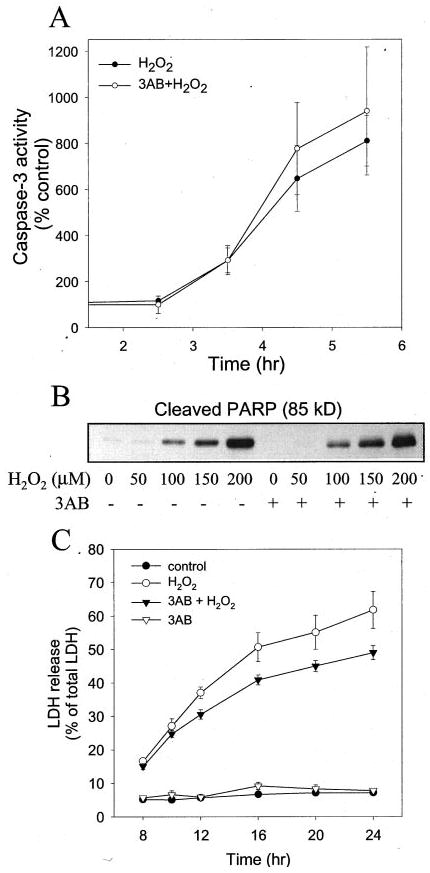
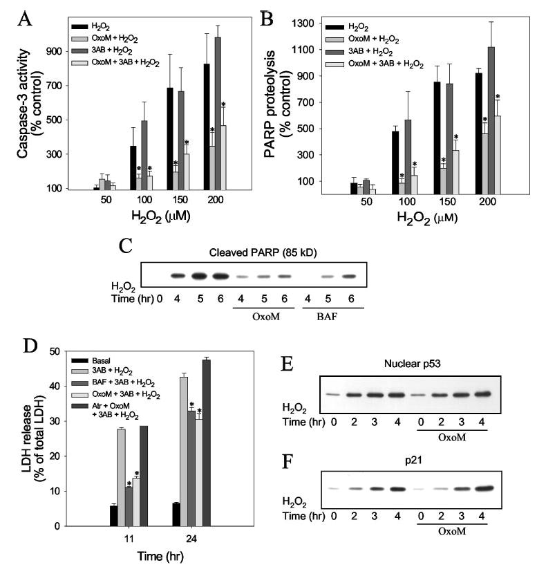
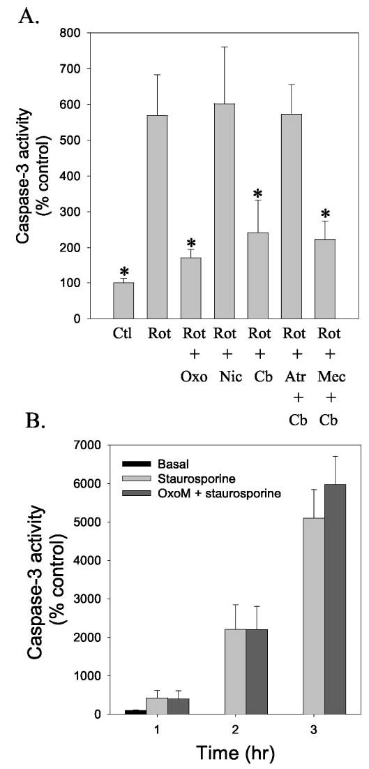
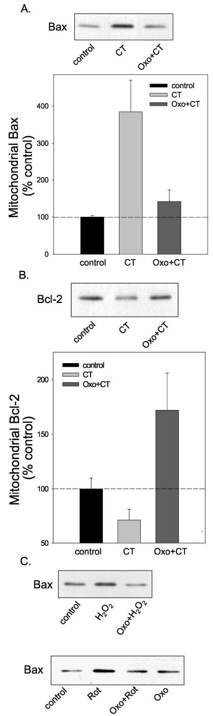
The impact of muscarinic receptor stimulation was examined on apoptotic signaling induced by DNA damage, oxidative stress, and mitochondrial impairment. Exposure of human neuroblastoma SH-SY5Y cells to the DNA-damaging agent camptothecin increased p53 levels, activated caspase-3, and caused cell death. Pretreatment with oxotremorine-M, a selective agonist of muscarinic receptors that are expressed endogenously in these cells, did not affect the accumulation of p53 but greatly attenuated caspase-3 activation and protected from cell death to nearly the same extent as treatment with a general caspase inhibitor. Treatment with 50-200 microm H(2)O(2) caused the activation of caspase-3 beginning after 2-3 h, followed by eventual cell death. Oxotremorine-M pretreatment protected cells from H(2)O(2)-induced caspase-3 activation and death, and this was equivalent to protection afforded by a caspase inhibitor. Muscarinic receptor stimulation also protected cells from caspase-3 activation induced by exposure to rotenone, a mitochondrial complex 1 inhibitor, but no protection was evident from staurosporine-induced caspase-3 activation. The mechanism of protection afforded by muscarinic receptor activation from camptothecin-induced apoptotic signaling involved blockade of mitochondrial cytochrome c release associated with a bolstering of mitochondrial bcl-2 levels and blockade of the translocation of Bax to mitochondria. Likely the most proximal of these events to muscarinic receptor activation, mitochondrial Bax accumulation, also was attenuated by oxotremorine-M treatment after treatment with H(2)O(2) or rotenone. These results demonstrate that stimulation of muscarinic receptors provides substantial protection from DNA damage, oxidative stress, and mitochondrial impairment, insults that may be encountered by neurons in development, aging, or neurodegenerative diseases. These findings suggest that neurotransmitter-induced signaling bolsters survival mechanisms, and inadequate neurotransmission may exacerbate neuronal loss.


Figures

Fig. 1
Fig. 1. Oxotremorine-M protects cells from camptothecin-induced caspase-3 activation and cell death
Caspase-3 activity (A) and PARP proteolysis (B) were measured in cells treated with 1 μm camptothecin (CT), 300 μm oxotremorine-M (OxoM) 30 min before camptothecin, and 1 μm atropine (Atr) 15 min before oxotremorine-M followed by camptothecin. C, p53 and p21 levels were measured in cells treated with camptothecin alone or with pretreatment with 300 μm oxotremorine-M for 30 min. D, lactate dehydrogenase (LDH) release into the media was measured 4 –10 h after treatment with camptothecin with or without pretreatments with 100 μm BAF or 300 μm oxotremorine-M and in cells treated with 1 μm atropine before oxotremorine-M treatment. Quantitative values are means ±S.E.; n =4 –5; *, p < 0.05.
Fig. 2
Fig. 2. H2O2 treatment induces apoptosis
A, the time course of caspase-3 activation was measured in cells treated with 100 μm H2O2 with or without a 30-min pretreatment with 100 μm 3AB. (B) PARP proteolysis was measured in cells treated with 50 to 200 μm H2O2 for 5 h, with or without a 30 min pretreatment with 100 μm 3AB, by immunoblot analysis. The immunoblot shows the 85-kDa product of PARP cleaved by caspase-3. C, the time course of lactate dehydrogenase release was measured in cells treated with 100 μmH2O2 with or withouta 30-min pretreatment with 100 μm 3AB, in cells treated with only 3AB, and in control untreated cells. Quantitative data are means ±S.E.; n =3–4.
Fig. 3
Fig. 3. Oxotremorine-M treatment protects cells from H2O2-induced apoptosis
SH-SY5Y cells were pretreated with 100 μm 3-AB for 30min, 100 μm BAF for 60 min, or 300 μm oxotremorine-M (OxoM) (with or without a 15-min pretreatment with 1 μm atropine (Atr)) for 30 min wherem indicated followed by treatment with H2O2. Caspase-3 activity (A) and PARP cleavage (B) were measured 6 h after treatment with 50 –200 μM H2O2. C, proteolyzed PARP was measured 4, 5, and 6 h after treatment with 100 μmH2O2 in cells pretreated with 3AB and, where indicated, with oxotremorine-M or BAF. D, lactate dehydrogenase (LDH) release into the media was measured 11 and 24 h after treatment with 100 μm H2O2 and the indicated agents. Nuclear p53 levels (E) and p21 levels (F) were measured 2, 3, and 4 h after treatment with 100 μm H2O2 without or with a 30-min pretreatment with 300 μM oxotremorine-M. Quantitative values are the means ±S.E.; n = 3–4; *, p < 0.05.
Fig. 4
Fig. 4. Muscarinic receptor stimulation attenuates caspase-3 activation induced by rotenone but not staurosporine
A, caspase-3 activity was measured in cells 16 h after treatment with 5 μm rotenone (Rot). Where indicated, cells were pretreated for 30 min with 300 μm oxotremorine-M (Oxo), 300 μm carbachol (Cb), or 10 μm nicotine (Nic). Cells were treated with the antagonists 10 μm atropine (Atr) or 10 μm mecamylamine (Mec) 15 min before treatment with agonists. Values are presented as the percent of caspase-3 activity in untreated control (Ctl) cells and are the means ±S.E.; n = 3–4; *, p < 0.05 compared with values from samples treated with rotenone alone. B, caspase-3 activity was measured in cells treated with 0.5 μm staurosporine alone or after a 30-min pretreatment with 300 μm oxotremorine-M. Values are presented as the percent of caspase-3 activity in untreated control cells (means ±S.E.; n = 3).
Fig. 5
Fig. 5. Muscarinic receptor stimulation blocked cytochrome c release from the mitochondria caused by camptothecin treatment
Cells were treated with 1 μm camptothecin (CT) or with 300 μm oxotremorine-M (Oxo) 30 min before camptothecin, and cytosolic and mitochondrial fractions were immunoblotted for cytochrome c (Cyt c). Quantitative values are the means ±S.E.; n = 3.
Fig. 6
Fig. 6. Mitochondrial Bax and bcl-2 levels
Cells were treated with 1 μm camptothecin (CT) or with 300 μm oxotremorine-M (Oxo) 30 min before camptothecin, and mitochondrial fractions were immunoblotted for Bax (A) or bcl-2 (B). Quantitative values are the means ±S.E.; n = 3. C, mitochondrial Bax levels were measured after treatment with 100 μm H2O2 (5 h) or 5μm rotenone (16 h) with or without a 30-min pretreatment with 300 μm oxotremorine-M.

References

    1. Zornig M, Hueber A, Baum W, Evan G. Biochim Biophys Acta. 2001;1551:1–37. - PubMed
    1. Bortner CD, Cidlowski JA. Annu Rev Pharmacol Toxicol. 2002;42:259–281. - PubMed
    1. Jacobs BL, van Praag H, Gage FH. Mol Psychiatry. 2000;5:262–269. - PubMed
    1. Kempermann G. Bipolar Disord. 2002;4:17–33. - PubMed
    1. Miller FD, Pozniak CD, Walsh GS. Cell Death Differ. 2000;7:880–888. - PubMed

Publication types

MeSH terms