. 2003 Jan 20;197(2):153-62.
doi: 10.1084/jem.20021109.

Vaginal submucosal dendritic cells, but not Langerhans cells, induce protective Th1 responses to herpes simplex virus-2

Affiliations

Vaginal submucosal dendritic cells, but not Langerhans cells, induce protective Th1 responses to herpes simplex virus-2

Xinyan Zhao et al. J Exp Med. .

Abstract

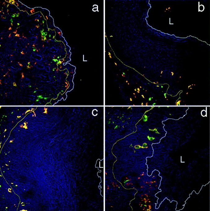
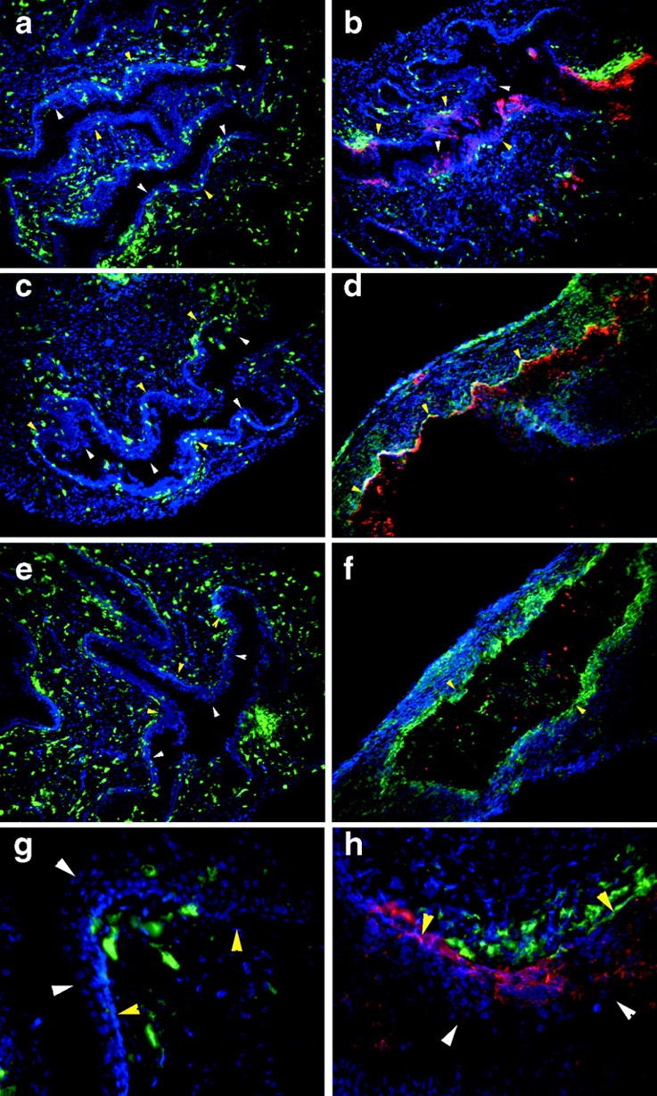
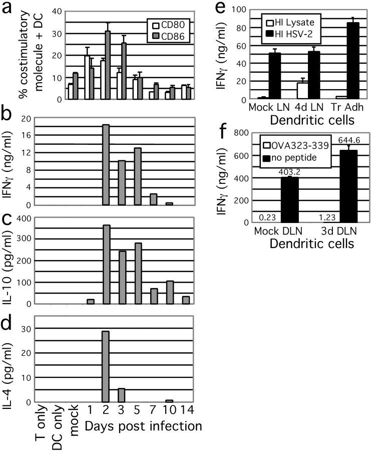
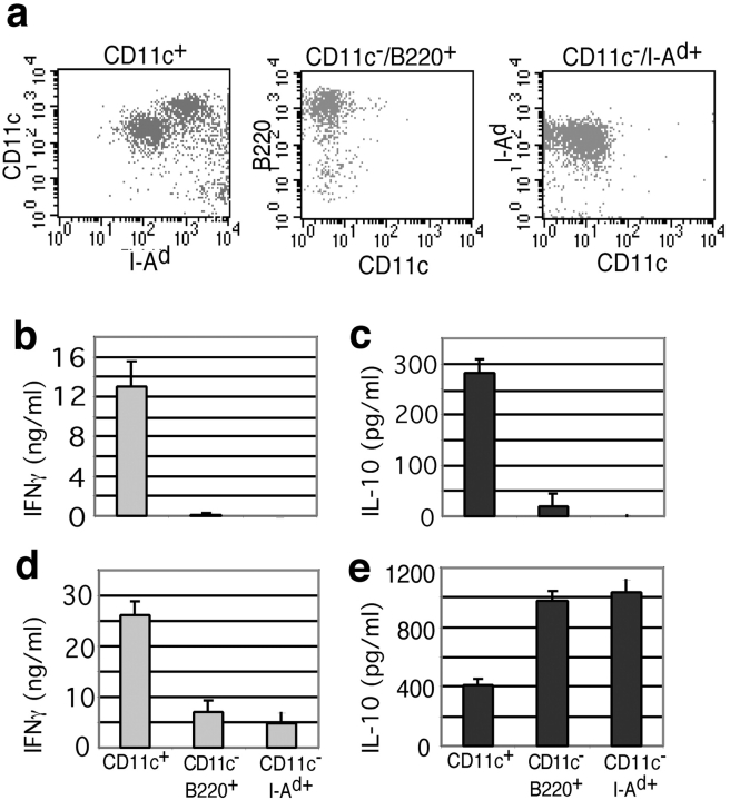
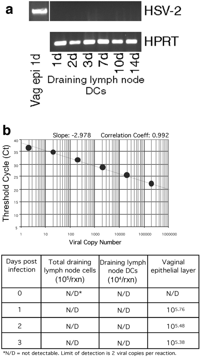
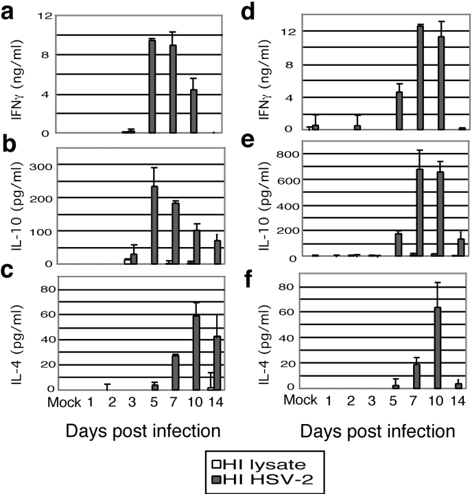
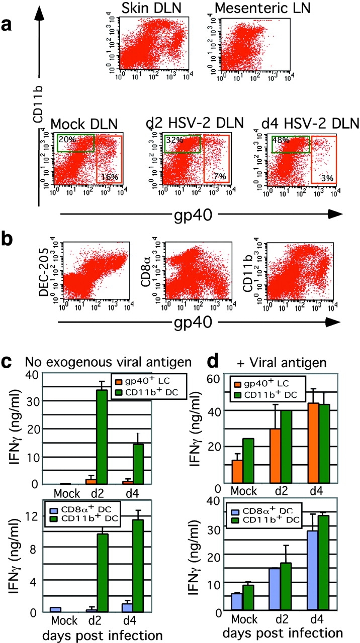
Herpes simplex virus (HSV) type 2 infection occurs primarily at the genital mucosal surfaces and is a leading cause of ulcerative lesions. Despite the availability of animal models for HSV-2 infection, little is known regarding the mechanism of immune induction within the vaginal mucosa. Here, we examined the cell types responsible for the initiation of protective Th1 immunity to HSV-2. Intravaginal inoculation of HSV-2 led to a rapid recruitment of submucosal dendritic cells (DCs) to the infected epithelium. Subsequently, CD11c(+) DCs harboring viral peptides in the context of MHC class II molecules emerged in the draining lymph nodes and were found to be responsible for the stimulation of IFNgamma secretion from HSV-specific CD4(+) T cells. Other antigen-presenting cells including B cells and macrophages did not present viral peptides to T cells in the draining lymph nodes. Next, we assessed the relative contribution to immune generation by the Langerhans cells in the vaginal epithelium, the submucosal CD11b(+) DCs, and the CD8alpha(+) lymph node DCs. Analysis of these DC populations from the draining lymph nodes revealed that only the CD11b(+) submucosal DCs, but not Langerhans cell-derived or CD8alpha(+) DCs, presented viral antigens to CD4(+) T cells and induced IFNgamma secretion. These results demonstrate a previously unanticipated role for submucosal DCs in the generation of protective Th1 immune responses to HSV-2 in the vaginal mucosa, and suggest their importance in immunity to other sexually transmitted diseases.


Figures

Figure 1.
Figure 1.
LC distribution in the vaginal epithelium during the estrous cycle. Frozen sections of vaginal tissues from mice at diestrous (a), estrous (b), metestrous-1 (c), and metestrous-2 (d) phases were stained with antibodies against MHC class II (red) or CD11c (green) and analyzed by confocal microscopy. Confocal images are overlaid with light transmission microscopy images to denote tissue morphology. The white line indicates the luminal edge of the epithelium, whereas the yellow line indicates the basement membrane. The images were captured using objective lenses of 20× (a–c) or 40× (d). L, lumen.
Figure 2.
Figure 2.
DC recruitment to the HSV-2–infected vaginal epithelium. BALB/c mice pretreated with Depo-Provera® were inoculated with ivag TK mutant HSV-2 (b, d, f, and h), or with control cell lysate (a, c, e, and g), and vaginal tissues were collected for staining with antibodies against CD11c (green) and HSV-2 (red) at 24 h (a, b, g, and h), 48 h (c and d), or 5 d (e and f) a.i. The nucleus was visualized by staining with DAPI (blue). Images were captured using a 20× objective lens (a–f) or with 40x lens (g and h). The epithelial layer is indicated by the white arrowheads (luminal edge) and yellow arrowheads (basement membrane). Panels g and h represent higher magnification images of the selected areas in panels a and b, respectively.
Figure 3.
Figure 3.
DCs in the draining lymph nodes up-regulate co-stimulatory molecules and present viral antigen. DCs were isolated from the draining lymph nodes of mice inoculated ivag with HSV-2 or with control cell lysate (mock) using magnetic beads. (a) Co-stimulatory molecules CD80 and CD86 expression levels were determined by flow cytometry. (b–d) Cytokine secretion from HSV-2–specific CD4+ T cells co-cultured with draining lymph node DCs, or from CD4+ T cells (day 5 a.i.) or DCs (day 3 a.i.) alone, were analyzed by ELISA. CD4+ HSV-2–specific T cells were obtained from the draining lymph node of day 5 ivag HSV-2–infected mice by positive selection using magnetic selection. To demonstrate the antigen specificity of the T cell responses, (e) DCs isolated from the draining lymph nodes of either mock-infected or day 3 HSV-2–infected mice, or in vitro–activated transiently adherent splenic DCs, were coincubated with day 5 draining lymph node CD4+ T cells in the presence (filled) or absence (white) of exogenously added virus antigens. T cell IFNγ secretion was measured by ELISA. (f) To demonstrate the requirement of specific antigen for T cell activation by the draining lymph node DCs, in vitro–activated OVA-specific T cells were coincubated with DCs isolated from the draining lymph nodes of either mock-infected or day 4 HSV-2–infected mice in the presence (filled) or absence (white) of OVA323–339 peptide. T cell IFNγ secretion was measured by ELISA.
Figure 4.
Figure 4.
CD11c+ DCs are crucial for IFNγ secretion from HSV-2–specific T cells. Various APC populations were isolated from draining lymph nodes of day 5 infected mice by magnetic selection. CD11c fraction was obtained using the depletion column and was further subdivided into B220+ or I-Ad+ groups by magnetic selection. (a) A representative FACS® profile of these APC populations, gated on live cells, is shown. These APC groups were used to stimulate HSV-2–specific CD4+ T cells in the absence (b and c) or presence (d and e) of exogenously added heat-inactivated virus. Cytokines secreted from T cells were measured by ELISA. The data are representative of three similar experiments. Each experiment was conducted with three to four mice.
Figure 5.
Figure 5.
Draining lymph node DCs do not contain HSV-2 viral DNA. Total DNA was isolated from DCs from the draining lymph nodes of mice inoculated ivag with HSV-2 or with control cell lysate using magnetic beads. (a) HSV-2 viral DNA was amplified using specific primers by PCR. DNA isolated from 24 h HSV-2–infected vaginal epithelium was used as a positive control for HSV-2–specific gene amplification. The presence of genomic DNA in each sample is depicted by PCR amplification of the housekeeping HPRT gene. To further confirm the lack of viral DNA in the DCs and total draining lymph node cells, either 104 (DCs) or 105 (total lymph node cells) cell equivalents of DNA was subjected to Real-time PCR at days 0, 1, 2, or 3 a.i. (b). In parallel, DNA obtained from the vaginal epithelial layer (sites of viral replication) at the corresponding time points were also examined by Real-time PCR as a positive control for infection in these mice. These reactions were conducted twice with similar results.
Figure 6.
Figure 6.
Cytokine secretion from CD4+ T cells in draining lymph nodes and spleen of ivag HSV-2–infected mice. CD4+ T cells positively selected from draining lymph nodes (a–c) and spleen (d–f) of mice infected ivag with 186TKΔKpn for various time periods were co-cultured with irradiated syngeneic splenocytes in the presence of heat-inactivated HSV-2 (filled bars) or heat-inactivated control cell lysate (white bars) for 3 d and supernatants were analyzed for IFNγ (a and d), IL-10 (b and e), or IL-4 (c and f).
Figure 7.
Figure 7.
Submucosal DCs, but not Langerhans cells or CD8α+ DCs, present viral peptides to CD4+ T cells in the draining lymph nodes. To differentiate the contribution of LCs and submucosal DCs in the presentation of HSV-2 antigens in vivo, CD11c+ cells were stained with gp40 (LC) and CD11b (submucosal DC) after HSV-2 ivag infection at 2 or 4 d a.i. and were analyzed by flow cytometry (a). The DC markers, DEC-205, CD8α, and CD11b, on CD11c+ cells were analyzed (b). The LC-derived (gp40+), submucosal DCs (CD11b+), or the CD8α+ DCs were FACS®-sorted from draining lymph nodes at 2 or 4 d a.i. and were co-cultured with HSV-2–specific CD4+ T cells in the absence (c) or presence (d) of viral antigens for 72 h. IFNγ secreted from T cells were analyzed by ELISA (c and d).

References

    1. Lenz, A., M. Heine, G. Schuler, and N. Romani. 1993. Human and murine dermis contain dendritic cells. Isolation by means of a novel method and phenotypical and functional characterization. J. Clin. Invest. 92:2587–2596. - PMC - PubMed
    1. Meunier, L., A. Gonzalez-Ramos, and K.D. Cooper. 1993. Heterogeneous populations of class II MHC+ cells in human dermal cell suspensions. Identification of a small subset responsible for potent dermal antigen-presenting cell activity with features analogous to Langerhans cells. J. Immunol. 151:4067–4080. - PubMed
    1. Baker, D.A., and S.A. Plotkin. 1978. Enhancement of vaginal infection in mice by herpes simplex virus type II with progesterone. Proc. Soc. Exp. Biol. Med. 158:131–134. - PubMed
    1. Gallichan, W.S., and K.L. Rosenthal. 1996. Effects of the estrous cycle on local humoral immune responses and protection of intranasally immunized female mice against herpes simplex virus type 2 infection in the genital tract. Virology. 224:487–497. - PubMed
    1. Overall, J.C., Jr., E.R. Kern, R.L. Schlitzer, S.B. Friedman, and L.A. Glasgow. 1975. Genital herpesvirus hominis infection in mice. I. Development of an experimental model. Infect. Immun. 11:476–480. - PMC - PubMed

Publication types

MeSH terms