. 2003 Jan 20;197(2):163-8.
doi: 10.1084/jem.20021500.

Human T cell receptor gammadelta cells recognize endogenous mevalonate metabolites in tumor cells

Affiliations

Human T cell receptor gammadelta cells recognize endogenous mevalonate metabolites in tumor cells

Hans-Jürgen Gober et al. J Exp Med. .

Abstract

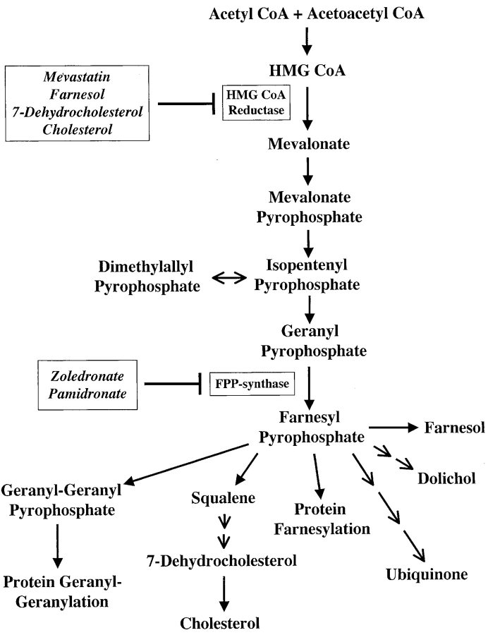
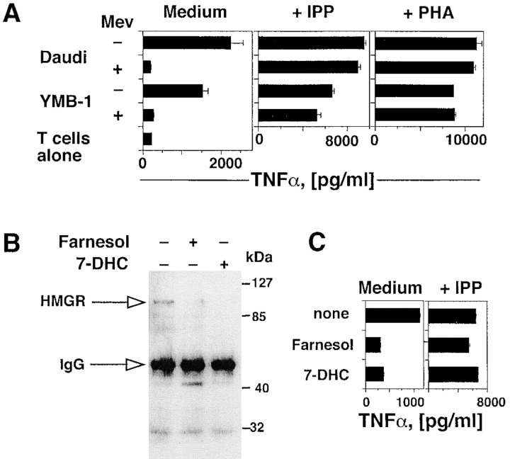
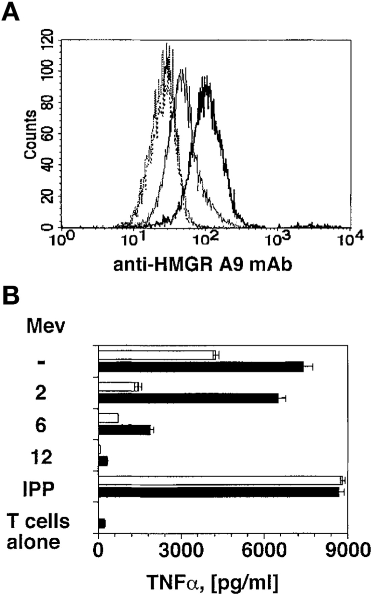
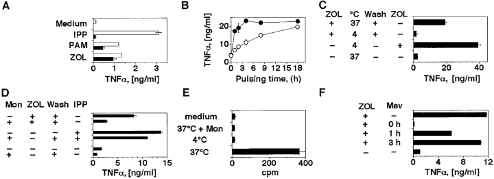
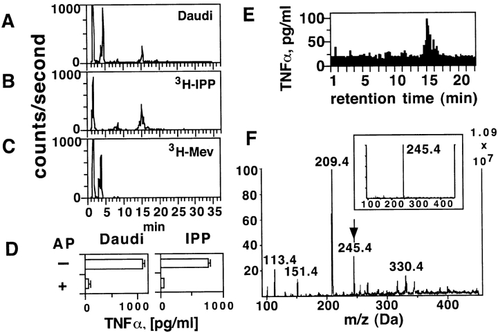
T lymphocytes expressing the T cell receptor (TCR)-gammadelta recognize unknown antigens on tumor cells. Here we identify metabolites of the mevalonate pathway as the tumor ligands that activate TCR-gammadelta cells. In tumor cells, blockade of hydroxy-methylglutaryl-CoA reductase (HMGR), the rate limiting enzyme of the mevalonate pathway, prevents both accumulation of mevalonate metabolites and recognition by TCR-gammadelta cells. When metabolite accumulation is induced by overexpressing HMGR or by treatment with nitrogen-containing bisphosphonate drugs, tumor cells derived from many tissues acquire the capacity to stimulate the same TCR-gammadelta population. Accumulation of mevalonate metabolites in tumor cells is a powerful danger signal that activates the immune response and may represent a novel target of tumor immunotherapy.


Figures

Figure 1.
Figure 1.
Mevalonate metabolic pathway. The compounds used in this study and the affected enzymes are indicated in frames.
Figure 2.
Figure 2.
Activation of TCR-γδ cells by tumor cells is dependent upon the intracellular levels and activity of HMGR. (A) Mevastatin prevents response of the TCR-γδ T cell clone G2B9 stimulated by Daudi and YMB-1 ligands, but has no effect on stimulation with IPP or PHA. Results show TNFα mean release ± SD. (B) HMGR protein levels are down-regulated by farnesol or 7-DHC as detected by immunoprecipitation and Western blot. (C) Treatment of Daudi cells with farnesol or 7-DHC prevents stimulation of the G2B9 cells. In control experiments the same treatments of Daudi cells did not affect stimulation with exogenous IPP.
Figure 3.
Figure 3.
HMGR-Daudi transfectants are potent stimulators. (A) Daudi cells transfected with the HMGR gene express higher levels of HMGR protein (bold line) than nontransfected Daudi cells (thin line). No difference between the two cell types was detected using an irrelevant mAb (dotted lines). (B) Daudi-HMGR cells (black bars) show increased capacity to stimulate Vγ9/Vδ2 cells as compared with nontransfected Daudi cells (white bars). Mevastatin was added at suboptimal doses (10 μM) 2, 6, or 12 h before incubation with T cells. Stimulation with IPP was used as positive control.
Figure 4.
Figure 4.
ZOL and PAM activate TCR-γδ cells after internalization in the APCs and accumulation of mevalonate metabolites. (A) THP-1 cells stimulate the T cell clone G2D7 after 2 h pulse (black bars) with ZOL or PAM, but not with IPP. Overnight incubation (white bars) with PAM and ZOL was used as positive control. Similar results were obtained by measuring IFN-γ release and using a second TCR Vγ9/Vδ2 clone (data not depicted). (B) ZOL (•) is faster than PAM (○) in inducing TCR-γδ cell stimulatory ligands in Daudi cells. (C) ZOL requires cellular internalization to become stimulatory. Daudi cells were pulsed at 4 or at 37°C. In one control group ZOL was added back to exclude inhibitory effects of incubation at 4°C. (D) Daudi cells have reduced stimulatory capacities when incubated with monensin (Mon) and ZOL. IPP stimulation was used as positive control. (E) Intracellular uptake by Daudi cells of 14C-ZOL. To exclude surface binding, pulse with 14C-ZOL was also performed at 4°C or in the presence of monensin. (F) Mevastatin completely inhibits activation only when added simultaneously with ZOL. Mevastatin was added to Daudi cells together with ZOL, 1 or 3 h later. 3 h after ZOL, Daudi cells were washed and T cells were added. Mevastatin was added back to keep its concentration constant during the entire culture period. Supernatants were harvested 12 h later.
Figure 5.
Figure 5.
The intracellular Daudi compounds activating TCR-γδ cells are metabolites of the mevalonate pathway. (A–C) The stimulatory Daudi ligands have the same retention time as IPP. Panel A shows the radioactive compounds generated with Daudi cell extracts after addition of 3H-mevalonic acid, panel B the reference 3H-IPP, panel C the reference 3H-mevalonic acid (3H-Mev). (D) The stimulatory ligands present in Daudi cell extracts are phosphatase sensitive. The semipurified material was treated with alkaline phosphatase (AP) and then tested in TCR-γδ cell stimulation assays. IPP treated identically was used as positive control for AP activity. (E) Bioactivity of TCR-γδ stimulatory ligands from Daudi cells. HPLC fractions were tested at a final dilution of 1:12 using THP-1 cells as APCs and the T cell clone G2B9. (F) Ion-spray Mass spectrometry of the most active fraction from panel E. The arrow indicates the mass of IPP. The insert shows the mass of reference IPP.

References

    1. Porcelli, S., M.B. Brenner, and H. Band. 1991. Biology of the human γδ T-cell receptor. Immunol. Rev. 120:137–183. - PubMed
    1. Burk, M.R., L. Mori, and G. De Libero. 1995. Human V gamma 9-V delta 2 cells are stimulated in a cross-reactive fashion by a variety of phosphorylated metabolites. Eur. J. Immunol. 25:2052–2058. - PubMed
    1. Tanaka, Y., C.T. Morita, Y. Tanaka, E. Nieves, M.B. Brenner, and B.R. Bloom. 1995. Natural and synthetic non-peptide antigens recognized by human gamma delta T cells. Nature. 375:155–158. - PubMed
    1. Belmant, C., E. Espinosa, R. Poupot, M.A. Peyrat, M. Guiraud, Y. Poquet, M. Bonneville, and J.J. Fournie. 1999. 3-Formyl-1-butyl pyrophosphate A novel mycobacterial metabolite-activating human gammadelta T cells. J. Biol. Chem. 274:32079–32084. - PubMed
    1. Feurle, J., E. Espinosa, S. Eckstein, F. Pont, V. Kunzmann, J.J. Fournie, M. Herderich, and M. Wilhelm. 2002. Escherichia coli produces phosphoantigens activating human gamma delta T cells. J. Biol. Chem. 277:148–154. - PubMed

Publication types

MeSH terms

Substances