. 2003 Jan 20;197(2):169-79.
doi: 10.1084/jem.20020978.

Differential regulation of cathepsin S and cathepsin L in interferon gamma-treated macrophages

Affiliations

Differential regulation of cathepsin S and cathepsin L in interferon gamma-treated macrophages

Courtney Beers et al. J Exp Med. .

Abstract

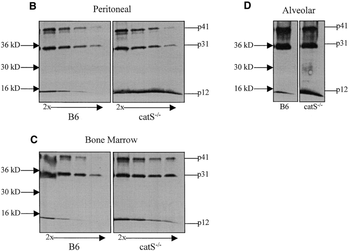
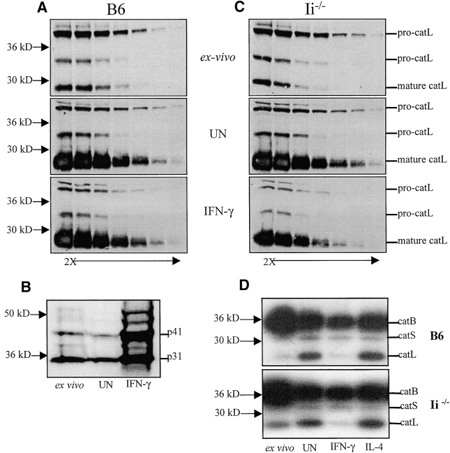
Cathepsin S (catS) and cathepsin L (catL) mediate late stages of invariant chain (Ii) degradation in discrete antigen-presenting cell types. Macrophages (Mphis) are unique in that they express both proteases and here we sought to determine the relative contribution of each enzyme. We observe that catL plays no significant role in Ii cleavage in interferon (IFN)-gamma-stimulated Mphis. In addition, our studies show that the level of catL activity is significantly decreased in Mphis cultured in the presence of IFN-gamma whereas catS activity increases. The decrease in catL activity upon cytokine treatment occurs despite the persistence of high levels of mature catL protein, suggesting that a specific inhibitor of the enzyme is up-regulated in IFN-gamma-stimulated peritoneal Mphis. Similar inhibition of activity is observed in dendritic cells engineered to overexpress catL. Such enzymatic inhibition in Mphis exhibits only partial dependence upon Ii and therefore, other mechanisms of catL inhibition are regulated by IFN-gamma. Thus, during a T helper cell type 1 immune response catL inhibition in Mphis results in preferential usage of catS, such that major histocompatibility complex class II presentation by all bone marrow-derived antigen-presenting cell is regulated by catS.


Figures

Figure 1.
Figure 1.
Ii degradation in cathepsin-deficient Mφs. (A) Thioglycollate-induced Mφs from B6, catS−/−, catL−/−, and catS−/− xcatL−/− mice were pulse labeled and chased for 0, 1, 3, and 6 h. Cell lysates were immunoprecipitated with the I-Ab-specific Ab M5/114 and immunoprecipitates were analyzed by 7.5–20% gradient SDS-PAGE under denaturing conditions. (B) Thioglycollate-induced pMφs, (C) BMMφs, and (D) alveolar Mφs from B6 and catS−/− mice were cultured for 48 h in the presence of IFN-γ and immunoblotted with the monoclonal Ab IN-1. Cells were solubilized in lysis buffer (1% NP-40/TBS in the presence of protease inhibitors) and lysates were then twofold serially diluted starting at 5 μg of total protein (alveolar Mφs are shown at 5 μg only). The position of Ii fragments p41, p31, and p12 are indicated.
Figure 1.
Figure 1.
Ii degradation in cathepsin-deficient Mφs. (A) Thioglycollate-induced Mφs from B6, catS−/−, catL−/−, and catS−/− xcatL−/− mice were pulse labeled and chased for 0, 1, 3, and 6 h. Cell lysates were immunoprecipitated with the I-Ab-specific Ab M5/114 and immunoprecipitates were analyzed by 7.5–20% gradient SDS-PAGE under denaturing conditions. (B) Thioglycollate-induced pMφs, (C) BMMφs, and (D) alveolar Mφs from B6 and catS−/− mice were cultured for 48 h in the presence of IFN-γ and immunoblotted with the monoclonal Ab IN-1. Cells were solubilized in lysis buffer (1% NP-40/TBS in the presence of protease inhibitors) and lysates were then twofold serially diluted starting at 5 μg of total protein (alveolar Mφs are shown at 5 μg only). The position of Ii fragments p41, p31, and p12 are indicated.
Figure 2.
Figure 2.
Active site labeling of cytokine-treated Mφ. (A) Thioglycollate-induced pMφs and (B) BMMφs from B6, catS−/−, and catL−/− mice were either taken directly ex vivo or activated for 48 h with or without IFN-γ. Cells were incubated for 2 h with the irreversible cysteine protease inhibitor Cbz- [125I]-Tyr-Ala-CN2. Cells were lysed and radiolabeled enzymes were analyzed on a 12% SDS-PAGE gel. Levels of active cathepsin in cells taken directly ex vivo (ex vivo) and after 48 h of plating with IFN-γ (IFN-γ) or without (UN) are shown. The position of cathepsin B (catB), cathepsin S (catS), and cathepsin L (catL) are indicated on the gel. C) The intensity of the active site labeling was quantified by the BioRad GS-700 Imaging Densitometer and analyzed by Multi-Analyzer software Version 1.0.2. The numbers correspond to the following: 1, B6 ex vivo; 2, B6 UN; 3, B6 IFN-γ; 4, catS−/− ex vivo; 5, catS−/− IFN-γ; 6, catS−/− UN; 7, catL−/− ex vivo; 8, catL−/− UN; 9, catL−/− IFN-γ.
Figure 3.
Figure 3.
Regulation of catL activity by mRNA. pMφs were purified by adherence to bacterial plastic plates. Cells were lysed directly on the plates 30 min after plating (ex vivo) and after 48 h of incubation in the presence or absence of IFN-γ. After RNA extraction and cDNA synthesis, quantitative PCR was performed in an ABI Prism 7700 sequence detector. Primers and probes for HPRT, catL and catS were designed using Primer Express software. The probes were labeled at the 5′ and 3′ ends with the fluorochromes FAM and TAMRA. mRNA levels were quantitated using the comparative CT method described in the ABI Prism 7700 sequence detector user bulletin number 2. The data is shown as the level of cathepsin mRNA in cells after 48 h of plating with IFN-γ (IFN-γ) or without (UN) compared with the level of mRNA ex vivo.
Figure 4.
Figure 4.
CatL and the effect of the p41 Ii isoform. The level of catL in (A) B6 and (C) Ii−/− thioglycollate-induced pMφs cultured for 48 h in the presence or absence of IFN-γ was detected by immunoblotting with a catL-specific polyclonal antiserum. Cells were solubilized in lysis buffer (1% NP-40/TBS in the presence of protease inhibitors) and lysates were then twofold serially diluted starting at 40 μg of total protein. Arrows indicate pro-catL and mature catL. These experiments were performed three times and yielded identical results. (B) Thioglycollate-induced pMφs from B6 mice were taken directly ex vivo or activated for 48 h in the presence or absence of IFN-γ. Cells were solubilized in lysis buffer (1% NP-40/TBS in the presence of protease inhibitors). 20 μg of lysate was separated on a 12% SDS-PAGE and immunoblotted with the IN-1 Ab. p41 and p31 are indicated. (D) Thioglycollate-induced pMφs and (E) BMMφs from B6 and Ii−/− mice were cultured for 48 h in the presence or absence of IFN-γ. 106 cells were incubated for 2 h with the irreversible cysteine protease inhibitor Cbz-[125I]-Tyr-Ala-CN2, lysed, and separated by 12% SDS-PAGE. Levels of active cathepsin in cells taken directly ex vivo (ex vivo) and after 48 h of plating with IFN-γ (IFN-γ) or without (UN) are shown. Arrows indicate the positions of cathepsin B (catB), cathepsin S (catS), and cathepsin L (catL).
Figure 4.
Figure 4.
CatL and the effect of the p41 Ii isoform. The level of catL in (A) B6 and (C) Ii−/− thioglycollate-induced pMφs cultured for 48 h in the presence or absence of IFN-γ was detected by immunoblotting with a catL-specific polyclonal antiserum. Cells were solubilized in lysis buffer (1% NP-40/TBS in the presence of protease inhibitors) and lysates were then twofold serially diluted starting at 40 μg of total protein. Arrows indicate pro-catL and mature catL. These experiments were performed three times and yielded identical results. (B) Thioglycollate-induced pMφs from B6 mice were taken directly ex vivo or activated for 48 h in the presence or absence of IFN-γ. Cells were solubilized in lysis buffer (1% NP-40/TBS in the presence of protease inhibitors). 20 μg of lysate was separated on a 12% SDS-PAGE and immunoblotted with the IN-1 Ab. p41 and p31 are indicated. (D) Thioglycollate-induced pMφs and (E) BMMφs from B6 and Ii−/− mice were cultured for 48 h in the presence or absence of IFN-γ. 106 cells were incubated for 2 h with the irreversible cysteine protease inhibitor Cbz-[125I]-Tyr-Ala-CN2, lysed, and separated by 12% SDS-PAGE. Levels of active cathepsin in cells taken directly ex vivo (ex vivo) and after 48 h of plating with IFN-γ (IFN-γ) or without (UN) are shown. Arrows indicate the positions of cathepsin B (catB), cathepsin S (catS), and cathepsin L (catL).
Figure 5.
Figure 5.
Secretion of catL upon IFN-γ treatment. B6 thioglycollate-induced pMφs taken directly ex vivo or cultured for 48 h in the presence or absence of IFN-γ or IL-4 were incubated serum free with the cysteine proteinase inhibitor biotin-Tyr-Ala-FMK for 2 h. Labeled cathepsin present (A) intracellularly and (B) in the supernatant were detected by immunoblotting. (C) Cells were incubated for 2 h in serum-free medium, the cells were removed, and the inhibitor biotin-Tyr-Ala-FMK was added for 20 min before analysis of labeled cathepsins by immunoblotting. (D) Thioglycollate-elicited pMφs cultured for 48 h in the presence or absence of IFN-γ were incubated for 2 h in serum-free medium. 5 μg of total supernatant protein were titrated and separated before catL immunoblotting.
Figure 6.
Figure 6.
Inhibition of catL activity in CD11c-catL tg mice. DCs purified from Flt3 ligand–treated CD11c-catL tg mice were twofold serially diluted starting at 106 cells. Transformed murine embryonic fibroblasts were twofold serially diluted starting at 2.5 × 10⁵ cells. For Western blot analysis, cells were solubilized in lysis buffer (1% NP-40/TBS in the presence of protease inhibitors) and lysates were separated by 12% SDS-PAGE and immunoblotted for catL. Pro-catL and mature catL are indicated. For active site labeling, cells were incubated for 2 h with the irreversible cysteine protease inhibitor Cbz-[125I]-Tyr-Ala-CN2. Cells were lysed and radiolabeled enzymes were analyzed on a 12% SDS-PAGE gel. Arrows indicate active catL.

References

    1. Cresswell, P. 1994. Assembly, transport, and function of MHC class II molecules. Annu. Rev. Immunol. 12:259–293. - PubMed
    1. Anderson, M.S., and J. Miller. 1992. Invariant chain can function as a chaperone protein for class II major histocompatibility complex molecules. Proc. Natl. Acad. Sci. USA. 89:2282–2286. - PMC - PubMed
    1. Cresswell, P. 1996. Invariant chain structure and MHC class II function. Cell. 84:505–507. - PubMed
    1. Bakke, O., and B. Dobberstein. 1990. MHC class II-associated invariant chain contains a sorting signal for endosomal compartments. Cell. 63:707–716. - PubMed
    1. Lotteau, V., L. Teyton, A. Peleraux, T. Nilsson, L. Karlsson, S.L. Schmid, V. Quaranta, and P.A. Peterson. 1990. Intracellular transport of class II MHC molecules directed by invariant chain. Nature. 348:600–605. - PubMed

Publication types

MeSH terms