. 2003 Feb 1;170(3):1579-85.
doi: 10.4049/jimmunol.170.3.1579.

Normal tissue depresses while tumor tissue enhances human T cell responses in vivo to a novel self/tumor melanoma antigen, OA1

Affiliations

Normal tissue depresses while tumor tissue enhances human T cell responses in vivo to a novel self/tumor melanoma antigen, OA1

Christopher E Touloukian et al. J Immunol. .

Abstract

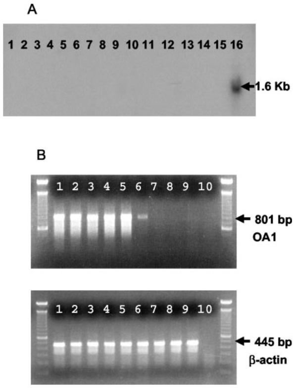
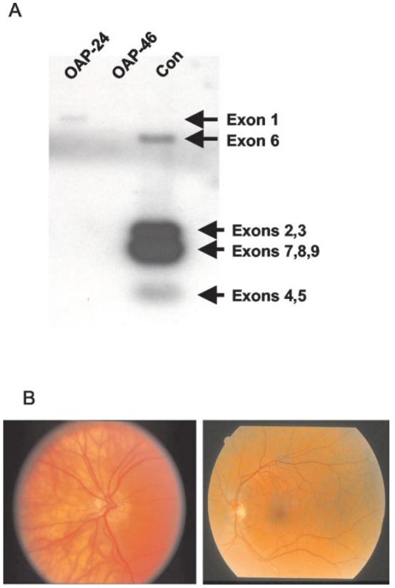
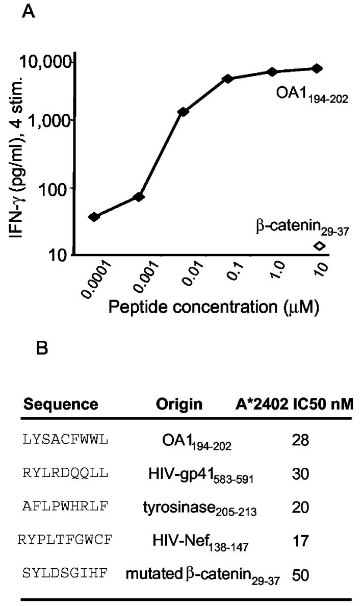
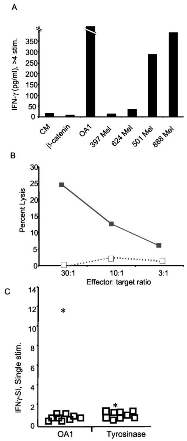
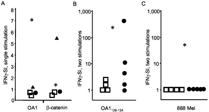
Antitumor T cells often recognize targets that are nonmutated "self" tissue differentiation Ags, but the relative impact of Ag expression by normal and transformed tissue for a human self/tumor Ag has not been studied. To examine the influence of self-tolerance mechanisms on the function of self/tumor-specific T cell responses in humans, we sought to identify an Ag that was expressed, processed, and presented in an MHC-restricted fashion by tumor cells, but for which there was the human equivalent of a "knockout." In this study, we report the first immunological characterization of a melanoma/melanocyte differentiation Ag, called OA1, which meets these criteria. This Ag, an X chromosome-encoded melanoma/melanocyte differentiation Ag, was completely deleted in a male patient. Using a newly identified HLA-A*2402-restricted epitope (LYSACFWWL) to study T cell tolerance, we found that OA1-specific T cell reactivity was more than five SD higher in the knockout patient that in normal controls. These data provide compelling evidence for T cell tolerance to OA1 in humans. Most surprisingly, we found elevated levels of OA1-specific T cells in patients with metastatic malignant melanoma, indicating that the tumor-bearing state partially reversed tolerance observed in normal (non-"knockout") individuals. Taken together, these findings indicated that tolerance can exist for self/tumor Ags in humans, and that this tolerance could be partially abrogated by the growth of the tumor, increasing the reactivity of tumor Ag-specific T cells. Thus, the tumor-bearing state reverses, in part, the tolerance of T cells that results from the normal expression of tissue differentiation Ags.


Figures

FIGURE 1
FIGURE 1
Analysis of OA1 in normal tissues and in tumor. A, Northern blot demonstrates specific expression of OA1 in melanocyte mRNA. Total RNA from 16 normal tissues (brain, heart, kidney, liver, lung, mam-mary gland, pancreas, placenta, prostate, skeletal muscle, spleen, stomach, testis, thymus, uterus, and melanocyte; lanes 1–16) was assayed with a full-length OA1 32P-labeled probe. Specific hybridization at 1.6 kb was observed for melanocyte mRNA only (lane 16). B, OA1 gene expression in tumor cells. RT-PCR was performed with OA1- and β-actin-specific primers on six different melanoma lines (397, 501, 526, 624, 888, 1102; lanes 1–6), one breast carcinoma line (MDA-231; lane 7), one colon carcinoma line (SW-480; lane 8), and one EBV-B line (1359 EBV-B; lane 9). Specific amplification of OA1 was observed (801-bp product) for all melanoma lines, but not for nonmelanoma lines and control water sample (lane 10). Control β-actin expression (445-bp product) was observed for all samples.
FIGURE 2
FIGURE 2
Analysis of genotype and phenotype of an Ag-loss variant patient. A, Volunteer OAP-46 has a full-length OA1 gene deletion. Southern blot of PstI-digested DNA from OA1 patients and a control male, separated electrophoretically, then hybridized with a full-length 32P-labeled OA1 cDNA probe. Patient OAP-46 had a full-length deletion spanning exons 1–9 confirming previous findings (17). All samples were positive for the β-actin probe (not shown). Note that hybridization with Exon 1 is only visible in the sample from patient OAP-24 (who was excluded from further immunological analysis), but not the sample from the control volunteer, presumably because the exons that are present on the blot compete for the labeled probe. The fragment from Exon 9 in the unused subject OAP-24, although present in this patient (as reported elsewhere, Ref.17), is not visible in the blot shown here. B, Fundoscopic photograph of an OA1-Ag-loss variant volunteer OAP-46 (left panel) showing characteristic hypopigmentation on retinal exam. That of a normal volunteer is shown on the right panel for comparison.
FIGURE 3
FIGURE 3
A, Recognition of OA1126–134 peptide-pulsed A*2402+ EBV-B cells (888 EBV-B) by CD8+ T cell line SE OA1-3 2/6, following the fourth stimulation. B, The binding affinity of the OA1 epitope to HLAA*2402 was equivalent to both HIV-derived peptides and both melanoma-derived peptides (mutated β-catenin and tyrosinase).
FIGURE 4
FIGURE 4
T cells from the OA1 Ag-loss variant patient avidly respond to OA1 and recognize melanoma. A, Long-term CD8+ T cells from line SE OA1-3 2/6, generated by four or more in vitro stimulations with OA1126–134, recognize OA1 peptide and A*2402+/OA1+ melanoma lines (501 and 888 Mel), but not A*2402/OA1+ melanoma lines (397 and 624 Mel), nor the control peptide (mutated β-catenin29–37) pulsed onto 888 EBV-B. B, T cell line obtained by repeated stimulation of PBMC from patient OAP-46 with peptide OA1126–134 lyse A*2402+/OA1+ melanoma 888 (■), but not A*2402/OA1+ melanoma 624 mel (□) in a 4-hour 51Cr-release assay. C, T cells from patient OAP-46 have superior reactivity to OA1. T cells from patient OAP-46 react with OA1126–134, but not tyrosinase 205–213, while T cells from normal volunteers and melanoma patients lack reactivity to both Ags. PBMC from nine normal volunteers (□) and OA1 patient OAP-46 (*) were stimulated for 7 days in vitro with either OA1126–134 or tyrosinase 205–213. To evaluate responses from all samples to both Ags, T cells were stimulated with OKT3 (anti-CD3), control peptide (mutated β-catenin 29–37 pulsed at 10 μM), or the specific peptide in question (either OA1126–134 or tyrosinase 205–213 also pulsed at 10 μM) both pulsed onto 888 EBV-B. All lines secreted IFN-γ in response to OKT3 (not shown). The IFN-γ-SI was generated by dividing the IFN-γ secretion produced to OA1126–134 or tyrosinase 205–213 against the secretion produced against mutated β-catenin 29–37. Experiments in all panels were performed three times with similar results, using different but comparable targets to demonstrate the generalizability of the findings. CM, culture media.
FIGURE 5
FIGURE 5
Ag expressed by tumor primes low-avidity T cells. A, T cell cultures generated after a single in vitro stimulation are peptide reactive: T cells from patient MG react with mutated β-catenin 29–37 and cells from patient OAP-46 react with OA1126–134. PBMC from melanoma patient MG (▲), OA1 KO patient OAP-46 (*), melanoma patient SE (●), and two normal volunteers (□) were stimulated for 7 days in vitro with mutated β-catenin 29–37. To evaluate responses from all samples, T cells were stimulated with either OKT3 1 μg/ml, control peptide (tyrosinase 205–213) or peptide. The IFN-γ-SI was generated by dividing the IFN-γ secretion produced to mutated β-catenin29–37against the secretion produced against the control peptide (tyrosinase 205–213). B, OA1-specific T cells generated by two in vitro OA1 peptide stimulations from melanoma patients can react to OA1 peptide. A comparative in vitro sensitization assay was performed with PBMC from four DR4+ normal volunteers (□), five DR4+ melanoma patients (●), and the OA1 KO patient (*) as a positive control. IFN-γ release was then measured in a 24-h coculture with EBV-B cells pulsed with β-catenin or OA1-peptide. The IFN-γ-SI was calculated based on the background reactivity to β-catenin at 10 μg/ml. C, The same OA1-specific T cells from melanoma patients, stimulated two times in vitro, do not recognize melanoma. T cells from the same individuals as above were stimulated in vitro with OA1126–134 as described in B and their reactivity to 888 mel was measured in a 24-h coculture assay followed by IFN-γ ELISA. The IFN-γ-SI was calculated based on the background-reactivity to the A*2402 line 624 mel. Experiments in all panels were performed between two and four times with similar results, using different but comparable targets to demonstrate the generalizability of the findings.

References

    1. Overwijk WW, Lee DS, Surman DR, Irvine KR, Touloukian CE, Chan CC, Carroll MW, Moss B, Rosenberg SA, Restifo NP. Vaccination with a recombinant vaccinia virus encoding a “self” antigen induces autoimmune vitiligo and tumor cell destruction in mice: requirement for CD4+ T lymphocytes. Proc. Natl. Acad. Sci. USA. 1999;96:2982. - PMC - PubMed
    1. Rosenberg SA. Progress in human tumour immunology and immuno-therapy. Nature. 2001;411:380. - PubMed
    1. Overwijk WW, Tsung A, Irvine KR, Parkhurst MR, Goletz TJ, Tsung K, Carroll MW, Liu C, Moss B, Rosenberg SA, Restifo NP. gp100/pmel 17 is a murine tumor rejection antigen: induction of “self”-reactive, tumoricidal T cells using high-affinity, altered peptide ligand. J. Exp. Med. 1998;188:277. - PMC - PubMed
    1. Staveley-O'Carroll K, Sotomayor E, Montgomery J, Borrello I, Hwang L, Fein S, Pardoll D, Levitsky H. Induction of antigen-specific T cell anergy: an early event in the course of tumor progression. Proc. Natl. Acad. Sci. USA. 1998;95:1178. - PMC - PubMed
    1. Mizoguchi H, O'Shea JJ, Longo DL, Loeffler CM, McVicar DW, Ochoa AC. Alterations in signal transduction molecules in T lymphocytes from tumor-bearing mice. Science. 1992;258:1795. - PubMed

MeSH terms