. 2003 Feb 4;100(3):916-21.
doi: 10.1073/pnas.242741799. Epub 2003 Jan 21.

Oncogenic targeting of an activated tyrosine kinase to the Golgi apparatus in a glioblastoma

Affiliations

Oncogenic targeting of an activated tyrosine kinase to the Golgi apparatus in a glioblastoma

Alan Charest et al. Proc Natl Acad Sci U S A. .

Abstract

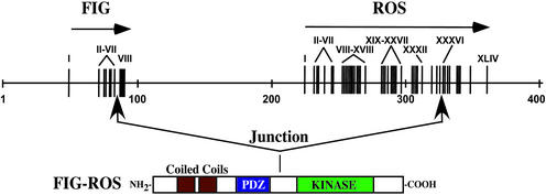
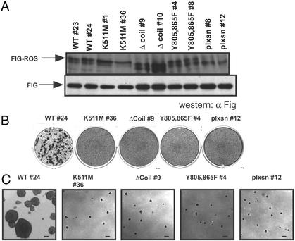
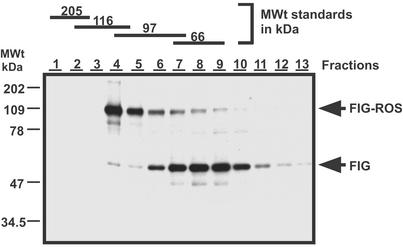
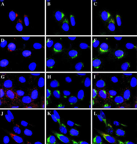
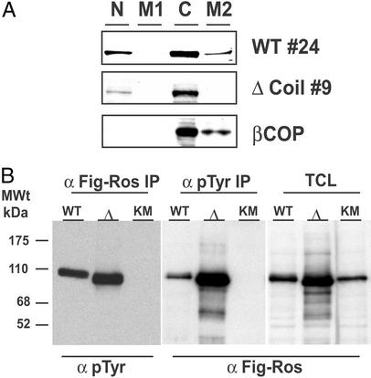
Activating oncogenic mutations of receptor tyrosine kinases (RTKs) have been reported in several types of cancers. In many cases, genomic rearrangements lead to the fusion of unrelated genes to the DNA coding for the kinase domain of RTKs. All RTK-derived fusion proteins reported so far display oligomerization sequences within the 5' fusion partners that are responsible for oncogenic activation. Here, we report a mechanism by which an altered RTK gains oncogenic potential in a glioblastoma cell line. A microdeletion on 6q21 results in the fusion of FIG, a gene coding for a Golgi apparatus-associated protein, to the kinase domain of the protooncogene c-ROS. The fused protein product FIG-ROS is a potent oncogene, and its transforming potential resides in its ability to interact with and become localized to the Golgi apparatus. Thus we have found a RTK fusion protein whose subcellular location leads to constitutive kinase activation and results in oncogenic transformation.


Figures

Figure 1
Figure 1
The ROS gene is rearranged in a human glioblastoma cell line. Shown is a schematic representation of FIG and ROS genomic DNA locus along with the predicted U118MG-derived FIG-ROS fusion hybrid protein. Numbered vertical bars represent exons for each gene.
Figure 2
Figure 2
FIG-ROS is an active oncogene. Rat1 cells infected with retroviral stocks carrying the indicated FIG-ROS variants were clonally derived after G418/neomycin drug selection. Two cell lines from each FIG-ROS isoform were selected for analysis. (A) Western blot of total cell lysates from cell lines expressing the indicated FIG-ROS variants incubated with an anti-FIG antibody recognizing p109FIG-ROS (Upper) and endogenous p59FIG (Lower). (B and C) Analysis of loss of contact inhibition (B) and anchorage-independent growth (C) of cell lines expressing FIG-ROS variants. Shown are single picture of triplicates for a single clone of each FIG-ROS variant. (Scale bar = 250 μm.)
Figure 3
Figure 3
Both p59FIG and p109FIG-ROS exist as monomers in vivo. Cells expressing FIG-ROS were lysed under nondenaturing conditions and proteins were separated on 5–20% sucrose gradients. Fractions were collected and FIG and FIG-ROS proteins were detected by Western blot analysis using an anti-FIG antibody as described in Materials and Methods. The numbers above each lane correspond to fraction numbers from the heaviest to the lightest portion of the gradient. Molecular size standards were included in a control gradient run in parallel and the fractions at which they eluted are indicated at the top.
Figure 4
Figure 4
The second coiled-coil domain of FIG-ROS is necessary for targeting to the Golgi apparatus. Shown are indirect immunofluorescence micrographs of clonally derived Rat1 cells expressing the following FIG-ROS variants: unmodified wild-type no. 24 (A–C), K511M no. 1 (D–F), Δcoil no. 4 (G–I), and Y805,865F no. 1 (J–L). Cells were fixed and costained with the anti-c-Myc 9E10 epitope tag antibody (A, D, G, and J) and a rabbit anti-β-COP polyclonal antibody (B, E, H, and K) as described in Materials and Methods. The two sets were merged (C, F, I, and L) to indicate colocalization (yellow) of FIG-ROS and the Golgi apparatus marker β-COP. Nuclei are stained with 4′,6-diamidino-2-phenylindole (DAPI; blue).
Figure 5
Figure 5
The Δcoil FIG-ROS mutant displays kinase activity and does not sediment with a Golgi membrane-containing fraction. Cell extracts from retrovirus-infected Rat1 clones were subjected to differential centrifugation cellular fractionation followed by Western blotting with an anti-FIG antibody as described in Materials and Methods (A) and immunoprecipitated with the indicated antibodies and Western blotted for the presence of phosphotyrosine residues (pTyr) and FIG-ROS as indicated (B). N, nuclear and unlysed low-speed fraction; M1, intermediate-speed plasma membrane-containing fraction; C, cytosolic fraction; M2, high-speed Golgi membrane-containing fraction; IP, immunoprecipitate; TCL, total cell lysate; Δ, Δcoil FIG-ROS isoform; KM, K511M FIG-ROS isoform.

References

    1. Blume-Jensen P, Hunter T. Nature. 2001;411:355–365. - PubMed
    1. Lamorte L, Park M. Surg Oncol Clin North Am. 2001;10:271–288. , viii. - PubMed
    1. Sonnenberg E, Godecke A, Walter B, Bladt F, Birchmeier C. EMBO J. 1991;10:3693–3702. - PMC - PubMed
    1. Sonnenberg-Riethmacher E, Walter B, Riethmacher D, Godecke S, Birchmeier C. Genes Dev. 1996;10:1184–1193. - PubMed
    1. Liu Z Z, Wada J, Kumar A, Carone F A, Takahashi M, Kanwar Y S. Dev Biol. 1996;178:133–148. - PubMed

Publication types

MeSH terms

LinkOut - more resources