. 2003 Feb;71(2):597-604.
doi: 10.1128/IAI.71.2.597-604.2003.

Plasmodium falciparum infection elicits both variant-specific and cross-reactive antibodies against variant surface antigens

Affiliations

Plasmodium falciparum infection elicits both variant-specific and cross-reactive antibodies against variant surface antigens

Rana Chattopadhyay et al. Infect Immun. 2003 Feb.

Abstract

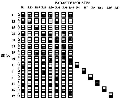
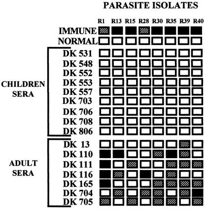
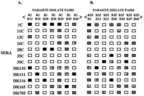
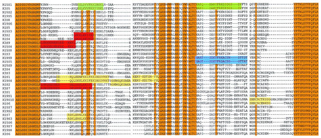
Naturally acquired antibodies to Plasmodium falciparum erythrocyte membrane protein-1 (PfEMP-1), the variant surface antigens expressed on the surface of infected erythrocytes, are thought to play a role in protection against P. falciparum malaria. Here, we have studied the development of antibodies to PfEMP-1 in adult malaria patients living in Rourkela, India, an area with a low malaria transmission rate, and prevalence of antibodies to PfEMP-1 in residents of San Dulakudar, India, a village in which P. falciparum malaria is hyperendemic. Convalescent-phase sera from adult malaria patients from Rourkela agglutinate homologous P. falciparum isolates as well as some heterologous isolates, suggesting that they develop partially cross-reactive antibodies to PfEMP-1 following infection. Adult sera from San Dulakudar agglutinate diverse P. falciparum isolates, suggesting that they have antibodies with wide recognition of diverse PfEMP-1. Mixed-agglutination assays using pairs of P. falciparum isolates confirm the presence of both variant-specific and partially cross-reactive antibodies in convalescent-phase sera from Rourkela and adult sera from San Dulakudar. Analysis of PfEMP-1 sequences suggests a molecular basis for the observed cross-reactivity.


Figures

FIG. 1.
FIG. 1.
Agglutination of P. falciparum field isolates with acute- and convalescent-phase sera from adult malaria patients residing in Rourkela, Orissa, India, a town with low malaria transmission rates. Acute-phase (A) and convalescent-phase (C) sera collected from adult malaria patients residing in Rourkela, a town with low malaria transmission, were used for agglutination assays with homologous and heterologous P. falciparum isolates. Blank boxes indicate that no agglutinates of three or more infected erythrocytes were seen. Hatched boxes indicate that on average five or less agglutinates containing five or more P. falciparum-infected red cells per agglutinate were seen per field. Black boxes indicate that on average greater than five agglutinates containing five or more infected red cells were seen per field. Twenty-five fields were examined at ×40 magnification for each agglutination assay.
FIG. 2.
FIG. 2.
Agglutination of P. falciparum field isolates with sera from children and adults from San Dulakudar, Orissa, India, a village where P. falciparum malaria is hyperendemic. Sera from children and adults residing in San Dulakudar, where P. falciparum malaria is hyperendemic, were used for agglutination assays with diverse P. falciparum isolates. Pooled sera from adults living in another area in Orissa in which P. falciparum was endemic were used as a positive control (Immune). Pooled sera from adults residing in regions of India where malaria was not endemic were used as a negative control (Normal). Twenty-five fields were examined at ×40 magnification for each agglutination assay. Blank, hatched, and black boxes indicate the frequency of agglutination as described for Fig. 1.
FIG. 3.
FIG. 3.
Mixed-agglutination assays with P. falciparum field isolates. (A and B) P. falciparum isolates were tested in mixed-agglutination assays in combination with parasite isolates R1 and R35 and convalescent-phase sera from adult malaria patients (1C, 11C, 13C, 16C, 28C, 35C, and 39C) residing in Rourkela, a town with a low malaria transmission rate, or with sera from adults residing in San Dulakudar (DK110, DK111, DK116, DK165, and DK705), a village where P. falciparum malaria was hyperendemic. Mixed-color agglutinates were defined as agglutinates with at least three infected erythrocytes, with at least one erythrocyte of each color. Blank boxes indicate cases where no mixed-color agglutinates were found. Blank boxes marked with an asterisk indicate cases where no mixed-color agglutinates were found, even though both isolates are individually recognized. Mixed-color agglutinates containing up to 20 P. falciparum-infected erythrocytes with at least two infected erythrocytes of each color labeled were scored in 25 fields at ×40 magnification. Hatched boxes indicate that on average one to five such mixed-color agglutinates are observed per field, and black boxes indicate that on average more than five such mixed-color agglutinates are observed per field. Mixed-color agglutinates constitute 20 to 30% of all agglutinates in cases where mixed-color agglutinates are observed.
FIG. 4.
FIG. 4.
Multiple alignment of DBLα sequences from Indian and Kenyan P. falciparum isolates. A multiple-sequence alignment of DBLα sequences derived from var genes from two Indian P. falciparum isolates from Rourkela, R1 (R1S1 to R1S7) and R15 (R15S1 to R15S9), and two Kenyan P. falciparum isolates, K3 (K3S1 to K3S9) and K19 (K19S1 to K19S10, is shown. Orange boxes indicate conserved stretches of either identical or highly conserved residues shared by all var genes. Colored boxes indicate short segments of at least 10-amino-acid residues with greater than 70% identity within hypervariable regions that are common to some var genes from heterologous Indian and Kenyan isolates. Blue boxes indicate sequences common to R15 and K19 and red boxes indicate sequences common to R1 and K3, yellow boxes indicate sequences common to R1 and K19, and green boxes indicate sequences common to R1 and K3, yellow boxes indicate sequences common to R15 and K19, and green boxes indicate sequences common to R15 and K3..

Comment in

References

    1. Aguiar, J. C., G. R. Albrecht, P. Cegielski, B. M. Greenwood, J. B. Jensen, G. Lallinger, A. Martinez, I. A. McGregor, J. N. Minjas, J. Neequaye, M. E. Patarroyo, J. A. Sherwood, and R. J. Howard. 1992. Agglutination of Plasmodium falciparum-infected erythrocytes from east and west African isolates by human sera from distant geographic regions. Am. J. Trop. Med. Hyg. 47:621-632. - PubMed
    1. Baruch, D., B. Pasloske, H. Singh, B. Xiahui, X. Ma, M. Feldman, T. Taraschi, and R. J. Howard. 1995. Cloning of the Plasmodium falciparum gene encoding PfEMP1, a malarial variant antigen and cytoadherence receptor on the surface of parasitised human erythrocytes. Cell 82:77-87. - PubMed
    1. Bull, P., B. Lowe, M. Kortok, C. Molyneux, C. Newbold, and K. Marsh. 1998. Parasite antigens on the infected red cell surface are targets for naturally acquired immunity to malaria. Nat. Med. 4:358-360. - PMC - PubMed
    1. Bull, P., B. Lowe, M. Kortok, and K. Marsh. 1999. Antibody recognition of Plasmodium falciparum erythrocyte surface antigens in Kenya: evidence for rare and prevalent variants. Infect. Immun. 67:733-739. - PMC - PubMed
    1. Chitnis, C. E., and L. H. Miller. 1994. Identification of the erythrocyte binding domains of Plasmodium vivax and Plasmodium knowlesi proteins involved in erythrocyte invasion. J. Exp. Med. 180:497-506. - PMC - PubMed

Publication types

MeSH terms

Substances