. 2003 Feb;71(2):717-25.
doi: 10.1128/IAI.71.2.717-725.2003.

Oral treponemes and their outer membrane extracts activate human gingival epithelial cells through toll-like receptor 2

Affiliations

Oral treponemes and their outer membrane extracts activate human gingival epithelial cells through toll-like receptor 2

Yasuyuki Asai et al. Infect Immun. 2003 Feb.

Abstract

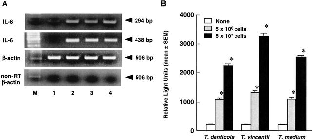
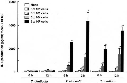
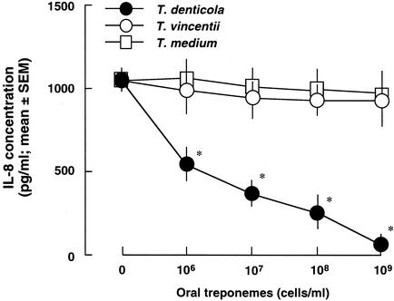
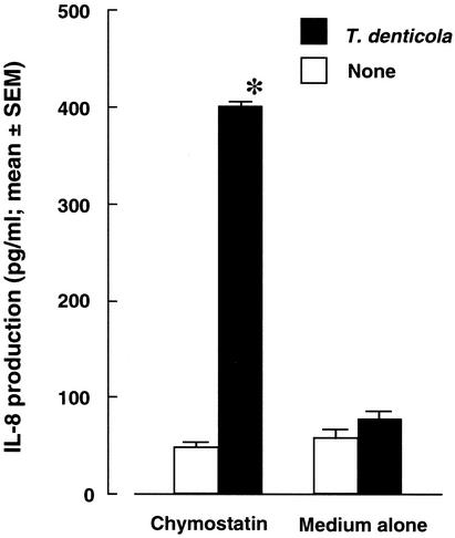
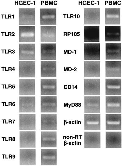
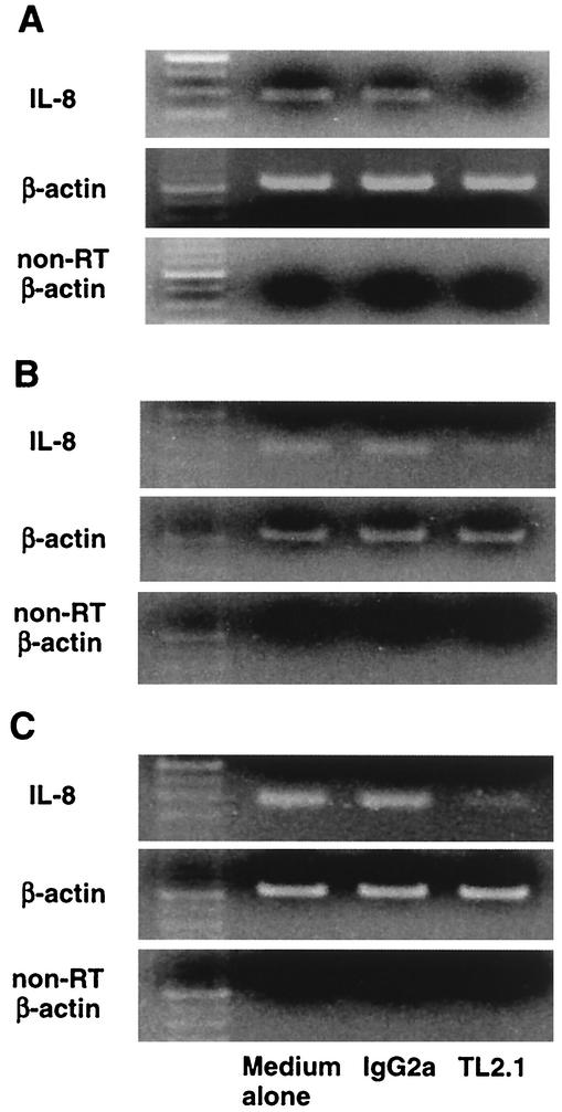
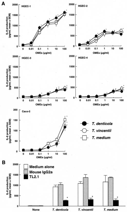
Oral treponemes are considered to be important in the development and progression of periodontal diseases. We investigated the mechanisms of recognition and activation of human gingival epithelial cells (HGEC) with the oral treponemes Treponema denticola, Treponema vincentii, and Treponema medium and their outer membrane extracts (OMEs). T. vincentii and T. medium but not T. denticola produced interleukin 8 (IL-8) in an HGEC culture. Further, all three treponemes induced IL-8 mRNA expression and NF-kappaB activation in HGEC. Among them, T. denticola especially exhibited trypsin- and chymotrypsin-like protease activities, and the addition of chymostatin, a chymotrypsin protease inhibitor, resulted in detectable IL-8 production by HGEC cultured with T. denticola. Additionally, IL-8 mRNA expression in HGEC cultured with the three treponemes and their OMEs was definitely inhibited by the mouse anti-human Toll-like receptor 2 (TLR2) monoclonal antibody TL2.1. These findings suggest that oral treponemes and their OMEs activate HGEC through TLR2.


Figures

FIG. 1.
FIG. 1.
Cytokine mRNA expression and NF-κB activation of HGEC after culturing with oral treponemes. (A) IL-8 and IL-6 mRNAs of HGEC-1 cultured with 5 × 10⁷ cells of oral treponemes for 2 h were analyzed by RT-PCR. β-Actin was assayed as a positive control. PCR products of non-reverse-transcribed (non-RT) samples were examined as a negative control. Lane 1, medium alone; lane 2, T. denticola; lane 3, T. vincentii; lane 4, T. medium; lane M, size marker. Experiments were done at least three times, and representative results are presented. (B) HGEC-1 were transfected with 1 μg of plasmid pNF-κB-Luc and then cultured with the indicated cells of oral treponemes in HuMedia-KG2 at 37°C for 4 h. After incubation, luciferase activity was estimated with a Luminometer. Experiments were done at least three times, and representative results are presented. Each assay was done with triplicate wells, and the data are expressed as the means ± SEM. Asterisks indicate significant differences seen between groups with and without the oral treponemes (P < 0.01).
FIG. 2.
FIG. 2.
IL-8 production by HGEC after culturing with oral treponemes. HGEC-1 were cultured with the indicated amounts of T. denticola, T. vincentii, or T. medium in HuMedia-KG2 at 37°C for 6 and 12 h. After incubation, supernatants were collected, and IL-8 production was determined by an ELISA. Experiments were done at least three times, and representative results are presented. Each assay was done with triplicate wells, and the data are expressed as the means ± SEM. Asterisks indicate significant differences seen between groups with and without oral treponemes (P < 0.01).
FIG. 3.
FIG. 3.
Degradation of PMA-induced IL-8 in HGEC. HGEC-1 were incubated with 10 ng of PMA/ml for 24 h, and supernatants were collected. The indicated doses of oral treponemes were cultured with 1 ml of these supernatants at 37°C for 24 h in humidified air containing 5% (vol/vol) CO2. After incubation, IL-8 production was determined by an ELISA. Experiments were done at least three times, and representative results are presented. Each assay was done with triplicate wells, and the data are expressed as the means ± SEM. Asterisks indicate significant differences seen between groups with and without oral treponemes (P < 0.01).
FIG. 4.
FIG. 4.
Effect of chymostatin on IL-8 production by HGEC after culturing with T. denticola. Bacterial cells were washed three times with or without 30 μg of chymostatin in PBS/ml and then suspended at a density of 107 cells/ml in HuMedia-KG2 with or without 30 μg of chymostatin/ml. HGEC-1 were cultured with 107 cells of T. denticola treated or not treated with chymostatin at 37°C for 12 h. After incubation, supernatants were collected, and IL-8 production was determined by an ELISA. Experiments were done at least three times, and representative results are presented. Each assay was done with triplicate wells, and the data are expressed as the means ± SEM. Asterisks indicate significant differences seen between groups with and without T. denticola (P < 0.01).
FIG. 5.
FIG. 5.
TLR mRNA expression of HGEC. The expression of human TLR mRNA was analyzed by RT-PCR as detailed in Materials and Methods. Human PBMC were used as a positive source of TLR mRNA expression to confirm the specificity of the primers and the accuracy of the assay. The β-actin gene was assayed as a positive control. PCR products of non-reverse-transcribed (non-RT) samples were examined as a negative control. M, size marker. Experiments were done at least three times, and representative results are presented.
FIG. 6.
FIG. 6.
Effect of a mouse anti-human TLR2 monoclonal antibody on the induction of IL-8 mRNA expression in HGEC after stimulation with oral treponemes. HGEC-1 were preincubated with or without a mouse monoclonal antibody to human TLR2 (TL2.1) or mouse IgG2a at 1 μg/ml for 30 min at room temperature and then cultured with 5 × 10⁷ cells of T. denticola (A), T. vincentii (B), or T. medium (C) for 2 h. β-Actin was assayed as a positive control. PCR products of non-reverse-transcribed (non-RT) samples were examined as a negative control. Experiments were done at least three times, and representative results are presented.
FIG.7.
FIG.7.
Induction of IL-8 production by HGEC stimulated with OMEs from oral treponemes. (A) HGEC and Caco-2 cells were stimulated with the indicated doses of the OMEs at 37°C for 12 h. After incubation, supernatants were collected, and IL-8 production was determined by an ELISA. (B) HGEC-1 were incubated with or without a mouse monoclonal antibody to human TLR2 (TL2.1) or mouse IgG2a at 1 μg/ml for 30 min at room temperature and then stimulated with 1 μg of the OMEs/ml for 12 h. After incubation, supernatants were collected, and IL-8 production was determined by an ELISA. Experiments were done at least three times, and representative results are presented. Each assay was done with triplicate wells, and the data are expressed as the means ± SEM. Asterisks indicate significant differences seen between groups with and without the test specimens (P < 0.01).

References

    1. Alexopoulou, L., A. C. Holt, R. Medzhitov, and R. A. Flavell. 2001. Recognition of double-stranded RNA and activation of NF-kappaB by Toll-like receptor 3. Nature 413:732-738. - PubMed
    1. Aliprantis, A. O., R. B. Yang, M. R. Mark, S. Suggett, B. Devaux, J. D. Radolf, G. R. Klimpel, P. Godowski, and A. Zychlinsky. 1999. Cell activation and apoptosis by bacterial lipoproteins through Toll-like receptor-2. Science 285:736-739. - PubMed
    1. Asai, Y., T. Jinno, H. Igarashi, Y. Ohyama, and T. Ogawa. 2002. Detection and quantification of oral treponemes in subgingival plaque by real-time PCR. J. Clin. Microbiol. 40:3334-3340. - PMC - PubMed
    1. Asai, Y., Y. Ohyama, K. Gen, and T. Ogawa. 2001. Bacterial fimbriae and their peptides activate human gingival epithelial cells through Toll-like receptor 2. Infect. Immun. 69:7387-7395. - PMC - PubMed
    1. Bartold, P. M., L. J. Walsh, and A. S. Narayanan. 2000. Molecular and cell biology of the gingiva. Periodontology 24:28-55. - PubMed

Publication types

MeSH terms

LinkOut - more resources