

Heterogeneity of mycolactones produced by clinical isolates of Mycobacterium ulcerans: implications for virulence
- PMID: 12540557
- PMCID: PMC145382
- DOI: 10.1128/IAI.71.2.774-783.2003
Heterogeneity of mycolactones produced by clinical isolates of Mycobacterium ulcerans: implications for virulence
Abstract
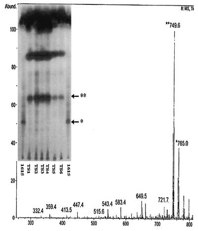
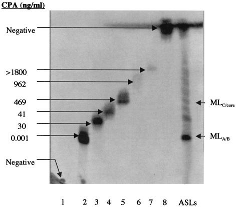
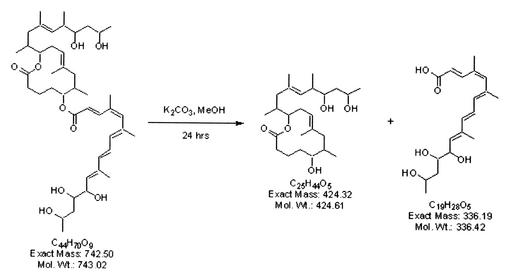
Mycobacterium ulcerans is the causative agent of Buruli ulcer, a severe necrotizing skin disease endemic in tropical countries. Clinical evidence suggests that M. ulcerans isolates from Asia, Mexico, and Australia may be less virulent than isolates from Africa. In vivo studies suggest that mycolactone, a polyketide-derived macrolide toxin, plays a major role in the tissue destruction and immune suppression which occur in cases of Buruli ulcer. Mycolactones were extracted from 34 isolates of M. ulcerans representing strains from Africa, Malaysia, Asia, Australia, and Mexico. Thin-layer chromatography, mass spectroscopic analysis, and cytopathic assays of partially purified mycolactones from these isolates revealed that M. ulcerans produces a heterogeneous mixture of mycolactone variants. Mycolactone A/B, the most biologically active mycolactone species, was identified by mass spectroscopy as [M(+)Na](+) at m/z 765.5 in all cytotoxic isolates except for those from Mexico. Mycolactone C [M+Na](+) at m/z 726.3 was the dominant mycolactone species in eight Australian isolates, and mycolactone D [M+Na](+) m/z 781.2 was characteristic of two Asian strains. Mycolactone species are conserved within specific geographic areas, suggesting that there may be a correlation between mycolactone profile and virulence. In addition, the core lactone, [M+Na](+) m/z 447.4, was identified as a minor species, supporting the hypothesis that mycolactones are synthesized by two polyketide synthases. A cytopathic assay of the core lactone showed that this molecule is sufficient for cytotoxicity, although it is much less potent than the complete mycolactone.
Figures

References
-
- Benowitz, A. B., S. Fidanze, P. L. Small, and Y. Kishi. 2001. Stereochemistry of the core structure of the mycolactones. J. Am. Chem. Soc. 123:5128-5129. - PubMed
-
- Cadapan, L. D., R. L. Arslanian, J. R. Carney, S. M. Zavala, P. L. Small, and P. Licari. 2001. Suspension cultivation of Mycobacterium ulcerans for the production of mycolactones. FEMS Microbiol. Lett. 205:385-389. - PubMed
-
- Connor, D. H., and H. F. Lunn. 1965. Mycobacterium ulcerans infection. Int. J. Lepr. 33:698-709. - PubMed
-
- Daffé, M. D., A. Varnerot, and V. V. Levy-Frebault. 1992. The phenolic mycoside of Mycobacterium ulcerans: structure and taxonomic implications. J. Gen. Microbiol. 138:131-137. - PubMed
-
- Dodge, O. G. 1964. Mycobacterial skin ulcers in Uganda; histopathological and experimental aspects. J. Pathol. Bacteriol. 88:167-174. - PubMed
Publication types
MeSH terms
Substances
LinkOut - more resources
Full Text Sources
Other Literature Sources
Medical
