. 2003 Feb 4;100(3):1352-7.
doi: 10.1073/pnas.242738899. Epub 2003 Jan 22.

Photochemical gating of heterologous ion channels: remote control over genetically designated populations of neurons

Affiliations

Photochemical gating of heterologous ion channels: remote control over genetically designated populations of neurons

Boris V Zemelman et al. Proc Natl Acad Sci U S A. .

Abstract

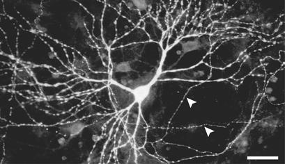
Heterologous proteins capable of transducing physical or chemical stimuli into electrical signals can be used to control the function of excitable cells in intact tissues or organisms. Restricted genetically to circumscribed populations of cellular targets, these selectively addressable sources of depolarizing current can supply distributed inputs to neural circuits, stimulate secretion, or regulate force and motility. In an initial demonstration of this principle, we have used elements of a G protein coupled signaling system, the phototransduction cascade of the fruit fly, to sensitize generalist vertebrate neurons to light [Zemelman, B. V., Lee, G. A., Ng, M. & Miesenböck, G. (2002) Neuron 33, 15-22]. We now describe the use of ectopically expressed ligand-gated ion channels as transducers of optical or pharmacological stimuli. When either the capsaicin receptor, TRPV1, the menthol receptor, TRPM8, or the ionotropic purinergic receptor P2X(2) was introduced into hippocampal neurons, the cells responded to pulsed applications of agonist with characteristic sequences of depolarization, spiking, and repolarization. Responses required cognate matches between receptor and agonist, peaked at firing frequencies of approximately 40 Hz, initiated and terminated rapidly, and did not attenuate. Precise dose-response relationships allowed current amplitudes and firing frequencies to be tuned by varying the concentration of ligand. Agonist could be administered either pharmacologically or, in the cases of TRPV1 and P2X(2), optically, through photorelease of the active compounds from the respective "caged" precursors, 4,5-dimethoxy-2-nitrobenzyl-capsaicin and P(3)-[1-(4,5-dimethoxy-2-nitrophenyl)ethyl]-ATP.


Figures

Figure 1
Figure 1
Synthesis and structure of DMNB-capsaicin. The DMNB chromophore is attached to the phenolic hydroxyl function of capsaicin, a molecular feature important for agonist activity.
Figure 2
Figure 2
Expression of TRPV1 in cultured hippocampal neurons. Dissociated cultures of hippocampal neurons were immunostained with an antibody against TRPV1. TRPV1 protein is detected on the soma and dendrites of a transfected neuron (identified by coexpression of GAP43-EGFP; not shown), as well as on several axons that traverse the field of view (white arrowheads). (Bar = 20 μm.)
Figure 3
Figure 3
Pharmacological stimulation of genetically designated target neurons. The membrane potentials of hippocampal neurons expressing TRPV1 (Top), TRPM8 (Middle), and P2X2 (Bottom) were recorded in whole-cell current-clamp mode. All neurons were challenged sequentially with 50 nM capsaicin (Left), 100 μM menthol (Center), and 50 μM ATP (Right); approximate periods of agonist application are indicated by horizontal bars. Only cognate matches between ionotropic receptor and agonist (shaded entries in the diagonal) elicit action potentials. Increased baseline noise of the TRPV1-expressing neuron after the application of capsaicin (Top, Center and Right) is caused by residual capsaicin in the perfusion system.
Figure 4
Figure 4
Dose dependence of the capsaicin response in TRPV1-expressing neurons. (A) Transmembrane currents of a TRPV1-expressing hippocampal neuron were recorded in whole-cell voltage-clamp mode, at a constant holding potential of −65 mV and variable concentrations of capsaicin. Charge transfers as a function of capsaicin concentration were calculated by integrating the agonist-evoked currents over time (Inset: approximate periods of agonist presence are indicated by a horizontal bar). Depolarizing currents are depicted as downward deflections from baseline; charge transfers and corresponding current traces are drawn in identical shades of gray. (B) Membrane potentials of a TRPV1-expressing hippocampal neuron were recorded in whole-cell current-clamp mode, at the indicated concentrations of capsaicin. Capsaicin was perfused into the recording chamber between 200 and 400 ms and remained present thereafter.
Figure 5
Figure 5
Photostimulation of genetically designated target neurons. Membrane potentials of hippocampal neurons expressing TRPV1 (A, black trace) or P2X2 (B, black trace), or of untransfected control neurons (A and B, gray traces), were recorded in whole-cell current-clamp mode in the presence of 50 μM DMNB-capsaicin (A) or 1 mM DMNPE-ATP (B), under safelight conditions (shaded backgrounds). The neurons were illuminated twice for 1 s (white backgrounds) with the unfiltered output of a mercury arc lamp, which delivered an optical energy of 5.1 mJ in the spectral band <400 nm. Optical stimuli were followed by stereotyped electrical responses in neurons that expressed the cognate ionotropic receptor.

References

    1. Zemelman B V, Miesenböck G. Curr Opin Neurobiol. 2001;11:409–414. - PubMed
    1. Zemelman B V, Lee G A, Ng M, Miesenböck G. Neuron. 2002;33:15–22. - PubMed
    1. Hille B. Ion Channels of Excitable Membranes. Sunderland, MA: Sinauer; 2001.
    1. Stryer L. J Biol Chem. 1991;266:10711–10714. - PubMed
    1. Wickman K, Clapham D E. Physiol Rev. 1995;75:865–885. - PubMed

Publication types

MeSH terms

LinkOut - more resources