. 2003 Feb;47(2):588-93.
doi: 10.1128/AAC.47.2.588-593.2003.

The antifungal protein from Aspergillus giganteus causes membrane permeabilization

Affiliations

The antifungal protein from Aspergillus giganteus causes membrane permeabilization

T Theis et al. Antimicrob Agents Chemother. 2003 Feb.

Abstract

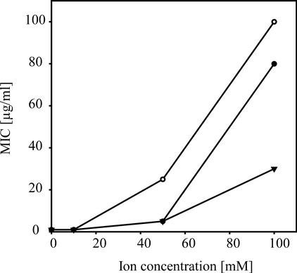
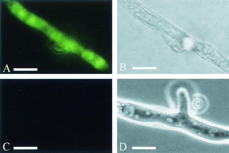
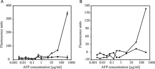
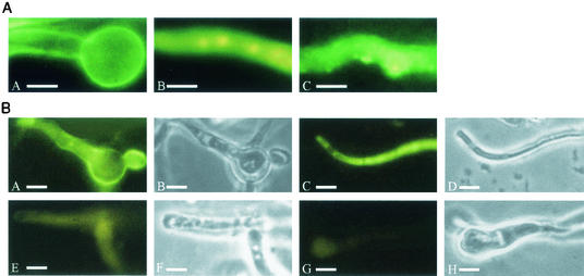
We investigated the inhibitory effects of the antifungal protein (AFP) from Aspergillus giganteus on the growth of several filamentous fungi. For this purpose, the MICs of AFP were determined and ranged from 0.1 micro g/ml for Fusarium oxysporum to 200 micro g/ml for Aspergillus nidulans. The antifungal activity of AFP was diminished in the presence of cations. We were able to show that incubation of AFP-sensitive fungi with the protein resulted in membrane permeabilization using an assay based on the uptake of the fluorescent dye SYTOX Green. No permeabilization by AFP could be detected at concentrations below the species-specific MIC. Furthermore, AFP-induced permeabilization could readily be detected after 5 min of incubation. Localization experiments with fluorescein isothiocyanate-labeled AFP and immunofluorescence staining with an AFP-specific antibody supported the observation that the protein interacts with membranes. After treatment of AFP-sensitive fungi with AFP, the protein was localized at the plasma membrane, whereas it was mainly detected inside the cells of AFP-resistant fungi. We conclude from these data that the growth-inhibitory effect of AFP is caused by permeabilization of the fungal membranes.


Figures

FIG. 1.
FIG. 1.
Effects of different ions on the MIC of AFP for A. niger. The MIC of AFP for A. niger was tested in the presence of different amounts of KH2PO4 (○), KCl (•), and NaCl (▴). The results of a representative experiment are shown.
FIG. 2.
FIG. 2.
Detection of AFP-induced SYTOX Green uptake by fluorescence microscopy. A. niger (A and B) and P. chrysogenum (C and D) were treated with 10 and 100 μg of AFP, respectively, in the presence of 0.2 μM SYTOX Green. After 1 h of incubation, SYTOX Green uptake was detected by fluorescence microscopy. (A and C) Fluorescence microscopy; (B and D) light-field microscopy. Bars, 15 μm.
FIG. 3.
FIG. 3.
Detection of AFP-induced SYTOX Green uptake. (A) A. niger (○) and P. chrysogenum (•) were incubated with different amounts of AFP in the presence of 0.2 μM SYTOX Green. Fluorescence was measured after 1 h of incubation. Data are averages of triplicate measures. (B) A. niger was incubated with different concentrations of AFP in the presence (•) or absence (○) of 100 mM KCl. Fluorescence was measured as described above. The results of a representative experiment are shown.
FIG. 4.
FIG. 4.
Localization of AFP within A. niger and P. chrysogenum. (A) FITC-labeled AFP. A. niger (panel A) and P. chrysogenum (panel B) were treated with 10 and 100 μg of FITC-labeled AFP ml, respectively. After 1 h of incubation, the labeled protein was detected by fluorescence microscopy. The control was A. niger treated with 10 μg of FITC-labeled α-sarcin per ml (panel C). Bars, 15 μm. (B) Immunofluorescence. A. niger (panels A and B) and P. chrysogenum (panels C and D) were treated with 10 μg of AFP per ml for 1 h. The protein was detected by immunofluorescence staining with an AFP-specific antibody. As negative controls, P. chrysogenum (panels E and F) and A. niger (panels G and H) were treated with the AFP-specific antibody in the absence of AFP. Panels A, C, E, and G, fluorescence microscopy; panels B, D, F, and H, light-field microscopy. Bars, 15 μm.

Similar articles

Cited by

References

    1. Bormann, C., D. Baier, I. Horr, C. Raps, J. Berger, G. Jung, and H. Schwarz. 1999. Characterization of a novel, antifungal, chitin-binding protein from Streptomyces tendae Tu901 that interferes with growth polarity. J. Bacteriol. 181:7421-7429. - PMC - PubMed
    1. Campos-Olivas, R., M. Bruix, J. Santoro, A. Martinez del Pozo, J. Lacadena, J. G. Gavilanes, and M. Rico. 1996. Structural basis for the catalytic mechanism and substrate specificity of the ribonuclease alpha-sarcin. FEBS Lett. 399:163-165. - PubMed
    1. Campos-Olivas, R., M. Bruix, J. Santoro, J. Lacadena, A. Martinez del Pozo, J. G. Gavilanes, and M. Rico. 1995. NMR solution structure of the antifungal protein from Aspergillus giganteus: evidence for cysteine pairing isomerism. Biochemistry 34:3009-3021. - PubMed
    1. Cociancich, S., A. Ghazi, C. Hetru, J. A. Hoffmann, and L. Letellier. 1993. Insect defensin, an inducible antibacterial peptide, forms voltage-dependent channels in Micrococcus luteus. J. Biol. Chem. 268:19239-19245. - PubMed
    1. De Lucca, A. J., and T. J. Walsh. 1999. Antifungal peptides: novel therapeutic compounds against emerging pathogens. Antimicrob. Agents Chemother. 43:1-11. - PMC - PubMed

Publication types

MeSH terms

LinkOut - more resources