

Verteporfin photodynamic therapy in highly myopic subfoveal choroidal neovascularisation
- PMID: 12543746
- PMCID: PMC1771483
- DOI: 10.1136/bjo.87.2.173
Verteporfin photodynamic therapy in highly myopic subfoveal choroidal neovascularisation
Abstract
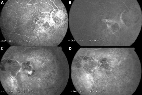
Aims: To analyse the visual and angiographic results of photodynamic therapy (PDT) with verteporfin in highly myopic patients with subfoveal choroidal neovascularisation (CNV).
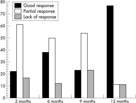
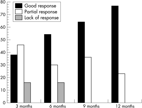
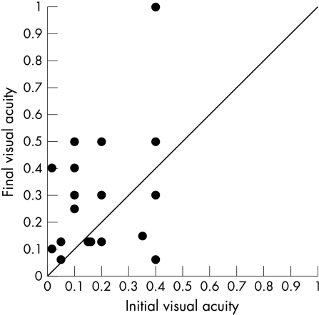
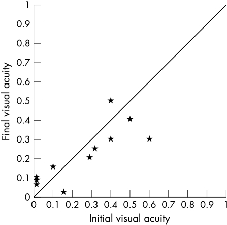
Methods: PDT was performed on highly myopic patients with subfoveal CNV. The patient population was divided into two groups according to age at onset of CNV (group 1 aged < or = 55 and group 2 aged >55 years old). Best corrected visual acuity (BCVA) and angiographic findings were considered.
Results: At the end of follow up group 2 had worse BCVA than group 1. 76% of patients in both groups showed a complete closure of CNV at the end of follow up.
Conclusion: Visual prognosis of myopic CNV treated by PDT is influenced by age at onset.
Figures
References
-
- Hotchkiss M, Fine S. Pathologic myopia and choroidal neovascularization. Am J Ophthalmol 1981;91:177–83. - PubMed
-
- Curtin B. The myopias: basic science and clinical management. Philadelphia: Harper and Row, 1985:7–10.
-
- Mondon H. Physiopathologie de la myopie forte. In: Mondon H, Metge P, eds. La myopie forte. Paris: Masson, 1994:29–57.
-
- Gass JDM. Stereoscopic atlas of macular diseases: diagnosis and treatment. 4th ed. St Louis: Mosby, 1997;1:126–8.
-
- Avila MP, Weiter JJ, Jalkh AE, et al. Natural history of choroidal neovascularization in degenerative myopia. Ophthalmology 1984;91:1573–81. - PubMed
Publication types
MeSH terms
Substances
LinkOut - more resources
Full Text Sources