

Optic disc morphology in south India: the Vellore Eye Study
- PMID: 12543749
- PMCID: PMC1771486
- DOI: 10.1136/bjo.87.2.189
Optic disc morphology in south India: the Vellore Eye Study
Abstract
Aim: To evaluate the morphology of the optic nerve head in an unselected population group in south India.
Methods: The study included 70 subjects forming a population based sample, selected in a random manner. Mean age was 47.5 (SD 8.7) years, mean refractive error measured -0.07 (1.11) dioptres (range -4.50 to +2.50 dioptres). Optic disc slides were morphometrically analysed.
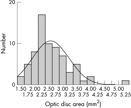
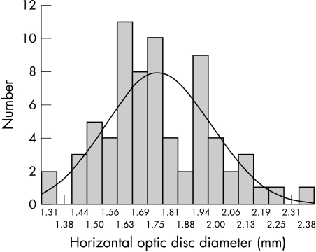
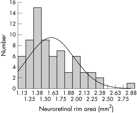
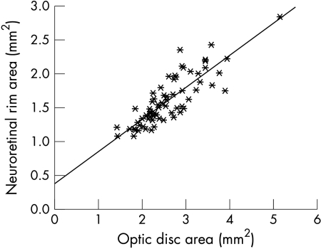
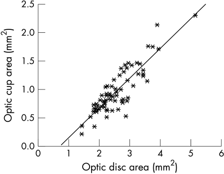
Results: Mean optic disc area measured 2.58 (0.65) mm(2). It was statistically independent of age and refractive error. Optic disc shape was slightly vertically oval. Mean neuroretinal rim area was 1.60 (0.37) mm(2). It was significantly and positively correlated with optic disc size and optic cup size. It was independent of age, sex, refractive error, and axial length. In all subjects included in the study, the rim was smallest in the temporal horizontal optic disc sector. Mean horizontal cup/disc diameter ratio (0.66 (0.07)) was significantly (p<0.001) higher than the mean vertical cup/disc diameter ratio (0.56 (0.08)). Both ratios were highly significantly (p <0.001) and positively correlated with optic disc size. The alpha zone of parapapillary atrophy (0.84 (0.29) mm(2)), and beta zone (0.13 (0.38) mm(2)), respectively, occurred in 69 (98.6%) subjects and in eight (11.4%) subjects, respectively. They were significantly larger in the temporal horizontal sector. The alpha zone was significantly (p<0.001) larger and occurred significantly more often than beta zone. Retinal arterioles and venules were wider, and in spatial correlation, the visibility of the retinal nerve fibre layer was significantly better, in the temporal inferior disc arcade and the temporal superior arcade than in the nasal superior arcade and the nasal inferior vessel arcade. Except for the absolute size measurements these optic nerve head parameters did not differ markedly (p >0.05) from the values found in white people.
Conclusions: South Indians and white people do not show marked differences in the morphology of the optic nerve head as measured by morphometric optic disc parameters, with the possible exception of the absolute optic disc dimensions.
Figures
References
-
- Chi T, Ritch R, Stickler D, et al. Racial differences in optic nerve head parameters. Arch Ophthalmol 1989;107:836–9. - PubMed
-
- Mansour AM. Racial variation of optic disc size. Ophthalmic Res 1991;23:67–72. - PubMed
-
- Tsai CS, Zangwill L, Gonzales C, et al. Ethnic differencs in optic nerve head topography. J Glaucoma 1995;4:248–57. - PubMed
-
- Varma R, Tielsch JM, Quigley HA, et al. Race-, age-, gender-, and refractive error-related differences in the normal optic disc. Arch Ophthalmol 1994;112:1068–76. - PubMed
-
- Jacob A, Thomas R, Koshi SP, et al. Prevalence of primary glaucoma in an urban south Indian population. Ind J Ophthalmol 1998;46:81–6. - PubMed
Publication types
MeSH terms
LinkOut - more resources
Full Text Sources