

Trypan blue staining of internal limiting membrane and epiretinal membrane during vitrectomy: visual results and histopathological findings
- PMID: 12543755
- PMCID: PMC1771493
- DOI: 10.1136/bjo.87.2.216
Trypan blue staining of internal limiting membrane and epiretinal membrane during vitrectomy: visual results and histopathological findings
Abstract
Aims: To report on the use of trypan blue (TB) 0.06% for staining the internal limiting membrane (ILM) and epiretinal membrane (ERM) during vitrectomy and report on their histology.
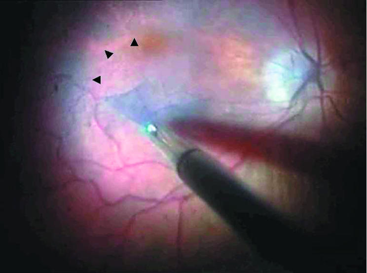
Method: 14 consecutive patients with idiopathic macular hole or macular pucker (seven patients each) were prospectively recruited for ILM or ERM peel respectively. After pars plana vitrectomy and induction of posterior vitreous detachment, 0.5 ml TB 0.06% in phosphate buffered saline (VisonBlue) was injected over the posterior pole in an air filled eye and left for 2 minutes. The stained tissue was peeled with intraocular forceps. Specimens were evaluated using histochemical and immunohistochemical methods.
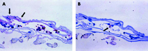
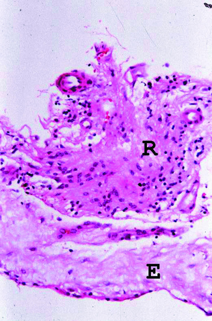
Results: The average follow up was 4.4 months. Internal limiting membranes and epiretinal membranes were stained satisfactorily in all cases and removed successfully. Eight patients (57%) had improvement of 2 or more Snellen lines. All seven macular holes closed. In the ERM cases, no residual membranes were observed clinically, at the latest follow up. No complications relating to the use of the dye were encountered intraoperatively or postoperatively. Of the 14 procedures, nine (four macular hole and five macular pucker) yielded sufficient tissue for histopathological evaluation. Histological and immunohistological assessment revealed that the morphology of these specimens was similar to that observed in macular hole ILM and macular pucker ERM removed without the aid of dye.
Conclusion: TB staining facilitated the identification and delineation of ILM and ERM removal during the surgical management of macular holes and macular pucker. The visual outcome of this series and the specimens removed suggest they are no different from those without TB staining. Its use in posterior segment appears to be safe but further studies are required to investigate its long term safety.
Figures
Comment in
-
Trypan blue stains the epiretinal membrane but not the internal limiting membrane.Br J Ophthalmol. 2003 Nov;87(11):1431-2. doi: 10.1136/bjo.87.11.1431-a. Br J Ophthalmol. 2003. PMID: 14609857 Free PMC article. No abstract available.
-
Staining of the ILM in macular surgery.Br J Ophthalmol. 2003 Dec;87(12):1530; author reply 1530. doi: 10.1136/bjo.87.12.1530. Br J Ophthalmol. 2003. PMID: 14660466 Free PMC article. No abstract available.
References
-
- Da Mata AP, Burk SE, Riemann CD, et al. Indocyanine green-assisted peeling of the retinal internal limiting membrane during vitrectomy surgery for macular hole repair. Ophthalmology 2001;108:1187–92. - PubMed
-
- Gandorfer A, Messmer EM, Ulbig MW, et al. Indocyanine green selectively stains the internal limiting membrane. Am J Ophthalmol 2001;131:387–8. - PubMed
-
- Gandorfer A, Haritoglou C, Gass CA, et al. Indocyanine green-assisted peeling of the internal limiting membrane may cause retinal damage. Am J Ophthalmol 2001;132:431–3. - PubMed
-
- Kwok AK, Li WW, Pang CP, et al. Indocyanine green staining and removal of internal limiting membrane in macular hole surgery: histology and outcome. Am J Ophthalmol 2001;132:178–83. - PubMed
-
- Kadonosono K, Itoh N, Uchio E, et al. Staining of internal limiting membrane in macular hole surgery. Arch Ophthalmol 2000;118:1116–8. - PubMed
MeSH terms
Substances
LinkOut - more resources
Full Text Sources
Other Literature Sources
Medical
Miscellaneous