Comparative Study
. 2003 Feb;162(2):413-25.
doi: 10.1016/S0002-9440(10)63836-9.

Apolipoprotein B in cholesterol-containing drusen and basal deposits of human eyes with age-related maculopathy

Affiliations
Comparative Study

Apolipoprotein B in cholesterol-containing drusen and basal deposits of human eyes with age-related maculopathy

Goldis Malek et al. Am J Pathol. 2003 Feb.

Abstract

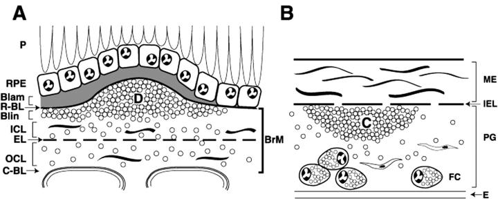
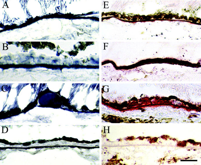
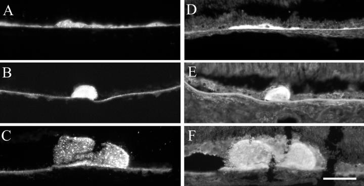
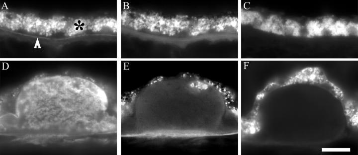
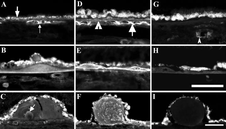
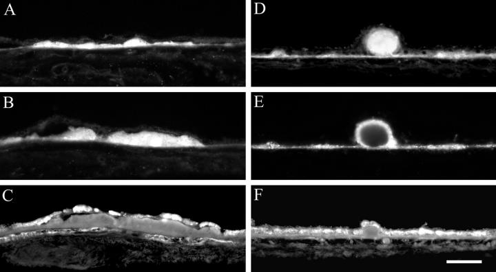
Lipids accumulate in Bruch's membrane (BrM), a specialized vascular intima of the eye, and in extracellular lesions associated with aging and age-related maculopathy (ARM). We tested the hypothesis that ARM and atherosclerotic cardiovascular disease share molecules and mechanisms pertaining to extracellular lipid accumulation by localizing cholesterol and apolipoprotein B (apo B) in BrM, basal deposits, and drusen. Human donor eyes were preserved <4 hours postmortem and cryosectioned. Sections were stained with traditional lipid stains and filipin for esterified and unesterified cholesterol or probed with antibodies to apo B, apo E, and apo C-III. Normal adult retinal pigment epithelium (RPE) was subjected to RT-PCR and Western blot analysis for apolipoprotein mRNA and protein. Esterified and unesterified cholesterol was present in all drusen and basal deposits of ARM and normal eyes. Both apo B and apo E but not apo C-III were found in BrM, drusen, and basal deposits. Fewer macular drusen were stained by traditional lipid stains and apolipoprotein antibodies than peripheral drusen. RPE contained apo B and apo E mRNA and protein. Finding cholesterol and apo B in sub-RPE deposits links ARM with important molecules and mechanisms in atherosclerosis initiation and progression. The combination of apo B mRNA and protein in RPE raises the possibility that intraocular assembly of apo B-containing lipoproteins is a pathway involved in forming cholesterol-enriched lesions in ARM.


Figures

Figure 1.
Figure 1.
Schematic cross-sections of Bruch’s membrane (BrM) from an eye with ARM (A) and atherosclerotic arterial intima (B). Endothelium and vascular lumina (choriocapillary, A; arterial, B) are at the bottom. Drawings are not at scale. For reference, the thickness of normal BrM and intima is 4 to 6 μm and 100 to 300 μm, respectively. Small circles in BrM (A) and PG layer (B) indicate esterified cholesterol-containing particles. A: P, photoreceptors; RPE, retinal pigment epithelium; R-BL, RPE basal lamina; Blam, basal laminar deposit; Blin, basal linear deposit; D, druse; ICL, inner collagenous layer; EL, elastic layer; OCL, outer collagenous layer; C-BL, choriocapillaris basal lamina. In normal eyes, BrM (thick bracket at right edge of panel A) consists of R-BL, ICL, EL, OCL, and C-Cl. Blam and Blin together comprise basal deposits, and basal deposits and drusen together comprise sub-RPE deposits. B: ME, musculoelastic layer; IEL, internal elastic layer; C, lipid-rich core; PG, proteoglycan layer; FC, foam cells; E, endothelium.
Figure 2.
Figure 2.
Lipid histochemistry of macula in eyes with ARM. Cryosections were stained with bromine sudan black B (A–D) and oil red O (E–H). Bruch’s membrane (A and E). Basal deposits (B and F). Drusen (C and G), Control sections (D and H) were lipid-extracted before staining. 79-year-old man (A, C, E, and F). 77-year-old man (B). 86-year-old woman (D). 95-year-old woman (G). 85-year-old woman (H). Bar, 0.05 mm.
Figure 3.
Figure 3.
Esterified and unesterified cholesterol in macular basal deposits and drusen of ARM eyes. Cryosections from ARM eyes were stained with filipin. Esterified cholesterol (A–C), unesterified cholesterol (D–F). A and D: Thin basal deposit, 73-year-old man. B and E: Small druse, 73-year-old man. C and F: Medium druse, 85-year-old man. Bar, 0.05 mm.
Figure 4.
Figure 4.
Apo B immunofluorescence and autofluorescence in cryosections of normal and ARM eyes. Sections were probed with polyclonal anti-apo B (A, B, D, and E) or non-immune serum (C and F). Images were obtained with a 40X plan fluor oil objective and either a rhodamine filter set (A, C, D and F) or an autofluorescence filter set (B and E). Exposure times were 0.1 second in panels A–C and 0.07 seconds in panels D–F. A–C: Normal, macula, 87-year-old man. D–F, ARM, periphery, 73-year-old-man. Bar in F, 0.15 mm. A: Apo B immunoreactivity is present in Bruch’s membrane (BrM), especially on the inner aspect (arrowhead). RPE is autofluorescent (*). B: In the same section as A, autofluorescence in BrM is distributed differently from the specific fluorescence in A. C: No fluorescence is detected in BrM of a control sections at the same exposure time as A. D: Apo B immunoreactivity is present in a druse. E: In the same section as D, autofluorescence is present in RPE and BrM and within the druse, in a different pattern from D. F: No fluorescence is detected in a druse in a control section at the same exposure time as D.
Figure 5.
Figure 5.
Indirect immunofluorescent localization of apo B, apo E, and apo C-III in cryosections of normal (A and G) and ARM (B–F, H, and I) eyes. Bruch’s membrane (BrM; A, D, and G), basal deposits (BD, B, E, and H), drusen (C, F, and I). Apo B, apo E, and apo C-III immunoreactivity is seen in the left, middle, and right columns respectively. A: Apo B is present in the vessels of the choroid (small arrow), and RPE is autofluorescent (large arrow); periphery of 73-year-old man. B: Apo B in BD; periphery, 78-year-old woman. C: Apo B in druse, white arrow points to apo B material within druse; macula, 81-year-old woman. D: Apo E was found in BrM (arrowhead) and intercapillary pillars (arrow); macula, 88-year-old woman. E: Apo E seen in thin BD; periphery, 85-year-old man. F: Apo E is heterogeneously present in druse; macula, 88-year-old woman. G: Apo C-III is absent from BrM, but present in vessels of the choroid (arrow); periphery, 73-year-old man. H: Apo C-III is absent from BD; periphery, 85-year-old man. I: Apo C-III is absent from druse; macula, 88-year-old woman. RPE, retinal pigment epithelium. Bar in H, 0.05 mm, applies to panels A–B, D–E, G–H; bar in I, 0.08 mm, applies to panels C, F, and I.
Figure 6.
Figure 6.
Co-localization of apo B, EC, and UC in sub-RPE deposits of normal and ARM eyes. Panels A–C represent the same basal deposit and D–F represent the same druse, on the basis of consistent location across adjacent cryosections. Basal deposits with apo B immunoreactivity (A) also contain EC (B) and UC (C). Drusen with apo B immunoreactivity (D) also contain EC (E) and UC (F). The UC-rich druse in F contains EC on the superficial rim only (E). RPE autofluorescence (A and D) is not visible at wavelengths used to visualize filipin (B, C, E, and F). A–C, Periphery, normal, 87-year-old woman. D–F, Periphery; ARM, 81-year-old woman. EC, esterified cholesterol; UC, unesterified cholesterol; RPE, retinal pigment epithelium. Bar, 0.05 mm.
Figure 7.
Figure 7.
Apolipoprotein mRNA and protein in native human RPE and HepG2 cells. A: RT-PCR analysis of apo B and apo E mRNA transcript in human RPE. The total RNA was isolated from human RPE. The mRNA of HepG2 cells was used as positive control. RT-PCR was carried out with primers specific for human apo B and apo E, and resolved by electrophoresis on 1.5% agarose gel. The expected sizes of the RT-PCR product for apo B and apo E are 614 bp and 163 bp. RT-PCR amplification products representing: lane 1, 100 bp DNA molecular weight ladder; lanes 2 and 3, apo B mRNA (614 bp); lanes 4 and 5, apo E mRNA (163 bp); lanes 2 and 4, human RPE; and lanes 3 and 5, HepG2. B: Partial nucleic acid sequence and deduced amino acid sequence of human RPE apo B. The result shows that the apo B RT-PCR fragment contains a CAA condon, encoding Gln-2153 in apo B-100, not TAA, the stop codon in apo B-48. An asterisk shows the putative editing site of apo B-48. C: Western blot of human RPE extract and HepG2 cells. Detergent-extracted proteins from freshly isolated human RPE (65-year-old male, lane 1, 60 μg and lane 3, 9 μg) and HepG2 cells (lane 2, 6 μg and lane 4, 6 μg) were separated by SDS-PAGE and then transferred to nitrocellulose. Lanes were probed with the following primary antibodies, each at 1:1000 dilutions: monoclonal anti-human apo B (1D1) (lanes 1 and 2) and apo E (lanes 2 and 3). Primary antibodies were detected with HRP-conjugated secondary antibodies at 1:2000 dilution (see Methods). Position and vendor estimated standard molecular weights are indicated at the left. RPE, retinal pigment epithelium; HRP, horseradish peroxidase-conjugated.

References

    1. Klein R, Klein BEK, Linton KLP: Prevalence of age-related maculopathy. Ophthalmology 1992, 99:933-943 - PubMed
    1. Mitchell P, Smith W, Attebo K, Wang JJ: Prevalence of age-related maculopathy in Australia: The Blue Mountains Eye Study. Ophthalmology 1995, 102:1450-1460 - PubMed
    1. Vingerling JR, Dielemans I, Hofman A, Grobbee DE, Hijmering M, Kramer CFL, de Jong PTVM: The prevalence of age-related maculopathy in the Rotterdam study. Ophthalmology 1995, 102:205-210 - PubMed
    1. Sarks SH: Ageing and degeneration in the macular region: a clinico-pathological study. Br J Ophthalmol 1976, 60:324-341 - PMC - PubMed
    1. Green WR, Enger C: Age-related macular degeneration histopathologic studies: The 1992 Lorenz E. Zimmerman Lecture. Ophthalmology 1993, 100:1519-1535 - PubMed

Publication types

MeSH terms