

The role of ultraviolet irradiation and heparin-binding epidermal growth factor-like growth factor in the pathogenesis of pterygium
- PMID: 12547714
- PMCID: PMC1851157
- DOI: 10.1016/S0002-9440(10)63850-3
The role of ultraviolet irradiation and heparin-binding epidermal growth factor-like growth factor in the pathogenesis of pterygium
Abstract
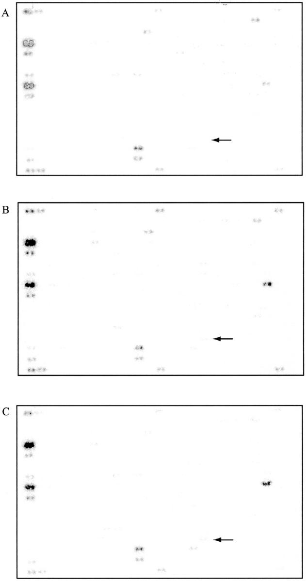
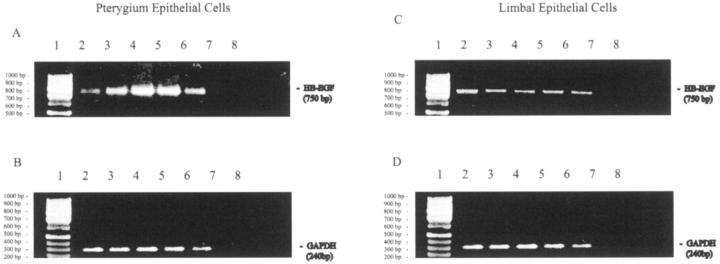
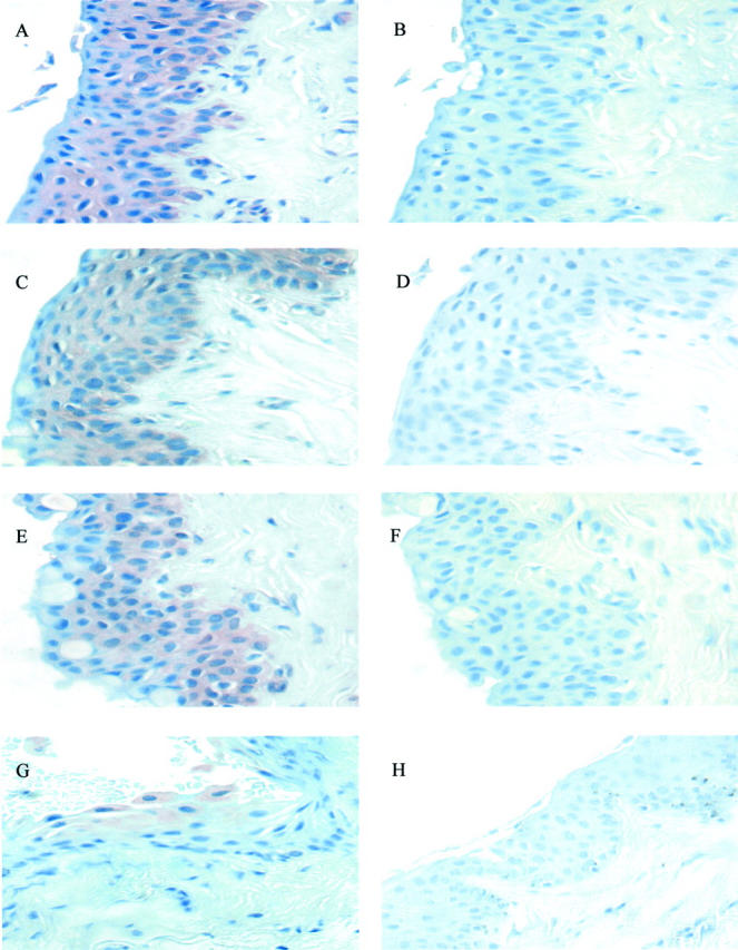
Ultraviolet (UV) light is one of the major factors implicated in the pathogenesis of pterygium. The mechanism by which UV light induces this disease remains elusive. The aim of this study was to evaluate the effects of UVB irradiation on the expression of growth factors in cultured pterygium epithelial cells and to demonstrate their distribution within pterygium. We cultured pterygial epithelial cells from pterygium explants and these cells were exposed to 20 mJ/cm(2) of UVB. Total RNA was extracted at 0, 6, and 12 hours after irradiation. (32)P-labeled cDNA was synthesized and analyzed using microarray technology to determine the differential expression of 268 growth factor and cytokine related genes. Semiquantitative reverse transcriptase-polymerase chain reaction was used to corroborate this data. Conditioned media derived from cells exposed to UVB irradiation was analyzed for protein expression by enzyme-linked immunosorbent assay. Immunohistochemistry was used to evaluate the distribution of heparin-binding epidermal growth factor-like growth factor (HB-EGF) in pterygium tissue. Analysis of the hybridization signals revealed that the genes encoding HB-EGF, fibroblast growth factor 3, and cytotoxic trail ligand receptor were consistently elevated at 6 and 12 hours after UVB treatment. HB-EGF mRNA was elevated 6.8-fold at 6 hours after irradiation and was augmented in culture supernatants after the same treatment. Furthermore, HB-EGF reactivity was identified in the epithelium and vasculature of pterygium by immunohistochemistry. HB-EGF was present in normal limbal epithelium, although it was not induced in cultured limbal epithelial cells by UV irradiation. HB-EGF is a potent mitogen, localized in pterygium tissue, and significantly induced by UVB in pterygium-derived epithelial cells. We postulate that this growth factor is a major driving force in the development of pterygia and a means by which UV irradiation causes the pathogenesis of pterygium.
Figures
References
-
- Coroneo MT, Di Girolamo N, Wakefield D: The pathogenesis of pterygia. Curr Opin Ophthalmol 1999, 10:282-288 - PubMed
-
- Taylor HR, West SK, Rosenthal FS, Munoz B, Newland HS, Emmett EA: Corneal changes associated with chronic UV irradiation. Arch Ophthalmol 1989, 107:1481-1484 - PubMed
-
- Threlfall TJ, English DR: Sun exposure and pterygium of the eye: a dose-response curve. Am J Ophthalmol 1999, 128:280-287 - PubMed
-
- Panchapakesan J, Hourihan F, Mitchell P: Prevalence of pterygium and pinguecula: the Blue Mountains Eye Study. Aust N Z J Ophthalmol 1998, 26:S2-S5 - PubMed
Publication types
MeSH terms
Substances
LinkOut - more resources
Full Text Sources
Other Literature Sources
