. 2003 Feb;162(2):597-608.
doi: 10.1016/s0002-9440(10)63853-9.

Transforming growth factor-beta mediates intestinal healing and susceptibility to injury in vitro and in vivo through epithelial cells

Affiliations

Transforming growth factor-beta mediates intestinal healing and susceptibility to injury in vitro and in vivo through epithelial cells

Paul L Beck et al. Am J Pathol. 2003 Feb.

Abstract

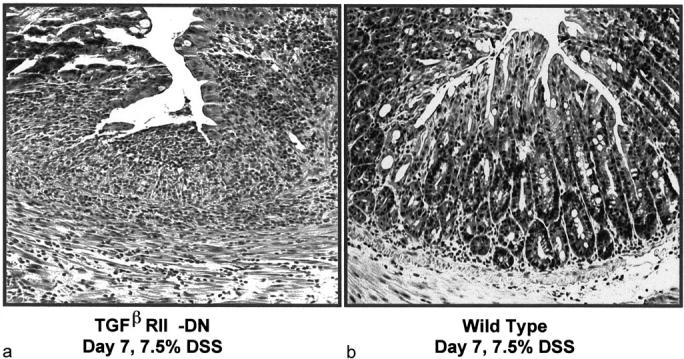
In vitro studies suggest that transforming growth factor (TGF)-beta has potent effects on gastrointestinal mucosal integrity, wound repair, and neoplasia. However, the multiplicity of actions of this peptide on many different cell types confounds efforts to define the role of TGF-beta within the intestinal epithelium in vivo. To delineate these effects selective blockade of intestinal epithelial TGF-beta activity was undertaken through targeted expression of a dominant-negative (DN) TGF-beta RII to intestinal epithelial cells in vitro and in vivo. Stable intestinal epithelial cell (IEC)-6 lines overexpressing TGF-beta RII-DN (nucleotides -7 to 573) were established. Transgenic mice overexpressing TGF-beta RII-DN under the regulation of a modified liver fatty acid-binding promoter (LFABP-PTS4) were constructed. In vitro healing was assessed by wounding of confluent monolayers. Colitis was induced by the addition of dextran sodium sulfate (2.5 to 7.5% w/v) to their drinking water. Overexpression of TGF-beta RII-DN in intestinal epithelial cell-6 cells resulted in a marked reduction in cell migration and TGF-beta-stimulated wound healing in vitro. TGF-beta RII-DN transgenic mice did not exhibit baseline intestinal inflammation or changes in survival, body weight, epithelial cell proliferation, aberrant crypt foci, or tumor formation. TGF-beta RII-DN mice were markedly more susceptible to dextran sodium sulfate-induced colitis and exhibited impaired recovery after colonic injury. TGF-beta is required for intestinal mucosal healing and TGF-beta modulation of the intestinal epithelium plays a central role in determining susceptibility to injury.


Figures

Figure 1.
Figure 1.
a: Expression of TGF-β RII-DN blocks TGF-β inhibition of IEC-6 proliferation. Proliferation was assessed by determination of thymidine incorporation in IEC-6 cell lines stably transfected with either the TGF-β RII-DN or empty vector under regulation of the CMV promoter. Cells were exposed to a final concentration of 0, 4, or 40 pmol of TGF-β for 24 hours followed by incubation in tritium-labeled thymidine. All experiments contained a minimum of five plates/well per group and repeated a minimum of three times (**, P < 0.01). b: Confirmation of TGF-β RII-DN in stably transfected IEC-6 cells. c-myc Western blot showing expression of the TGF-β RII-DN (21 kd) in stably transfected IEC-6 cell lines.
Figure 2.
Figure 2.
a and b: Blockade of TGF-β signaling reduces intestinal epithelial cell migration and in vitro wound repair. Stable IEC-6 cell lines transfected with either the TGF-β RII-DN or empty vector under regulation of the CMV promoter were plated and allowed to grow to confluency and then wounded with a razor blade. The CMV-TGF-β RII-DN lines had reduced spontaneous and TGF-β-induced (40 pmol/L) migration across the wound edge at the 24 hour time point. Cells were assessed by direct inspection (a) and quantified by cell counting (b) as described in Materials and Methods. Each study consisted of minimum of 12 plates/well per group and the study was repeated three times on two different stable cell lines per group (***, P < 0.001).
Figure 3.
Figure 3.
Expression of TGF-β RII-DN in transgenic mice. Tissue expression of the mutant receptor construct was assessed by reverse transcriptase-PCR after DNase treatment was restricted to the colon, ileum, and jejunum. No expression was seen in duodenum, stomach, heart, kidney, liver, or skeletal muscle. Primers: coding, GAG CAG AAG CTG ATC TCT GAG; noncoding, GCC CGG ATC CAA GCG GCC GCT AAC GCG GTA GCA GTA GAA GAT; 38 cycles of 94°C for 1 minute, 61°C for 1 minute, 72°C for 1 minute.
Figure 4.
Figure 4.
Localization of TGF-β RII-DN protein expression in transgenic mice. The transgene-encoded protein was assessed by Western blot with anti-sera to the c-myc tag in protein extracts from the colonic epithelium of TGF-β RII-DN transgenic mice versus wild-type mice showing PTS4-LFAB directed expression of the TGF-β RII-DN (21 kd).
Figure 5.
Figure 5.
Immunohistochemical localization of the c-myc-tagged TGF-β RII-DN. Specific c-myc staining was localized to epithelial cells. Some specific staining was noted in epithelial cells at the base of the crypts but the highest degree of staining was present in epithelial cells closest to the luminal surface (a). No significant specific staining was noted in wild-type mice or in the TGF-β RII-DN mice when the primary antibody (anti-c-myc) was excluded, or an unrelated antibody was substituted for the primary antibody. The secondary antibody was labeled with CY3. Staining was localized to the cytoplasm and cell surface without specific localization to the nucleus as determined by the use of the DNA-specific dye Hoechst 33258 (b).
Figure 6.
Figure 6.
Blockade of TGF-β response does not alter proliferation of intestinal epithelial cells. Cell proliferation determined by BrdU staining. BrdU was given to wild-type and transgenic TGF-β RII-DN mice 1 hour before sacrifice at 3 and 12 months of age. Tissue was removed, processed, and stained with anti-BrdU antibodies. There was no difference in cell proliferation as determined by BrdU staining in the TGF-β RII-DN compared to wild-type animals. Expressed as BrdU-labeling index; BrdU-labeling index is defined as the number of positive staining cells × 100/total number of cells.
Figure 7.
Figure 7.
Blockade of TGF-β response in intestinal epithelial cells increases susceptibility to injury (a). Changes in baseline body weight during induction of colitis with 7.5% DSS. The TGF-β RII-DN mice were more susceptible to DSS-induced colitis than wild-type mice (n = 14 mice per group; *, P < 0.05; **, P < 0.01). Fecal blood loss during induction of colitis with 7.5% DSS. The TGF-β RII-DN mice were more susceptible to DSS-induced colitis than wild-type mice with significantly greater amounts of fecal blood loss (n = 14 mice per group; *, P < 0.05; **, P < 0.01). See Materials and Methods for scoring system.
Figure 8.
Figure 8.
The TGF-β RII-DN mice were more susceptible to DSS-induced colitis. a: TGF-β RII-DN mice. b: Wild-type mice. All of the TGF-β RII-DN mice had extensive areas of ulceration and transmural inflammation; in contrast only superficial ulceration and inflammation was noted in the wild-type mice.
Figure 9.
Figure 9.
TGF-β RII-DN mice are more susceptible to colonic injury. Survival curves for TGF-β RII-DN and wild-type (wt) mice during induction of colitis. The marked increased susceptibility of the TGF-β RII-DN to DSS-induced colitis resulted in a significant reduction in survival compared to wild-type mice. Survival curves were created using the Kaplan-Meier method and survival comparisons were performed using the log-rank or Mantel-Haenszel test, which generate a two-tailed P value.
Figure 10.
Figure 10.
Healing is impaired by blockade of intestinal epithelial responses to TGF-β. a: To assess effects on repair rather than primary injury, mice were given a low dose of DSS (2.5% w/v). Changes in baseline body weight. There were no significant differences in basal body weights of TGF-β RII-DN mice versus wild-type animals during the exposure to 2.5% DSS. Once the DSS was discontinued the wild-type mice rapidly returned to baseline whereas the TGF-β RII-DN mice failed to return to baseline (*, P < 0.05; **, P < 0.01). b: Clinical assessment (day 25). Ten days after discontinuation of DSS the wild-type mice had no fecal blood loss, minimal diarrhea, and on sacrifice had minimal adhesions compared to the TGF-β RII-DN mice. All assessments were done in a blinded manner on coded animals (*, P < 0.05; **, P < 0.01; ***, P < 0.001). c and d: Histology (day 25). Ten days after discontinuation of DSS the wild-type animals had no significant signs of ulceration and minimal superficial inflammation, TGF-β RII-DN mice exhibited large areas of ulceration and transmural inflammation. TGF-β RII-DN mice lack re-epithelialization of the ulcerated area (Figure 10c) ▶ . e: Tissue MPO. Colonic tissue MPO content was determined before induction of colitis (day 0), at the end of DSS exposure (day 15) and on sacrifice on day 25 (***, P < 0.001). f: Survival curves for TGF-β RII-DN and wild-type (wt) mice during induction of colitis and after discontinuation of DSS (day 15). The impaired recovery of TGF-β RII-DN mice resulted in a significant reduction in survival compared to wild-type mice. Survival curves were created using the Kaplan-Meier method and survival comparisons were performed using the log-rank or Mantel-Haenszel test, which generate a two-tailed P value.

References

    1. Chen RH, Derynck R: Homomeric interactions between type II transforming growth factor-beta receptors. J Biol Chem 1994, 269:22868-22874 - PubMed
    1. Derynck R, Feng XH: TGF-beta receptor signaling. Biochim Biophys Acta 1997, 1333:F105-F150 - PubMed
    1. Zhao Y, Young SL: Requirement of transforming growth factor-beta (TGF-beta) type II receptor for TGF-beta-induced proliferation and growth inhibition. J Biol Chem 1996, 271:2369-2372 - PubMed
    1. Wahl SM: Transforming growth factor beta: the good, the bad, and the ugly. J Exp Med 1994, 180:1587-1590 - PMC - PubMed
    1. Shull MM, Ormsby I, Kier AB, Pawlowski S, Diebold RJ, Yin M, Allen R, Sidman C, Proetzel G, Calvin D, Annunziata N, Doetschman T: Targeted disruption of the mouse transforming growth factor-beta 1 gene results in multifocal inflammatory disease. Nature 1992, 359:693-699 - PMC - PubMed

Publication types

MeSH terms

LinkOut - more resources