. 2003 Feb;84(2 Pt 1):1252-62.
doi: 10.1016/S0006-3495(03)74940-6.

Force measurements of the alpha5beta1 integrin-fibronectin interaction

Affiliations

Force measurements of the alpha5beta1 integrin-fibronectin interaction

Feiya Li et al. Biophys J. 2003 Feb.

Abstract

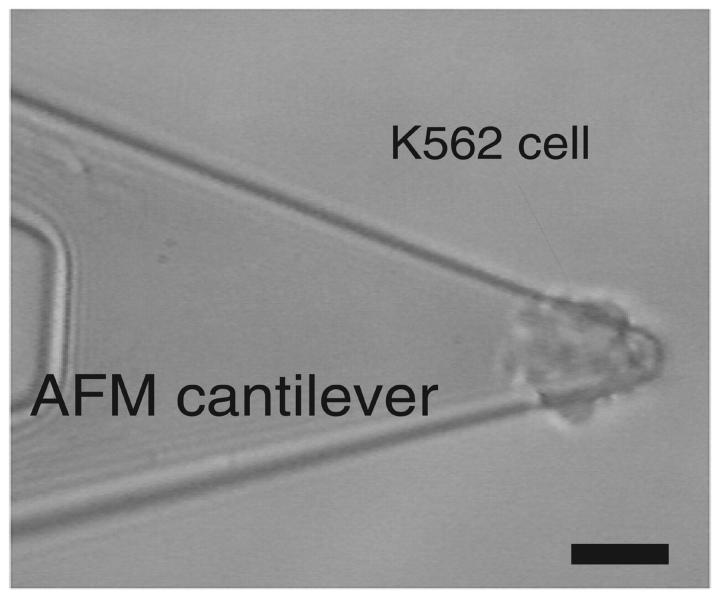
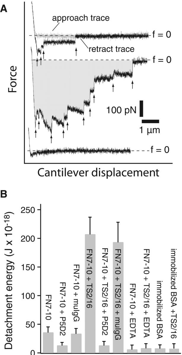
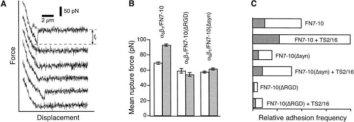
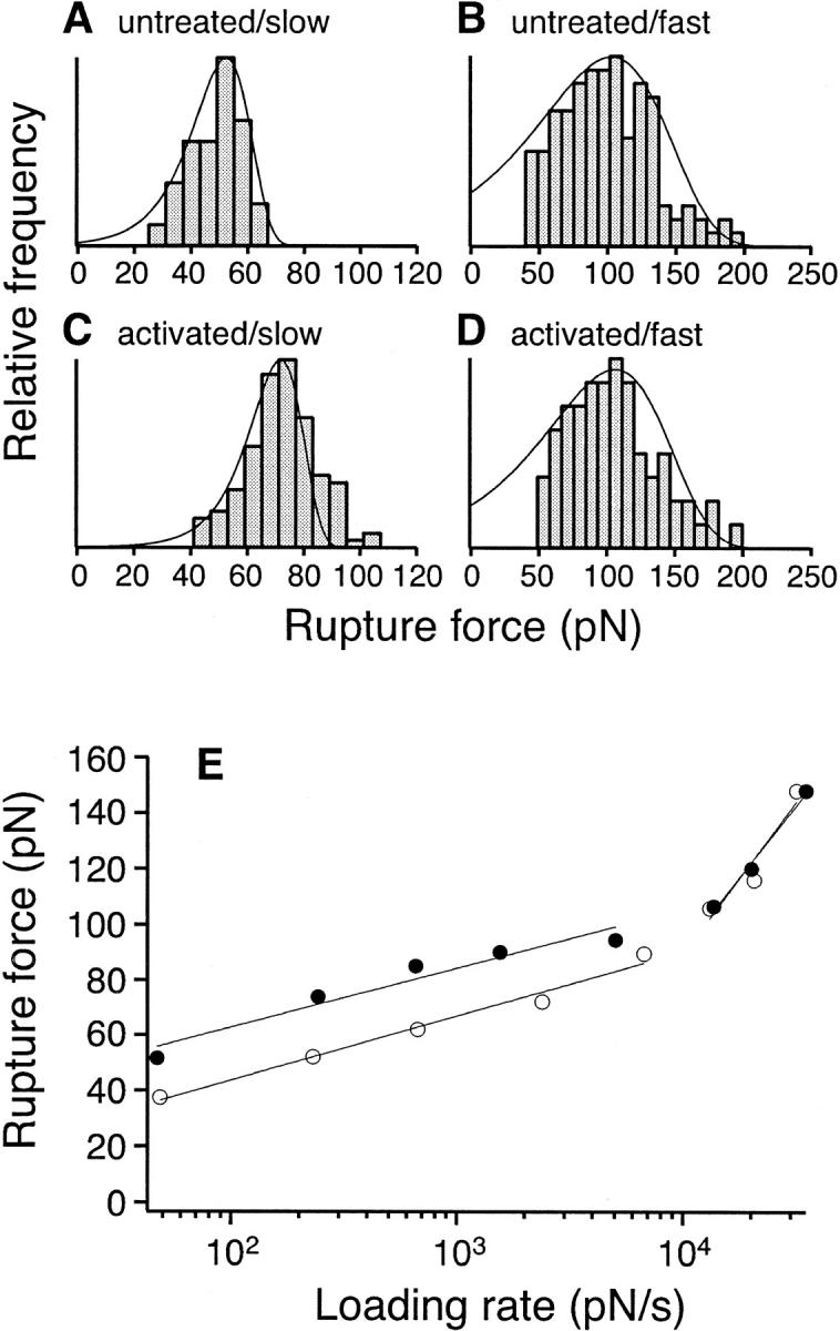
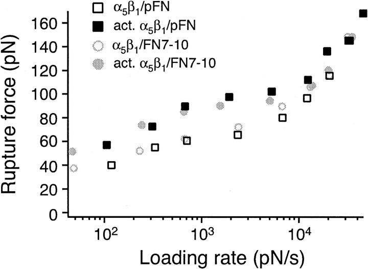
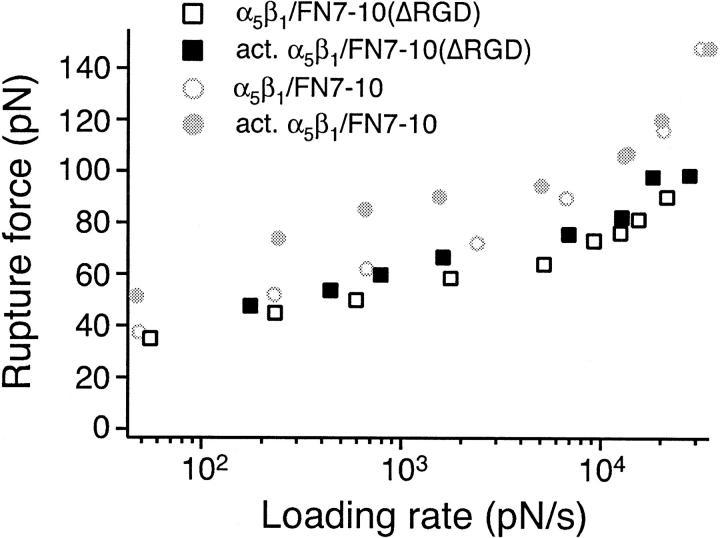
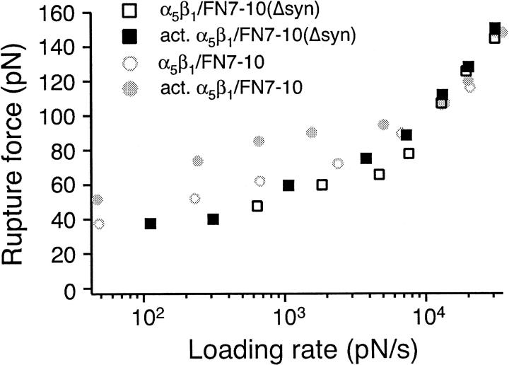
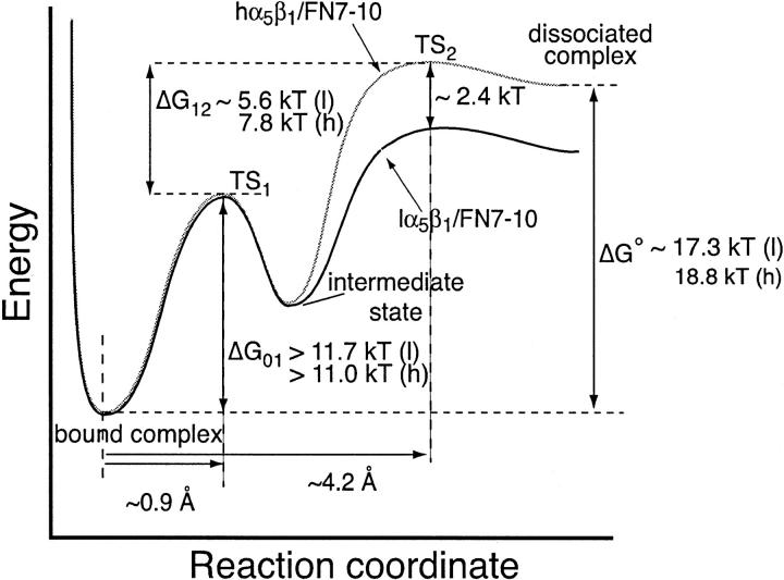
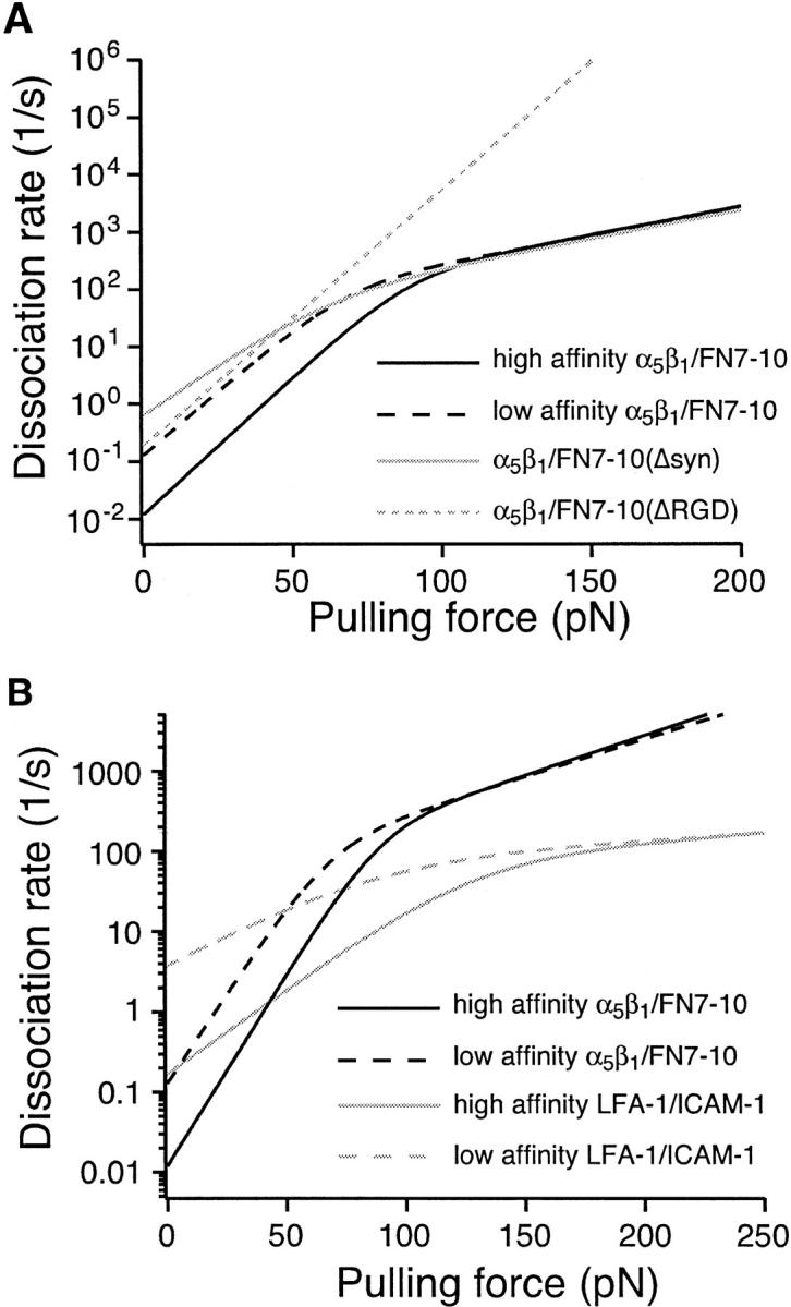
The interaction of the alpha(5)beta(1) integrin and its ligand, fibronectin (FN), plays a crucial role in the adhesion of cells to the extracellular matrix. An important intrinsic property of the alpha(5)beta(1)/FN interaction is the dynamic response of the complex to a pulling force. We have carried out atomic force microscopy measurements of the interaction between alpha(5)beta(1) and a fibronectin fragment derived from the seventh through tenth type III repeats of FN (i.e., FN7-10) containing both the arg-gly-asp (RGD) sequence and the synergy site. Direct force measurements obtained from an experimental system consisting of an alpha(5)beta(1) expressing K562 cell attached to the atomic force microscopy cantilever and FN7-10 adsorbed on a substrate were used to determine the dynamic response of the alpha(5)beta(1)/FN7-10 complex to a pulling force. The experiments were carried out over a three-orders-of-magnitude change in loading rate and under conditions that allowed for detection of individual alpha(5)beta(1)/FN7-10 interactions. The dynamic rupture force of the alpha(5)beta(1)/FN7-10 complex revealed two regimes of loading: a fast loading regime (>10,000 pN/s) and a slow loading regime (<10,000 pN/s) that characterize the inner and outer activation barriers of the complex, respectively. Activation by TS2/16 antibody increased both the frequency of adhesion and elevated the rupture force of the alpha(5)beta(1)/wild type FN7-10 complex to higher values in the slow loading regime. In experiments carried out with a FN7-10 RGD deleted mutant, the force measurements revealed that both inner and outer activation barriers were suppressed by the mutation. Mutations to the synergy site of FN, however, suppressed only the outer barrier activation of the complex. For both the RGD and synergy deletions, the frequency of adhesion was less than that of the wild type FN7-10, but was increased by integrin activation. The rupture force of these mutants was only slightly less than that of the wild type, and was not increased by activation. These results suggest that integrin activation involved a cooperative interaction with both the RGD and synergy sites.


Figures

FIGURE 1
FIGURE 1
Micrograph of a K562 cell attached to the end of an AFM cantilever. The bar is 20 μm.
FIGURE 2
FIGURE 2
AFM force measurements of α5β1-mediated cell adhesion. (A) A series of AFM force-displacement retract traces of the interaction between an unactivated K562 cell and FN7-10 (top record, approach and retract traces), after the K562 cell was activated by 10% TS2/16 (middle record, retract trace only) and in the presence of antibodies (P5D2, 20 μg/ml) against the β-subunit of α5β1 (bottom record, retract trace only). The arrows point to the positions in the traces where the α5β1/FN7-10 complex ruptured. The shaded area in the middle trace is the detachment energy. (B) Detachment energies of α5β1-mediated cell adhesion for different cell states. Higher detachment energies are due to both a larger number of adhesions and larger forces of detachment. The error bar is the standard deviation.
FIGURE 3
FIGURE 3
(A) Force-displacement traces between K562 and FN7-10 under conditions of minimal contact. Two of the six traces (first and fifth) revealed adhesion. fr is the rupture force of the α5β1 integrin/FN bond. ks is the system spring constant and is used to determine the loading rate of the measurement. The cantilever retraction rate of the measurements was 5 μm/s. (B) Mean rupture forces of individual α5β1 integrins complexed to FN7-10, FN7-10 (ΔRGD), and FN7-10(Δsyn). Measurements were acquired at a loading rate of 1,800 pN/s. Open and gray bars correspond to measurements from untreated and TS2/16 activated cells, respectively. The error bar is the standard error of the mean. (C) Adhesion frequency of K562/FN binding under conditions of limited tip–substrate contact. Open and gray bars correspond to adhesion frequencies before and after the addition of the function-blocking antibody, JBS5 (20 μg/ml), respectively.
FIGURE 4
FIGURE 4
Measurements of the rupture force of individual α5β1 integrin/fibronectin bonds at different loading rates. (A, B) Force histograms of α5β1/FN7-10 interaction at (A) slow (230 pN/s) and (B) fast (13,000 pN/s) loading rates. (C, D) Force histograms of the high affinity α5β1/FN7-10 interaction (activated by TS2/16) at (C) slow (240 pN/s) and (D) fast (13,500 pN/s) loading rates. The fitted probability density functions (Eq. 1) in AD were obtained using the Bell model parameters listed in Table 1. (E) Single molecule force measurements between FN7-10 and unactivated K562 cells (open circle) or 10% TS2/16 activated K562 cells (solid circle) as a function of loading rates. The best-fit curves (gray lines) were obtained using Eq. 2 (see Discussion).
FIGURE 5
FIGURE 5
Single molecule force measurements between human plasma fibronectin and unactivated K562 cells (open square) or K562 cells (solid square) activated by TS2/16. The mode rupture force is plotted as a function of loading rates. Measurements of the α5β1/FN7-10 interaction are plotted in open and closed circles.
FIGURE 6
FIGURE 6
Single molecule force measurements of the interactions between live K562 (open circle) or fixed K562 cells (solid circle) and FN7-10. The mode rupture force is plotted as a function of loading rates.
FIGURE 7
FIGURE 7
Measurements of the rupture force between individual FN7-10 (ΔRGDS) and α5β1 integrin in unactivated K562 (open square) or activated K562 cells (solid square). The mode rupture force is plotted as a function of loading rates. The data plotted in gray circles are measurements between FN7-10 and unactivated K562 (open circle) or activated K562 cells (solid circle).
FIGURE 8
FIGURE 8
Measurements of the rupture force between individual FN7-10(Δsyn) and unactivated K562 (open square) or activated K562 (solid square) cells. The mode rupture force is plotted as a function of loading rates. The data plotted in gray circles are measurements between FN7-10 and unactivated K562 (open circle) or activated K562 cells (solid circle).
FIGURE 9
FIGURE 9
Intermolecular potential of the α5β1/FN7-10 interaction. The forced dissociation of the α5β1/FN7-10 involves overcoming two transition states, TS1 and TS2. Estimates of the equilibrium free energies (ΔGO) were derived from the equilibrium affinity constants reported in Takagi et al. (2001). (l) and (h) denote energies corresponding to the low and high affinity α5β1/FN7-10 complexes, respectively. Estimates of the energy difference (ΔG12) between TS1 and TS2 were obtained using: ΔG12 = −kBT ln(k°1/k°2) where k°1 and k°2 are the dissociation rate constants of the inner and outer activation energy barriers, respectively. Estimates of the energy difference (ΔG01) between TS1 and the bound state were based on the inequality: ΔG12 + ΔG01 ≥ ΔG°.
FIGURE 10
FIGURE 10
Kinetic profiles of the α5β1/FN interaction. (A) Effects of FN7-10 mutations on the kinetic profile of the α5β1/FN interaction. (B) Comparison of the kinetic profiles of the α5β1/FN7-10 interaction with the LFA-1/ICAM-1 interaction.

References

    1. Akiyama, S. K., and S. S. Yamada. 1985. The interaction of plasma fibronectin with fibroblastic cells in suspension. J. Biol. Chem. 260:4492–4500. - PubMed
    1. Aota, S., M. Nomizu, and K. M. Yamada. 1994. The short amino acid sequence Pro-His-Ser-Arg-Asn in human fibronectin enhances cell-adhesive function. J. Biol. Chem. 269:24756–24761. - PubMed
    1. Arroyo, A. G., A. Garcia-Pardo, and F. Sanchez-Madrid. 1993. A high affinity conformational state on VLA integrin heterodimers induced by an anti-beta 1 chain monoclonal antibody. J. Biol. Chem. 268:9863–9868. - PubMed
    1. Aukhil, I., P. Joshi, Y. Yan, and H. P. Erickson. 1993. Cell- and heparin-binding domains of the hexabrachion arm identified by tenascin expression proteins. J. Biol. Chem. 268:2542–2553. - PubMed
    1. Barillari, G., L. Albonici, S. Incerpi, L. Bogetto, G. Pistritto, A. Volpi, B. Ensoli, and V. Manzari. 2001. Inflammatory cytokines stimulate vascular smooth muscle cells locomotion and growth by enhancing alpha5beta1 integrin expression and function. Atherosclerosis. 154:377–385. - PubMed

Publication types