. 2003 Feb;84(2 Pt 1):1337-43.
doi: 10.1016/S0006-3495(03)74949-2.

Structure and composition of the fusion pore

Affiliations

Structure and composition of the fusion pore

Bhanu P Jena et al. Biophys J. 2003 Feb.

Abstract

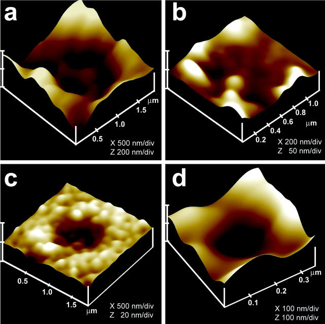
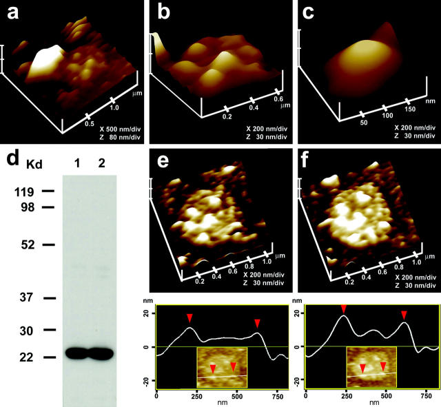
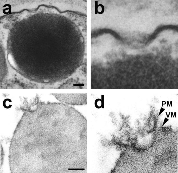
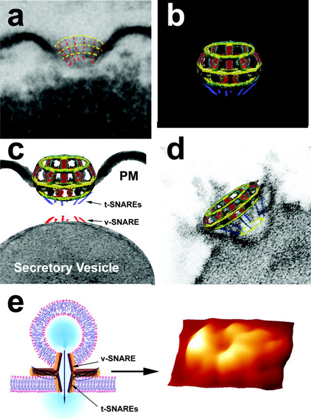
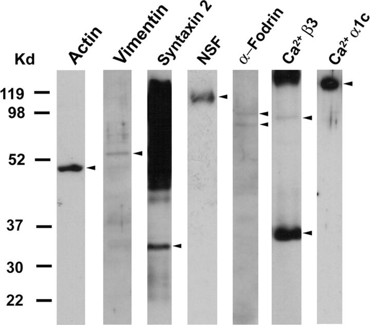
Earlier studies using atomic force microscopy (AFM) demonstrated the presence of fusion pores at the cell plasma membrane in a number of live secretory cells, revealing their morphology and dynamics at nm resolution and in real time. Fusion pores were stable structures at the cell plasma membrane where secretory vesicles dock and fuse to release vesicular contents. In the present study, transmission electron microscopy confirms the presence of fusion pores and reveals their detailed structure and association with membrane-bound secretory vesicles in pancreatic acinar cells. Immunochemical studies demonstrated that t-SNAREs, NSF, actin, vimentin, alpha-fodrin and the calcium channels alpha1c and beta3 are associated with the fusion complex. The localization and possible arrangement of SNAREs at the fusion pore are further demonstrated from combined AFM, immunoAFM, and electrophysiological measurements. These studies reveal the fusion pore or porosome to be a cup-shaped lipoprotein structure, the base of which has t-SNAREs and allows for docking and release of secretory products from membrane-bound vesicles.


Figures

FIGURE 1
FIGURE 1
AFM and immunoAFM micrographs of the fusion pore, demonstrating pore morphology and the release of secretory products at the site. (a) A pit with four fusion pores within, found at the apical surface in a live pancreatic acinar cell; (b) After stimulation of secretion, amylase-specific immunogold localizes at the pit and fusion pores within, demonstrating them to be secretory release sites; (c) Some fusion pores demonstrate greater immunogold localization, suggesting more release of amylase from them; (d) AFM micrograph of a single fusion pore in a live acinar cell.
FIGURE 2
FIGURE 2
Morphology of the cytosolic side of the fusion pore revealed in AFM studies on isolated pancreatic plasma membrane preparations. (a) This AFM micrograph of isolated plasma membrane preparation reveals the cytosolic end of a pit with inverted cup-shaped structures, the fusion pores. Note the 600 nm in diameter ZG at the left hand corner of the pit; (b) Higher magnification of the same pit showing clearly the 4–5 fusion pores within; (c) The cytosolic end of a single fusion pore is depicted in this AFM micrograph; (d) Immunoblot analysis of 10 μg and 20 μg of pancreatic plasma membrane preparations, using SNAP-23 antibody, demonstrates a single 23 kDa immunoreactive band; (e and f) The cytosolic side of the plasma membrane demonstrates the presence of a pit with a number of fusion pores within, shown (e) before and (f) after addition of the SNAP-23 antibody. Note the increase in height of the fusion pore cone base revealed by section analysis (bottom panel), demonstrating localization of SNAP-23 antibody to the base of the fusion pore.
FIGURE 3
FIGURE 3
Transmission electron micrograph of the fusion pore showing vertical and lateral structures, giving it a basket-like appearance. (a) Two fusion pores at the apical plasma membrane of a pancreatic acinar cell, with a docked zymogen granule; (b) High resolution image of one of the fusion pores clearly shows three lateral and a number of vertical ridges, giving it a basket-like appearance; (c) An isolated zymogen granule associated with a fusion pore, reveals (d) clearly the lateral and vertical structures of the fusion pore complex. Scale = 100 nm.
FIGURE 4
FIGURE 4
The fusion pore complex. (a) Electron micrograph of a fusion pore, with positions of the vertical and lateral structures depicted by red and yellow dashed lines for clarity; (b) A schematic model of the fusion pore showing (c) the presence of t-SNAREs at the base of the basket, where the v-SNARE associated ZG can dock; (d) Thus, the t- and v-SNAREs associated in opposing bilayers can interact in a circular array (yellow ring) to establish continuity between ZG contents and the fusion pore; (e) When v-SNARE-reconstituted artificial lipid vesicles are allowed to interact with a t-SNARE-reconstituted lipid support, the t-/v-SNAREs interact in a circular array as shown in the AFM micrograph on the right. The pore formed by t-/v-SNARE interaction is conducting, as demonstrated by electrophysiological measurements (Cho et al., 2002e).
FIGURE 5
FIGURE 5
SNAP-23 associated proteins in pancreatic acinar cells. Total pancreatic homogenate was immunoprecipitated using the SNAP-23 specific antibody. The precipitated material was resolved using 12.5% SDS-PAGE, electrotransferred to nitrocellulose membrane, and then probed using antibodies to a number of proteins. Association of SNAP-23 with syntaxin 2; with cytoskeletal proteins actin, α-fodrin, and vimentin; and with calcium channels β3 and α1c, together with the SNARE regulatory protein NSF, is demonstrated (arrowheads). Lanes showing more than one arrowhead suggest presence of isomers or possible proteolytic degradation of the specific protein.

References

    1. Bennett, V. 1990. Spectrin-based membrane skeleton: A multipotential adaptor between plasma membrane and cytoplasm. Physiol. Rev. 70:1029–1065. - PubMed
    1. Bradford, M. M. 1976. A rapid and sensitive method for the quantitation of microgram quantities of protein utilizing the principle of protein-dye binding. Anal. Biochem. 72:248–254. - PubMed
    1. Cho, S.-J., K. Jeftinija, A. Glavaski, S. Jeftinija, B. P. Jena, and L. L. Anderson. 2002a. Structure and dynamics of the fusion pores in live GH-secreting cells revealed using atomic force microscopy. Endocrinology. 143:1144–1148. - PubMed
    1. Cho, S.-J., A. Wakade, G. D. Pappas, and B. P. Jena. 2002b. New structure involved in transient membrane fusion and exocytosis. New York Acad. Sci. 971:254–256. - PubMed
    1. Cho, S.-J., A. S. Quinn, M. H. Stromer, S. Dash, J. Cho, D. J. Taatjes, and B. P. Jena. 2002c. Structure and dynamics of the fusion pore in live cells. Cell Biol. Int. 26:35–42. - PubMed

Publication types

MeSH terms

LinkOut - more resources