. 2003 Feb;84(2 Pt 1):1399-409.
doi: 10.1016/S0006-3495(03)74954-6.

Single channel analysis of the regulation of GIRK1/GIRK4 channels by protein phosphorylation

Affiliations

Single channel analysis of the regulation of GIRK1/GIRK4 channels by protein phosphorylation

Carmen Müllner et al. Biophys J. 2003 Feb.

Abstract

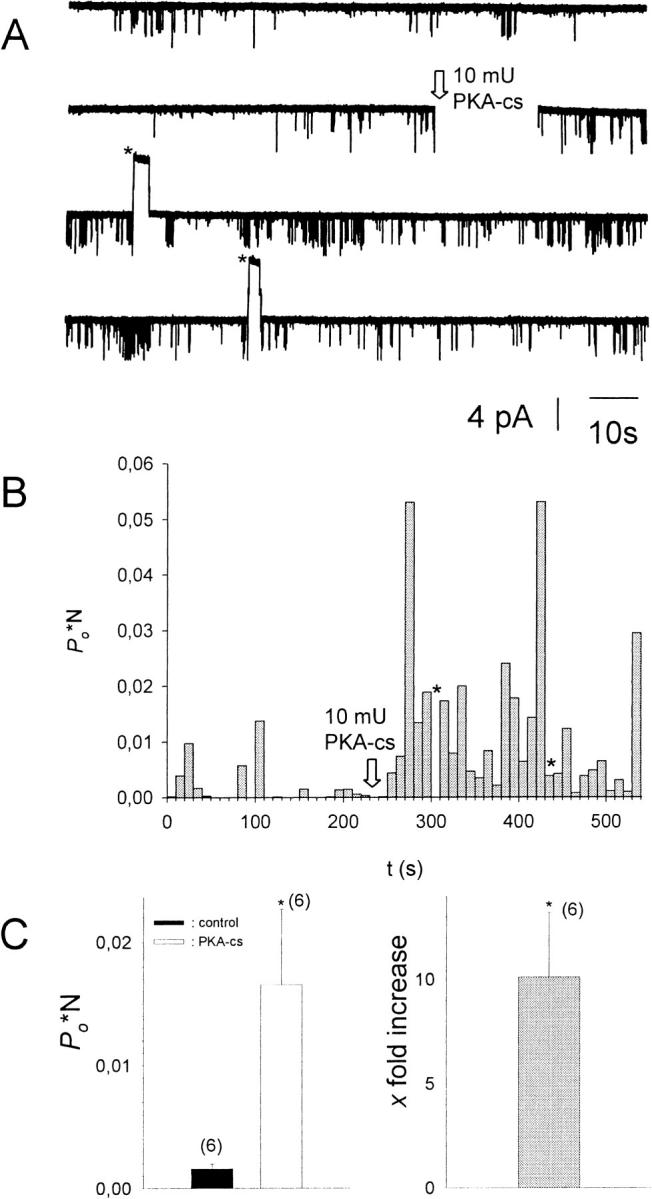
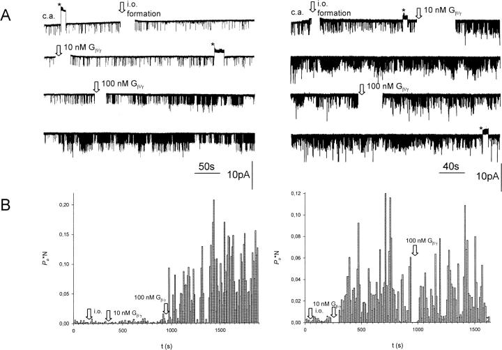
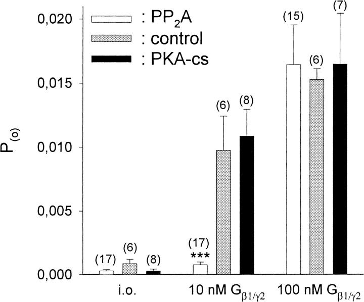
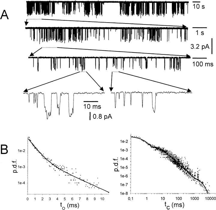
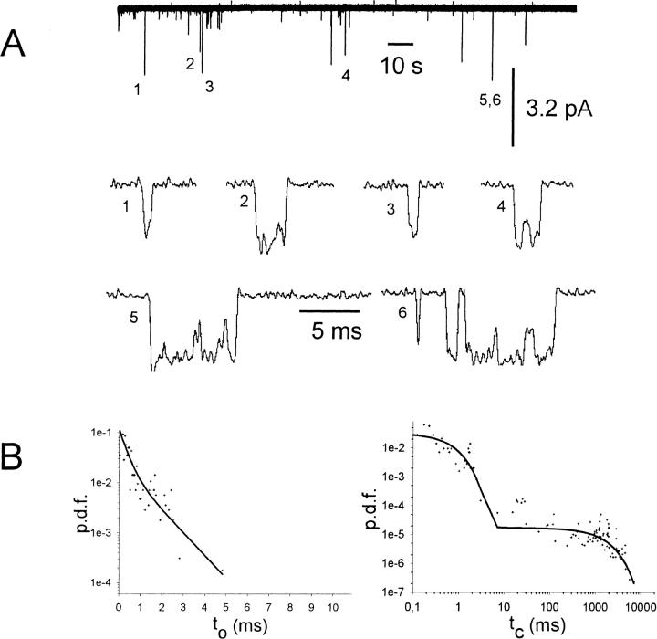
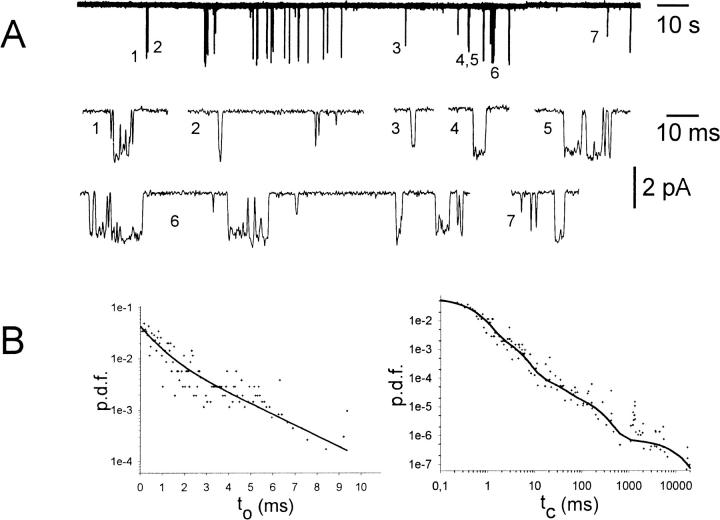
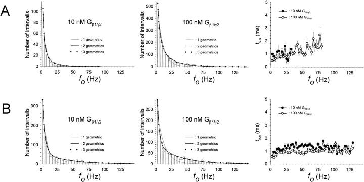
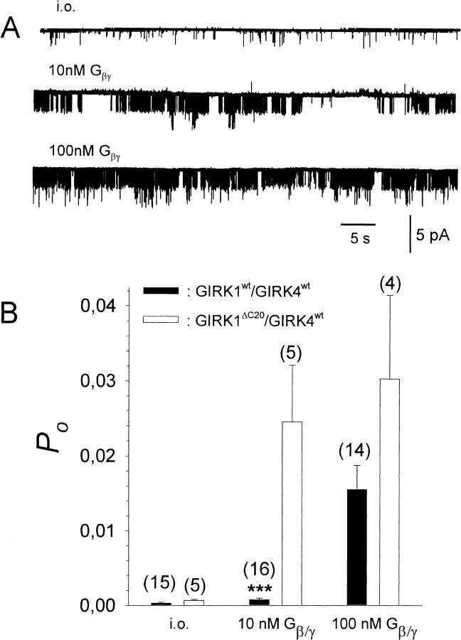
G-Protein activated, inwardly rectifying potassium channels (GIRKs) are important effectors of G-protein beta/gamma-subunits, playing essential roles in the humoral regulation of cardiac activity and also in higher brain functions. G-protein activation of channels of the GIRK1/GIRK4 heterooligomeric composition is controlled via phosphorylation by cyclic AMP dependent protein kinase (PKA) and dephosphorylation by protein phosphatase 2A (PP(2)A). To study the molecular mechanism of this unprecedented example of G-protein effector regulation, single channel recordings were performed on isolated patches of plasma membranes of Xenopus laevis oocytes. Our study shows that: (i) The open probability (P(o)) of GIRK1/GIRK4 channels, stimulated by coexpressed m(2)-receptors, was significantly increased upon addition of the catalytic subunit of PKA to the cytosolic face of an isolated membrane patch. (ii) At moderate concentrations of recombinant G(beta1/gamma2), used to activate the channel, P(o) was significantly reduced in patches treated with PP(2)A, when compared to patches with PKA-cs. (iii) Several single channel gating parameters, including modal gating behavior, were significantly different between phosphorylated and dephosphorylated channels, indicating different gating behavior between the two forms of the protein. Most of these changes were, however, not responsible for the marked difference in P(o) at moderate G-protein concentrations. (iv) An increase of the frequency of openings (f(o)) and a reduction of dwell time duration of the channel in the long-lasting C(5) state was responsible for facilitation of GIRK1/GIRK4 channels by protein phosphorylation. Dephosphorylation by PP(2)A led to an increase of G(beta1/gamma2) concentration required for full activation of the channel and hence to a reduction of the apparent affinity of GIRK1/GIRK4 for G(beta1/gamma2). (v) Although possibly not directly the target of protein phosphorylation/dephosphorylation, the last 20 C-terminal amino acids of the GIRK1 subunit are required for the reduction of apparent affinity for the G-protein by PP(2)A, indicating that they constitute an essential part of the off-switch.


Figures

FIGURE 1
FIGURE 1
Addition of PKA-cs to the cytosolic solution results in an increase of Po*N. (A) Current recorded from a multichannel i.o. patch at −80 mV (N = 3). 10−5 M acetylcholine was present in the pipette solution, 1 mM ATP, and 0.1 mM GTP in the bath solution. Channel openings are represented by downward deflections of the current trace at −80 mV. The arrow indicates addition of 20 mU/ml PKA-cs to the (intracellular) bath solution (total bath volume was 500 μl). Asterisks signify time spans when the membrane potential was switched to +80 mV to test for inward rectification of channel openings. (B) Po*N, calculated for 10-s intervals, versus time, shown for the entire current trace visible in A. (C, left) Average Po*N before (control) and after addition of 20 mU/ml PKA-cs (PKA-cs), obtained from six different patches. (C, right) Average increase in Po*N upon addition of PKA-cs (n = 6). *, The change in Po*N compared to control conditions is statistically significant at the p < 0.05 level.
FIGURE 2
FIGURE 2
Effect of dephosphorylation/phosphorylation by PP2A/PKA-cs treatment on the activation of GIRK1/GIRK4 channels by Gβ1/γ2. (A, left) Current recorded from a multichannel i.o. patch at −80 mV (N = 3). 1 mM ATP and 134 mU/ml PP2A were present in the bath solution. Channel openings are represented by downward deflections of the current trace at −80 mV. First, currents were recorded in the cell attached (c.a.) configuration. The arrows indicate formation of an isolated (i.o.) patch, addition of 10 nM Gβ1/γ2, and later 100 nM Gβ1/γ2 to the bathing solution. Asterisks signify time spans when the membrane potential was switched to +80 mV to test for inward rectification of channel openings. (A, right) Same experimental paradigm as for current trace shown in the left panel, but 20 mU/ml PKA-cs was present in the bath solution instead of PP2A (N = 2). (B) P(o)*N, calculated for 10-s intervals, versus time, shown for the entire current traces visible in A.
FIGURE 3
FIGURE 3
Channel activity (Po) at different concentrations of Gβ1/γ2 for patches treated with PP2A, PKA-cs, or no enzyme added. Number of experiments (n) is given in parentheses above each bar. Number of channels in the patch (N ≥ 1). ***, The average Po for PP2A-treated patches deviates significantly at the p < 0.001 level from Po in patches treated with PKA-cs.
FIGURE 4
FIGURE 4
Single channel trace recorded from an inside-out patch treated with PKA-cs and 100 nM Gβ1/γ2. (A, top) 200 s of history in the lifetime of a single GIRK1/GIRK4 channel, phosphorylated by PKA-cs and activated by Gβ1/γ2. (A, lower traces) Segments of the current trace shown on the top of A, expanded at increasing timescales, to demonstrate the multiplicity of channel closed time distributions over several orders of magnitude in durations. At the most expanded timescale, short and longer lived openings also can be seen. (B, left) Probability density function (p.d.f.) of open time distribution for the current trace shown in A. +, experimental data; (solid line), maximum likelihood regression obtained by fitting the data to the sum of two exponentials. (B, right) The p.d.f. of closed time distributions. Data were fitted by the sum of five exponentials.
FIGURE 5
FIGURE 5
Single channel trace recorded from an inside-out patch under basal conditions. (A, top) 200 s of history in the lifetime of a single GIRK1/GIRK4 channel excised into a bathing solution containing PKA-cs. No recombinant Gβ1/γ2 was present. (A, lower traces) Single openings of the current trace shown on top, shown at expanded timescale. Single (1,2,3,4) as well as multiple openings (5,6) could be detected within a burst, resulting in at least two different kinetically distinguishable closed states. (B) Probability density functions of open (left) and closed (right) time distributions. +, experimental data; solid line, maximum likelihood fit. Both probability density functions (p.d.f.) were fit by the sum of two exponentials.
FIGURE 6
FIGURE 6
Single channel trace recorded from an inside-out patch treated with PP2A and 10 nM Gβ1/γ2. (A, top) 200 s of history in the lifetime of a single GIRK1/GIRK4 channel, dephosphorylated by PP2A and activated by 10 nM Gβ1/γ2. (A, lower traces) Single and multiple openings of the current trace shown on top and shown below at expanded timescale. (B, left) Probability density function (p.d.f.) of open time distribution for the current trace shown in A. +, experimental data; (solid line), least square regression obtained by fitting the data to the sum of two exponentials. (B, right) The p.d.f. of closed time distribution. Data were fitted by the sum of five exponentials.
FIGURE 7
FIGURE 7
Modal gating of single phosphorylated/dephosphorylated GIRK1/GIRK4 channels. (A) Frequency distributions obtained from patches treated with PP2A at 10 nM Gβ1/γ2 (left) and at 100 nM Gβ1/γ2 (middle). Mean channel open time (to,s) as a function of fo is shown for both 10 nM and 100 nM Gβ1/γ2 (right). (B) Frequency distributions and distributions of to,s, but for patches treated with PKA-cs.
FIGURE 8
FIGURE 8
Effect of deletion of the last 20 C-terminal amino acids on the activation by Gβ1/γ2 in the presence of PP2A. (A) Current trace recorded at −80 mV from an i.o. patch containing multiple GIRK1ΔC20/GIRK4wt channels (N = 3) at increasing concentrations of Gβ1/γ2. (B) Comparisons of Po at different concentrations of recombinant Gβ1/γ2 for GIRK1wt/GIRK4wt and GIRK1ΔC20/GIRK4wt channels. ***, The average Po for patches containing GIRK1wt/GIRK4wt channels deviates significantly (p < 0.001) from patches containing channels of the GIRK1ΔC20/GIRK4wt composition.

References

    1. Bard, J., M. T. Kunkel, and E. G. Peralta. 2000. Single channel studies of inward rectifier potassium channel regulation by muscarinic acetylcholine receptors. J. Gen. Physiol. 116:645–651. - PMC - PubMed
    1. Dascal, N., and I. Lotan. 1992. Expression of exogenous ion channels and neurotransmitter receptors in RNA-injected Xenopus oocytes. In Methods in Molecular Neurobiology, Vol. 13. A. Longstaff and P. Revest, editors. Humana Press, Totowa, New Jersey. 205–225.
    1. Dascal, N. 1997. Signalling via the G protein-activated K+ channels. Cell. Signal. 9:551–573. - PubMed
    1. Dascal, N., C. A. Doupnik, T. Ivanina, S. Bausch, W. Wang, C. Lin, J. Garvey, C. Chavkin, H. A. Lester, and N. Davidson. 1995. Inhibition of function in Xenopus oocytes of the inwardly rectifying G-protein-activated atrial K channel (GIRK1) by overexpression of a membrane-attached form of the C-terminal tail. Proc. Natl. Acad. Sci. USA. 92:6758–6762. - PMC - PubMed
    1. Dascal, N., W. Schreibmayer, N. F. Lim, W. Wang, C. Chavkin, L. DiMagno, C. Labarca, B. L. Kieffer, C. Gaveriaux-Ruff, D. Trollinger, H. A. Lester, and N. Davidson. 1993. Atrial G protein-activated K+ channel: expression cloning and molecular properties. Proc. Natl. Acad. Sci. USA. 90:10235–10239. - PMC - PubMed

Publication types

MeSH terms

Substances

LinkOut - more resources