. 2003 Feb 4;100(3):1298-303.
doi: 10.1073/pnas.0337541100. Epub 2003 Jan 23.

Abrogation of postentry restriction of HIV-1-based lentiviral vector transduction in simian cells

Affiliations

Abrogation of postentry restriction of HIV-1-based lentiviral vector transduction in simian cells

Neeltje A Kootstra et al. Proc Natl Acad Sci U S A. .

Abstract

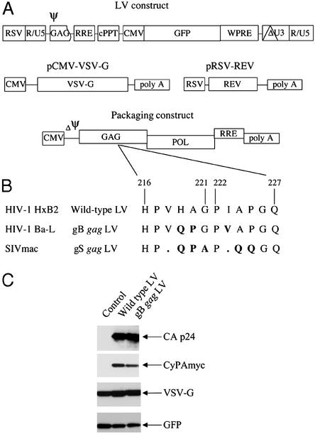
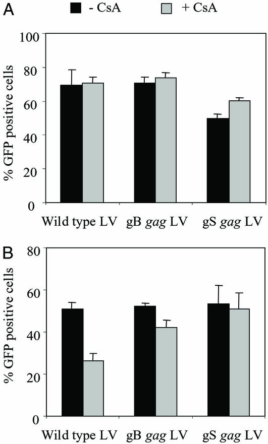
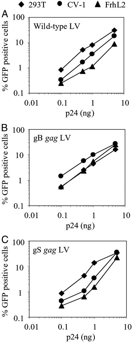
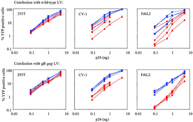
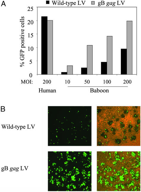
HIV-1 replication in simian cells is restricted at an early postentry step because of the presence of an inhibitory cellular factor. This block reduces the usefulness of HIV-1-based lentiviral vectors in primate animal models. Here, we demonstrate that substitution of the cyclophilin A (CyPA) binding region in the capsid of an HIV-1-based lentiviral vector (LV) with that of the macrophage tropic HIV-1 Ba-L resulted in a vector that was resistant to the inhibitory effect and efficiently transduced simian cells. Notably, the chimeric gag LV efficiently transduced primary simian hematopoietic progenitor cells, a critical cellular target in gene therapy. The alterations in the CyPA binding region did not affect CyPA incorporation; however, transduction by the gag chimeric LV seemed to be relatively insensitive to cyclosporin A, indicating that it does not require CyPA for early postentry steps. In dual infection experiments, the gag chimeric LV failed to remove the block to transduction of the WT LV, suggesting that the gag chimeric LV did not saturate the inhibitory simian cellular factor. These data suggest that the CyPA binding region of capsid contains a viral determinant involved in the postentry restriction of HIV-1-based lentiviral vectors. Overall, the findings demonstrate that the host range of HIV-1-based LV can be altered by modifications in the packaging construct.


Figures

Figure 1
Figure 1
(A) Schematic presentation of the HIV-1-based LV packaging construct (27). LVs are produced by cotransfections of four different plasmid constructs into 293T cells. The viral RNA genome is produced from the LV construct and contains the promoter and transgene sequences. In addition, the LV construct contains the following cis-acting sequences: packaging signal (Ψ) comprising the 5′ UTR and the 5′ sequence of the gag ORF, the RRE, the cPPT, and the woodchuck hepatitis virus posttranscriptional regulatory element. The 3′ LTR contains a large deletion in the U3 region (depicted as ΔU3). The LV packaging system consists of three constructs: the packaging construct, pRSV-rev, and pCMV-VSV-G. The packaging construct contains in addition the cis-acting RRE and lacks a packaging signal (ΔΨ). (B) Amino acid sequence alignment of the gag CyPA binding region of the WT, gB, and gS gag LV packaging constructs. The WT LV was generated from HIV-1 HxB2. The gB gag LV contains the CyPA binding region of the macrophage tropic HIV-1 Ba-L, and the gS gag LV contains the corresponding region of SIVmac. (C) Western blot analysis for CyPA incorporation into LV virions. LVs were produced in 293T cells by transient transfection of the LV construct expressing GFP, the packaging construct for WT or gB gag LV, pRSV-rev, and pCMV-VSV-G. Myc-tagged CyPA was expressed from an additional construct. In the control cells, the packaging construct was not cotransfected. Virions were isolated on a sucrose gradient, and virion-associated proteins were analyzed by using antibodies against p24, myc, VSV-G, and GFP.
Figure 2
Figure 2
The effect of CsA treatment on the transduction efficiency of the WT and gag chimeric LVs. (A) Producer cells. WT and chimeric gag LVs expressing GFP were produced in the presence of CsA (5 μg/ml), and their transduction efficiency was analyzed in 293T cells at an inoculum of 5 ng of p24. The average transduction efficiency of two independent experiments is given. Black bar, LVs produced in the absence of CsA; gray bar, LVs produced in the presence of CsA. (B) Target cells. 293T cells were inoculated with 5 ng of p24 of GFP expressing LV in the absence or presence of CsA (1 μg/ml). Five days after inoculation, transduction efficiencies were analyzed by FACS. The average inhibition observed in six independent experiments is given. Black bar, no CsA treatment; gray bar, CsA treatment.
Figure 3
Figure 3
Efficient transduction of simian cells by gB gag LV. 293T (⧫), CV-1 (●), and FrhL2 (▴) cells were transduced with increasing amounts of WT (A), gB (B), and gS (C) gag LVs expressing GFP. Six days after inoculation, transduction efficiencies were analyzed by FACS.
Figure 4
Figure 4
The reduced transduction of WT LV in simian cells cannot be overcome by coinfection with the gB gag LV. 293T, CV-1, and FrhL2 cells were inoculated with increasing amounts of WT (red) and gB gag (blue) LV expressing YFP. Coinfection was performed with WT (Upper) and gB gag (Lower) LV expressing CFP at the following concentrations: 1 ng of p24 (■), 5 ng of p24 (⧫), 10 ng of p24 (●), and no coinfection (▴). Six days after inoculation, YFP expression was analyzed by FACS.
Figure 5
Figure 5
Enhanced transduction of primary baboon hematopoietic progenitor cells by gB gag LV. (A) Baboon CD34+ hematopoietic progenitor cells were inoculated with the WT (black bar) and the gB gag LV (gray bar) at an moi of 10, 50, 100, and 200. In parallel, human CD34+ hematopoietic progenitor cells were inoculated with the WT (black bar) and the gB gag LV (gray bar) at an moi of 200. GFP expression was analyzed at day 5 after inoculation by FACS. (B) GFP expression in baboon CD34+ hematopoietic progenitor cell cultures transduced with WT or gB gag LV at an moi of 200. (Left) Fluorescence microscope image. (Right) Merge of fluorescence and light microscopy image (×100).

References

    1. Deng H, Liu R, Ellmeier W, Choe S, Unutmaz D, Burkhart M, Di Marzio P, Marmon S, Sutton R E, Hill C M, et al. Nature. 1996;381:661–666. - PubMed
    1. Dragic T, Litwin V, Allaway G P, Martin S R, Huang Y, Nagashima K A, Cayanan C, Maddon P J, Koup R A, Moore J P, Paxton W A. Nature. 1996;381:667–673. - PubMed
    1. Zack J A, Arrigo S J, Weitsman S R, Go A S, Haislip A, Chen I S. Cell. 1990;61:213–222. - PubMed
    1. Korin Y D, Zack J A. J Virol. 1998;72:3161–3168. - PMC - PubMed
    1. Schuitemaker H, Kootstra N A, Fouchier R A, Hooibrink B, Miedema F. EMBO J. 1994;13:5929–5936. - PMC - PubMed

Publication types

MeSH terms

LinkOut - more resources