. 2003 Jan-Feb;10(1):26-39.
doi: 10.1101/lm.51103.

A role for ERK MAP kinase in physiologic temporal integration in hippocampal area CA1

Affiliations

A role for ERK MAP kinase in physiologic temporal integration in hippocampal area CA1

Joel C Selcher et al. Learn Mem. 2003 Jan-Feb.

Abstract

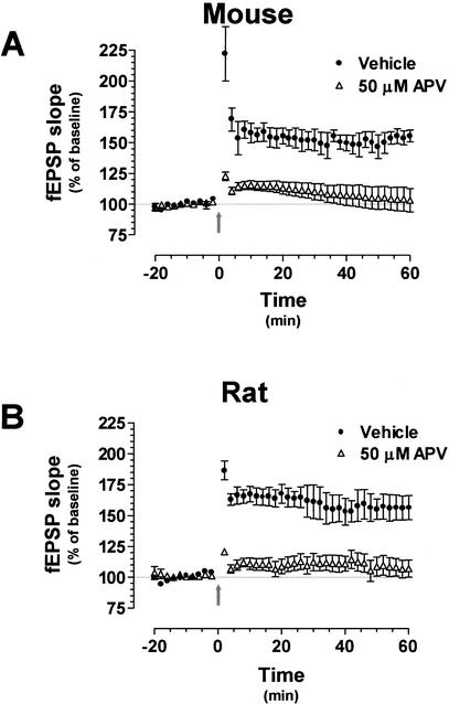
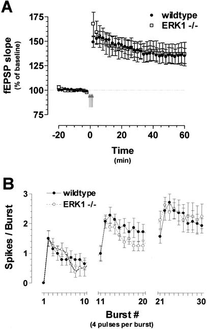
Recent studies demonstrate a requirement for the Extracellular signal Regulated Kinase (ERK) mitogen-activated protein kinase (MAPK) cascade in both the induction of long-lasting forms of hippocampal synaptic plasticity and in hippocampus-dependent associative and spatial learning. In the present studies, we investigated mechanisms by which ERK might contribute to synaptic plasticity at Schaffer collateral synapses in hippocampal slices. We found that long-term potentiation (LTP) induced with a pair of 100-Hz tetani does not require ERK activation in mice whereas it does in rats. However, in mice, inhibition of ERK activation blocked LTP induced by two LTP induction paradigms that mimicked the endogenous theta rhythm. In an additional series of studies, we found that mice specifically deficient in the ERK1 isoform of MAPK showed no impairments in tests of hippocampal physiology. To investigate ERK-dependent mechanisms operating during LTP-inducing stimulation paradigms, we monitored spike production in the cell body layer of the hippocampus during the period of theta-like LTP-inducing stimulation. Theta-burst stimulation (TBS) produced a significant amount of postsynaptic spiking, and the likelihood of spike production increased progressively over the course of the three trains of TBS independent of any apparent increase in Excitatory Post-Synaptic Potential (EPSP) magnitude. Inhibition of ERK activation dampened this TBS-associated increase in spiking. These data indicate that, for specific patterns of stimulation, ERK may function in the regulation of neuronal excitability in hippocampal area CA1. Overall, our data indicate that the progressive increase in spiking observed during TBS represents a form of physiologic temporal integration that is dependent on ERK MAPK activity.


Figures

Figure 1
Figure 1
100-Hz long-term potentiation (LTP) does not require ERK activation in mouse hippocampus. (A) LTP induced with a pair of 100-Hz tetani in mouse hippocampal slices in the presence of either vehicle (n = 19), 10 μM SL327 (n = 11), or 20 μM U0126 (n = 10). Inset, representative traces from vehicle- and SL327-treated mouse slices before (gray) and after (black) tetanization. Scale bars are 1 mV by 8 msec. (B) LTP induced with this same induction protocol in rat hippocampal slices in the presence of either vehicle (n = 10), 10 μM SL327 (n = 7), or 20 μM U0126 (n = 5). Inset, representative traces from vehicle- and SL327-treated mouse slices before (gray) and after (black) tetanization. Scale bars are 1 mV by 8 msec. fEPSP = field Excitatory Postsynaptic Potential
Figure 2
Figure 2
100-Hz long-term potentiation (LTP) is NMDA receptor-dependent in the mouse hippocampus. (A) LTP induced with a pair of 100-Hz tetani in the presence of either vehicle (n = 5) or 50 μM APV (n = 4). High-frequency stimulation (HFS)–LTP was completely blocked by the NMDA receptor antagonist in both sets of hippocampal slices. (B) Hippocampal slices prepared from rat brains were exposed to the same LTP-induction paradigm in the presence of either vehicle (n = 6) or 50 μM APV (n = 5). HFS–LTP was completely blocked by the NMDA receptor antagonist in both sets of hippocampal slices. fEPSP = field Excitatory Postsynaptic Potential
Figure 3
Figure 3
MEK inhibitors effectively block ERK activation in the mouse hippocampus. Representative Western blots and densitometric analysis of ERK2 (p42 mitogen-activated protein kinase) activation in area CA1 of mouse hippocampal slices treated with vehicle (C; gray bar, n = 8), 20 μM U0126 (+U0; white bar, n = 9), 50 μM forskolin (FSK; black bar, n = 6), or forskolin plus U0126 (FSK+U0; striped bar, n = 6). **P < 0.001.
Figure 4
Figure 4
U0126 impairs long-term potentiation (LTP) induced with θ frequency stimulation (TFS) in the mouse hippocampus. (A) Schematic depicting TFS. This LTP induction paradigm consists of 150 single pulses delivered at 5 Hz. (B) LTP induced with TFS (TFS–LTP) is impaired in the presence of the MEK inhibitor (n = 11 slices) compared with vehicle-treated control slices (n = 11 slices). The thick arrow represents the 30 sec of TFS. fEPSP = field Excitatory Postsynaptic Potential
Figure 5
Figure 5
Long-term potentiation (LTP) initiated by θ-burst stimulation (TBS) requires ERK activation in the mouse hippocampus. (A) Schematic depicting TBS. This LTP induction paradigm consists of three trains of 10 high-frequency bursts delivered at 5 Hz. (B) LTP induced with TBS (TBS–LTP is significantly impaired in the presence of 20 μM U0126 (n = 13 slices) compared with vehicle-treated control slices (n = 12 slices). The three red arrows represent the three TBS trains. fEPSP = field Excitatory Postsynaptic Potential
Figure 6
Figure 6
Increased action potential firing over the course of θ-burst stimulation (TBS) is blocked by MEK inhibitors. (A) Representative traces in response to TBS from a vehicle-treated slice. Note the profound difference in spiking between the first and last bursts of the stimulation paradigm. Scale bars (bottom right corner of shaded box) are 1 mV by 5 msec. (B) Representative traces in response to TBS from a slice treated with U0126. Compared with controls, there is much less difference in spiking between the first and last bursts of the stimulation paradigm. Scale bars are 1 mV by 5 msec. (C) Increased spiking during TBS is modulated by ERK. Population spike counts recorded in stratum pyramidale of hippocampal area CA1 during TBS in slices pretreated with vehicle (n = 13 slices) or 20μM U0126 (n = 11 slices). Slices exposed to vehicle showed a progressive increase in spike generation during TBS; administration of U0126 impaired this enhanced spiking.
Figure 6
Figure 6
Increased action potential firing over the course of θ-burst stimulation (TBS) is blocked by MEK inhibitors. (A) Representative traces in response to TBS from a vehicle-treated slice. Note the profound difference in spiking between the first and last bursts of the stimulation paradigm. Scale bars (bottom right corner of shaded box) are 1 mV by 5 msec. (B) Representative traces in response to TBS from a slice treated with U0126. Compared with controls, there is much less difference in spiking between the first and last bursts of the stimulation paradigm. Scale bars are 1 mV by 5 msec. (C) Increased spiking during TBS is modulated by ERK. Population spike counts recorded in stratum pyramidale of hippocampal area CA1 during TBS in slices pretreated with vehicle (n = 13 slices) or 20μM U0126 (n = 11 slices). Slices exposed to vehicle showed a progressive increase in spike generation during TBS; administration of U0126 impaired this enhanced spiking.
Figure 7
Figure 7
Effects of APV and bicuculline on increased action potential firing with θ-burst stimulation (TBS). (A) Population spike counts recorded in stratum pyramidale of hippocampal area CA1 during TBS in slices pretreated with vehicle (n = 6 slices), 30 μM bicuculline (n = 6 slices), 5 μM APV (n = 7 slices), or 5 μM APV/20 μM U0126 (n = 14 slices). All slices showed a progressive increase in spike generation during TBS. (B) Results from panel A showing the average of each set of bursts. Compared with vehicle treated slices for the first set of bursts (bursts 1–10), bicuculline treated slices showed an increase in spike generation. Slices treated with APV showed a significant reduction in spike generation, which was attenuated further with the combination of APV and U0126. (*P = 0.0001).
Figure 8
Figure 8
Increased population spike amplitude over the course of θ frequency stimulation (TFS). (A) Representative traces in response to TFS (5 Hz, 30 sec) from a vehicle-treated slice. During TFS, the amplitude of the population spike grows, whereas the slope of the EPSP gradually decreases. (B) Representative traces in response to TFS (5 Hz, 30 sec) from a U0126-treated slice. Although U0126 impairs TFS-long-term potentiation (LTP), the drug appears to have little effect on the enhancement of the population spike during the stimulation paradigm. (C) Transient E-S potentiation during TFS in the mouse hippocampus. The ratio of the population spike (measured in stratum pyramidale) to the EPSP slope (measured in stratum radiatum) was investigated during TFS. Over the course of the 30 sec of stimulation, the pSpike:EPSP ratio was enhanced. This alteration in E-S coupling was characterized by a gradual decline in the EPSP slope in conjunction with increasing population spike amplitude during TFS. The data was normalized to the initial S-E ratio measured 1 sec into the LTP-inducing stimulation.
Figure 8
Figure 8
Increased population spike amplitude over the course of θ frequency stimulation (TFS). (A) Representative traces in response to TFS (5 Hz, 30 sec) from a vehicle-treated slice. During TFS, the amplitude of the population spike grows, whereas the slope of the EPSP gradually decreases. (B) Representative traces in response to TFS (5 Hz, 30 sec) from a U0126-treated slice. Although U0126 impairs TFS-long-term potentiation (LTP), the drug appears to have little effect on the enhancement of the population spike during the stimulation paradigm. (C) Transient E-S potentiation during TFS in the mouse hippocampus. The ratio of the population spike (measured in stratum pyramidale) to the EPSP slope (measured in stratum radiatum) was investigated during TFS. Over the course of the 30 sec of stimulation, the pSpike:EPSP ratio was enhanced. This alteration in E-S coupling was characterized by a gradual decline in the EPSP slope in conjunction with increasing population spike amplitude during TFS. The data was normalized to the initial S-E ratio measured 1 sec into the LTP-inducing stimulation.
Figure 9
Figure 9
Long-term potentiation (LTP) is unimpaired in ERK1 knockout mice. High-frequency stimulation-LTP is unimpaired in ERK1 knockout mice. (A) Mice lacking the ERK1 isoform display normal θ-burst stimulation (TBS)-LTP. θ-burst stimulation consists of three trains of 10 high-frequency bursts delivered at 5 Hz. LTP induced with TBS (TBS–LTP) was normal in ERK1 knockout mice (n = 10 slices) compared with wild-type controls (n = 14 slices). The three arrows represent the three TBS trains. fEPSP = field Excitatory Postsynaptic Potential (B) ERK1 null mice exhibit normal postsynaptic spiking during TBS. Population spike counts recorded in stratum pyramidale of hippocampal area CA1 during TBS in slices taken from ERK1-deficient mice (n = 8 slices) or wild-type controls (n = 14 slices). Both sets of slices showed significant increases in action potential firing, and there was no difference in spike counts between the two strains.

References

    1. Adams JP, Anderson AE, Varga AW, Dineley KT, Cook RG, Pfaffinger PJ, Sweatt JD. The A-type potassium channel Kv4.2 is a substrate for the mitogen-activated protein kinase ERK. J Neurochem. 2000;75:2277–2287. - PubMed
    1. Atkins CM, Chen SJ, Klann E, Sweatt JD. Increased phosphorylation of myelin basic protein during hippocampal long-term potentiation. J Neurochem. 1997;68:1960–1967. - PubMed
    1. Atkins CM, Selcher JC, Petraitis JJ, Trzaskos JM, Sweatt JD. The MAPK cascade is required for mammalian associative learning. Nat Neurosci. 1998;1:602–609. - PubMed
    1. Bland BH, Seto MG, Sinclair BR, Fraser SM. The pharmacology of hippocampal θ cells: Evidence that the sensory processing correlate is cholinergic. Brain Res. 1984;299:121–131. - PubMed
    1. Bliss TVP, Lomo T. Long-lasting potentiation of synaptic transmission in the dentate area of the anaesthetized rabbit following stimulation of the perforant path. J Physiol. 1973;232:331–356. - PMC - PubMed

Publication types

MeSH terms

LinkOut - more resources