. 2003 Feb;77(4):2385-99.
doi: 10.1128/jvi.77.4.2385-2399.2003.

Characterization of a cytolytic strain of equine infectious anemia virus

Affiliations

Characterization of a cytolytic strain of equine infectious anemia virus

Wendy Maury et al. J Virol. 2003 Feb.

Abstract

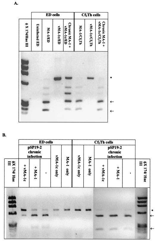
A novel strain of equine infectious anemia virus (EIAV) called vMA-1c that rapidly and specifically killed infected equine fibroblasts (ED cells) but not other infectible cell lines was established. This strain was generated from an avirulent, noncytopathic strain of EIAV, MA-1. Studies with this new cytolytic strain of virus have permitted us to define viral parameters associated with EIAV-induced cell killing and begin to explore the mechanism. vMA-1c infection resulted in induction of rapid cell death, enhanced fusogenic activity, and increased rates of spread in equine fibroblasts compared to other strains of EIAV. The highly cytolytic nature of vMA-1c suggested that this strain might be superinfecting equine fibroblasts. Receptor interference studies demonstrated that prior infection of equine fibroblasts with EIAV did not alter the ability of vMA-1c to infect and kill these cells. In similar studies in a canine fibroblast cell line, receptor interference did occur. vMA-1c infection of equine fibroblasts was also associated with large quantities of unintegrated viral DNA, a well-established hallmark of retroviral superinfection. Cloning of the vMA-1c genome identified nucleotide changes that would result in at least one amino acid change in all viral proteins. A chimeric infectious molecular clone containing the vMA-1c tat, S2, and env open reading frames recapitulated most of the characteristics of vMA-1c, including superinfection, fibroblast killing, and fusogenic activity. In summary, in vitro selection for a strain of EIAV that rapidly killed cells resulted in the generation of a virus that was able to superinfect these cells, presumably by the use of a novel mechanism of cell entry. This phenotype mapped to the 3' half of the genome.


Figures

FIG. 1.
FIG. 1.
vMA-1c infection of equine dermal fibroblasts (ED cells). All cells were immunostained for EIAV antigens with horse anti-EIAV antiserum. (A) An uninfected ED cell culture. (B) vMA-1c-infected ED cell culture fixed and stained 2 days postinfection. Syncytia are prominent at this time. (C) vMA-1c-infected ED cell culture fixed and stained 5 days postinfection. (D) Immunostaining of ED cells chronically infected with parental MA-1 virus. Magnification, ×100.
FIG. 2.
FIG. 2.
Characterization of vMA-1c infection. (A) vMA-1c spreads faster in ED cells than MA-1. Equivalent infectious units of vMA-1c or MA-1 were added to duplicate wells of ED cells (≈2 × 10⁴/well), and infected wells were fixed at the times noted. Fixed cells were immunostained for EIAV antigens. (B) vMA-1c does not spread faster in Cf2Th cells than MA-1. Duplicate wells of Cf2Th cells (≈2 × 10⁴/well) were infected with vMA-1c or MA-1, and wells were fixed at the times noted. Fixed cells were immunostained for EIAV antigens, and the numbers of infected cells/well were determined. (C) vMA-1c killing of ED cells. A total of 1,000 infectious units of vMA-1c, MA-1, or pSP19-2 were added to triplicate cultures (≈2 × 10⁴/well) and maintained for 6 days. At the termination of the infection, cells were washed with phosphate-buffered saline and lysed. Detection of cellular ATP levels was determined with the ATPLite-M assay as a measure of cell viability. (D) Absence of vMA-1c cytolysis of Cf2Th cells. Assays were performed as described for C. Experiments were performed at least three times, and all treatments were performed in duplicate or triplicate. Shown are means of representative experiments ± standard deviation or range.
FIG. 3.
FIG. 3.
Nucleotide sequence of LTR and deduced amino acid sequences of vMA-1c open reading frames. Two long-template PCR-generated clones of the 5′ (5′ LTRPCR-2 and -3) and the 3′ (7886-1 and 7886-6) halves of the genome were sequenced and compared to parental MA-1 sequences. An NcoI site present in the integrase gene is the 3′ terminus for the 5′-half clones and the 5′ terminus for the 3′ clones. Dashes represent the identical amino acid (or nucleic acid within the LTR), and an asterisk represents a stop codon in the clones that is not present in MA-1. The number symbol represents the location of the NcoI site in the integrase gene. The amino acids, proline and tryptophan, which are located at the end of the 5′ clones and the beginning of the 3′ clones were duplicated during cloning and are shown in both sets of clones.
FIG. 3.
FIG. 3.
Nucleotide sequence of LTR and deduced amino acid sequences of vMA-1c open reading frames. Two long-template PCR-generated clones of the 5′ (5′ LTRPCR-2 and -3) and the 3′ (7886-1 and 7886-6) halves of the genome were sequenced and compared to parental MA-1 sequences. An NcoI site present in the integrase gene is the 3′ terminus for the 5′-half clones and the 5′ terminus for the 3′ clones. Dashes represent the identical amino acid (or nucleic acid within the LTR), and an asterisk represents a stop codon in the clones that is not present in MA-1. The number symbol represents the location of the NcoI site in the integrase gene. The amino acids, proline and tryptophan, which are located at the end of the 5′ clones and the beginning of the 3′ clones were duplicated during cloning and are shown in both sets of clones.
FIG. 4.
FIG. 4.
vMA-1c kills ED cells that are chronically infected with wild-type EIAV but not chronically infected Cf2Th cells. MA-1, vMA-1c, or pSP19-2 was incubated with (A) uninfected ED cells, (B) uninfected Cf2Th cells, (C) chronically pSP19-2-infected ED cells, (D) chronically pSP19-2-infected Cf2Th cells, (E) chronically MA-1-infected ED cells, or (F) chronically MA-1-infected Cf2Th cells. All chronically infected cultures were >98% EIAV antigen positive at the time of superinfection. At the termination of the infections, cells were lysed and cellular ATP levels were determined with ATPLite-M as a measure of cell viability. Experiments were performed at least three times, and all treatments were performed in triplicate in each experiment. Shown are means of a representative experiment ± standard deviation of the mean. The percentage of the uninfected-control value is shown above each bar.
FIG. 5.
FIG. 5.
Detection of vMA-1c superinfection of ED cells. (A) Elevated levels of unintegrated DNA in vMA-1c-infected ED cells detected by Southern blotting. ED cells were acutely infected with virus, and Hirt supernatants were extracted at day 4 postinfection. In addition, chronically infected (>98% EIAV antigen positive) pSP19-2-infected ED cells were superinfected with either MA-1 or vMA-1c for 4 days prior to Hirt supernatant extraction. Southern blot analysis was performed on 5 μg of undigested low-molecular-weight DNA. DNA from a full-length clone of EIAV, p29A, was used as the probe. A total of 2.4 ng (not shown), 7.2 ng, and 28.2 ng of EcoRI-digested p29a that is 8.3 kb in length (6) was run in parallel to identify full-length linear viral sequences.
FIG. 6.
FIG. 6.
Restriction site polymorphism analysis of PCR-amplified DNA from Hirt supernatants demonstrated that vMA-1c is able to superinfect ED cells but not Cf2Th cells. Naïve or chronically infected ED cells were infected with MA-1 or vMA-1c for 4 days. Low-molecular-weight (Hirt) DNA was isolated from infected cells, and DNA was amplified with EIAV env primers 6743 and 7426C′. (A) vMA-1c is able to superinfect MA-1-infected ED cells but not MA-1-infected Cf2Th cells. env DNA was amplified from MA-1-infected cells and contained two MscI sites producing 286-, 200-, and 194-bp bands upon MscI digestion. The sites were not present in vMA-1c, resulting in a 683-bp band. MscI did not cutthe MA-1-amplified DNA to completion, presumably due to restriction site polymorphisms present in the MA-1 stock, and partially digested bands are evident in the gel. An asterisk indicates the 683-bp band, and the arrows identify the completely digested fragments. (B) vMA-1c is able to superinfect pSP19-2-infected ED cells but not pSP19-2-infected Cf2Th cells. The parental MA-1 virus was unable to superinfect either of the chronically pSP19-2-infected cell populations. Low-molecular-weight DNA was PCR amplified with primers 6743 and 7426C′ from chronically pSP19-2-infected ED and Cf2Th cells superinfected with vMA-1c or MA-1. Restriction site polymorphism analysis was then performed with BstEII digestion. pSP19-2 contains a BstEII site, producing 439- and 244-bp bands upon digestion; the site was not present in vMA-1c or MA-1, resulting in a 683-bp band. An asterisk indicates the 683-bp band, and the arrows identify the completely digested fragments.
FIG. 7.
FIG. 7.
Neutralizing antibodies block vMA-1c-induced ED killing. (A) Serial dilutions of horse anti-EIAV antibodies (AB) (6) directed against the MA-1 strain of virus were incubated with vMA-1c for 30 min prior to addition to ED cells. A multiplicity of infection of either 0.0035 (1×) or 0.035 (10×) was used to determine virus neutralization. Cells were fixed and immunostained for virus antigens at 40 h postinfection. (B) Either 0, 500, or 5,000 infectious units of vMA-1c were incubated for 30 min with a 1:256 dilution of neutralizing antiserum and then added to 2 × 10⁴ ED cells. At 7 days postinfection, cells were washed and lysed, and cellular ATP levels were determined by ATPLite-M as a measure of cell viability. (C) Either 50, 500, or 5,000 infectious units of vMA-1c were incubated with ED cells (2 × 10⁴) for 2 h. Medium was removed, and medium containing a 1:256 dilution of horse anti-EIAV was added for the remainder of the study. At 7 days postinfection, cells were washed and lysed, and cellular ATP levels were determined by ATPLite-M as a measure of cell viability. Experiments were performed at least three times, and infections were performed in quadruplicate in each experiment. Shown are means of a representative experiment ± standard deviation of the mean.
FIG. 8.
FIG. 8.
Chimeric EIAV infectious clones containing vMA-1c tat, S2, and env sequences resulted in increased syncytium formation within the ED cell culture, decreased cell numbers, and EIAV-infected ED cell superinfection. (A) Schematic diagrams of the chimeric clones p19/vEnvltr and p19/vEnv. (B) A photomicrograph of p19/vEnv fusogenic activity. Magnification, ×200. Syncytia were readily apparent in cultures infected with the chimeric virus 4 days postinfection. (C) Cell death resulted from p19/vEnv infection of ED cells. ED cells were infected at 24-h intervals with either vMA-1c or p19/vEnv. MTT assays were performed at the termination of the experiment as a measure of cell viability. Cell killing by p19/vEnv lagged behind vMA-1c killing by approximately 2 days. (D) Chronic EIAV infection does not protect ED cells from p19/vEnv-induced killing. ED cells or ED cells that were chronically infected with either MA-1 or pSP19-2 were infected with vMA-1c or p19/vEnv. At 8 days postinfection, cell viability was determined by ATPLite-M assay. The percentage of the uninfected-control value is shown above each bar.

Similar articles

Cited by

References

    1. Adkins, H. B., S. C. Blacklow, and J. A. Young. 2001. Two functionally distinct forms of a retroviral receptor explain the nonreciprocal receptor interference among subgroups B, D, and E avian leukosis viruses. J. Virol. 75:3520-3526. - PMC - PubMed
    1. Anderson, M. M., A. S. Lauring, C. C. Burns, and J. Overbaugh. 2000. Identification of a cellular cofactor required for infection by feline leukemia virus. Science 287:1828-1830. - PubMed
    1. Berlioz-Torrent, C., B. L. Shacklett, L. Erdtmann, L. Delamarre, I. Bouchaert, P. Sonigo, M. C. Dokhelar, and R. Benarous. 1999. Interactions of the cytoplasmic domains of human and simian retroviral transmembrane proteins with components of the clathrin adaptor complexes modulate intracellular and cell surface expression of envelope glycoproteins. J. Virol. 73:1350-1361. - PMC - PubMed
    1. Brojatsch, J., J. Naughton, H. B. Adkins, and J. A. Young. 2000. TVB receptors for cytopathic and noncytopathic subgroups of avian leukosis viruses are functional death receptors. J. Virol. 74:11490-11494. - PMC - PubMed
    1. Brojatsch, J., J. Naughton, M. M. Rolls, K. Zingler, and J. A. Young. 1996. CAR1, a TNFR-related protein, is a cellular receptor for cytopathic avian leukosis-sarcoma viruses and mediates apoptosis. Cell 87:845-855. - PubMed

Publication types

LinkOut - more resources