. 2003 Feb;77(4):2469-76.
doi: 10.1128/jvi.77.4.2469-2476.2003.

Lack of tumor necrosis factor alpha induces impaired proliferation of hepatitis B virus-specific cytotoxic T lymphocytes

Affiliations

Lack of tumor necrosis factor alpha induces impaired proliferation of hepatitis B virus-specific cytotoxic T lymphocytes

Senji Kasahara et al. J Virol. 2003 Feb.

Abstract

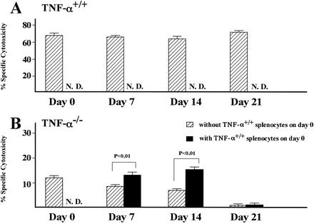
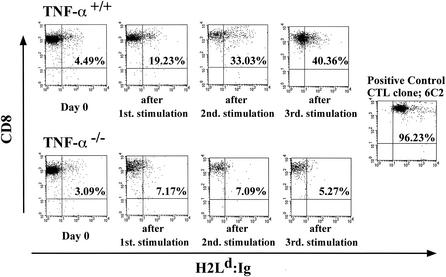
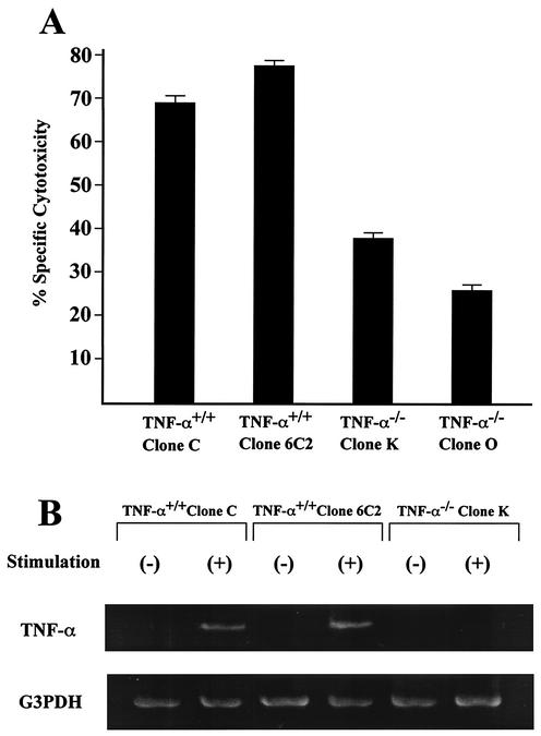
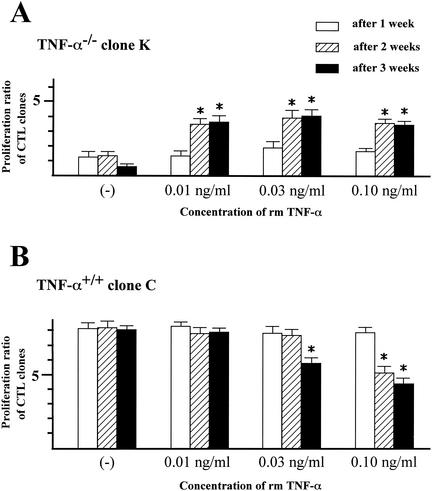
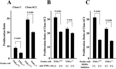
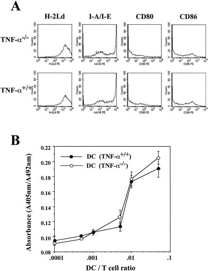
Recent studies have shown that tumor necrosis factor alpha (TNF-alpha) plays critical roles in not only viral clearance but also lymphoid tissue development and stem cell differentiation. In this study, we attempted to induce hepatitis B virus (HBV)-specific cytotoxic T lymphocytes (CTLs) by immunization of TNF-alpha knockout (TNF-alpha(-/-)) mice with HBsAg-encoding plasmid DNA. An immunization with the HBV plasmid failed to induce CTL responses in TNF-alpha(-/-) mice, although CTLs were readily induced in wild-type mice by the same protocol. Weak CTL responses were produced in TNF-alpha(-/-) mice after two sessions of immunization with the HBV plasmid; however, TNF-alpha was required to maintain the responses of these CTL lines to in vitro stimulation and, even then, the responses were lost after 3 weeks. Interestingly, a limiting dilution of a CTL line showed that HBV-specific CTL clones with high specific cytotoxicity were present in TNF-alpha(-/-) mice, but these clones again failed to proliferate for more than 3 weeks. Furthermore, since exogenously added TNF-alpha enhanced the proliferation of a TNF-alpha(-/-) clone but suppressed that of a TNF-alpha(+/+) clone in vitro, TNF-alpha also has a direct effect on the proliferation of CTLs. In conclusion, TNF-alpha is essential rather than important for the proliferation of HBV-specific CTLs both in vivo and in vitro and this effect is not only due to the activation of dendritic cells but is also induced by the direct effect on CTLs.


Figures

FIG. 1.
FIG. 1.
Cytotoxic activity of HBsAg-specific CTLs induced in TNF-α+/+ and TNF-α−/− mice. (A) TNF-α+/+ mice; (B) TNF-α−/− mice. Mice were immunized by two cycles of injection of plasmid into the bilateral quadriceps muscles 5 days after cardiotoxin injection. Splenocytes prepared 2 weeks after the final immunization were incubated with or without MMC-treated TNF-α+/+ splenocytes on day 0. For the assay, effector cells were incubated for 4 h with Eu-labeled target cells at an effector-to-target cell ratio of 20, and the percent specific cytotoxicity was calculated by subtracting the percent cytotoxicity for P815 cells (HBsAg negative) from that for P815preS1 cells (HBsAg positive). Spontaneous release was always less than 20% of the total. Each data point represents the mean of triplicate samples. N.D., not done.
FIG. 2.
FIG. 2.
Flow cytometric analysis of the frequency of HBsAg-specific CD8+ cells in TNF-α+/+ and TNF-α−/− mice. Mice were immunized by two cycles of injection of plasmid into the bilateral quadriceps muscles 5 days after cardiotoxin injection, and splenocytes were prepared 2 weeks after the final injection. In order to evaluate differences in HBsAg-specific cells in immunized splenocytes, the CD8+ H-2Ld-Ig+ fraction (upper right) was used as the HBsAg-specific range. The HBsAg-specific range of the TNF-α+/+ CTL line gradually increased by more than 40% after the third stimulation in vitro. On the other hand, the HBsAg-specific range of the TNF-α−/− CTL line did not expand after stimulation in vitro. The HBsAg-specific range of the TNF-α+/+ HBsAg-specific CTL clone 6C2 (established after immunization with recombinant vaccinia virus) was more than 95%. Similar results were obtained in three separate experiments. In this experiment, immunized TNF-α−/− splenocytes were restimulated with MMC-treated TNF-α−/− splenocytes and P815preS1 and TNF-α+/+ spleen cells were restimulated with MMC-treated TNF-α+/+ splenocytes and P815preS1 every 7 days, respectively.
FIG. 3.
FIG. 3.
Generation of HBsAg-specific TNF-α−/− CTL clones. Immunized splenocytes were incubated with MMC-treated TNF-α+/+ splenocytes on day 0 and stimulated once a week with MMC-treated TNF-α+/+ splenocytes (feeder cells) and P815preS1 cells (specific stimulator). After the second stimulation, the primed splenocytes were cloned at a rate of 1 cell/well. (A) Results of a cytotoxicity assay performed as described in the legend to Fig. 1, at an effector-to-target cell ratio of 20. Each data point represents the mean of triplicate samples. (B) Expression of TNF-α mRNA confirmed by RT-PCR. To avoid detecting TNF-α mRNA expression in feeder and P815preS1 cells, the CTL clones were stimulated with plate-bound anti-CD3ɛ antibody. G3PDH, glyceraldehyde-3-phosphate dehydrogenase.
FIG. 4.
FIG. 4.
Effect of exogenous sTNF-α on the proliferation of TNF-α−/− (A) and TNF-α+/+ (B) CTL clones. At each weekly stimulation with irradiated TNF-α+/+ splenocytes (feeder cells) and P815preS1 (specific stimulator), HBsAg-specific TNF-α−/− CTL clones were incubated with sTNF-α at concentrations of 0.01 to 0.1 ng/ml and the number of CTLs was counted 1 week after stimulation. The proliferative ratio was calculated as the number of CTLs 1 week after stimulation/the number of CTLs before stimulation. Each bar and error bar represents the mean and the SD, respectively, of results for triplicate samples. *, P < 0.001 versus the control group; rmTNF-α, recombinant mouse TNF-α; (−), no TNF-α was used.
FIG. 5.
FIG. 5.
Effect of TNF-α on the proliferation of TNF-α+/+ CTL clones. (A) The TNF-α+/+ CTL clones C and 6C2 were incubated with MMC-treated TNF-α+/+ or TNF-α−/− splenocytes (feeder cells) and P815preS1 cells (specific stimulator). Then 1 week after the second weekly stimulation, the number of CTLs was counted and the proliferative ratio was calculated as the number of CTLs 1 week after the last stimulation/the number of CTLs before the last stimulation. (B) The TNF-α+/+ CTL clone 6C2 was stimulated twice at weekly intervals with MMC-treated TNF-α+/+ or TNF-α−/− splenocytes (feeder cells) and P815preS1 cells (specific stimulator) in the presence of 5 μg of anti-TNF-α antibody (Ab) or hamster IgG/ml. Then 1 week later, the number of CTLs was counted and the proliferative ratio was calculated as described above. Purified hamster IgG was used as a control antibody. (C) Clone 6C2 was stimulated once with MMC-treated TNF-α+/+ feeder cells and P815preS1 cells alone or in the presence of MMP inhibitor (10 μM). Then 1 week later, the number of CTLs was counted and the proliferative ratio was measured as described above. MMP inhibitor was added on day 0. Each bar and error bar represent the mean and the SD, respectively, of results for triplicate samples. Statistically significantly differences between groups are indicated.
FIG. 6.
FIG. 6.
Comparison of DC function in TNF-α+/+ and TNF-α−/− mice. CD11c+ cell separation was carried out by using the VarioMACS system. (A) Flow cytometric analysis of the expression of the MHC-I molecule H-2Ld (restriction element for HBsAg-specific CTL), the MHC-II molecule I-A/I-E, CD80, and CD86 in TNF-α+/+ and TNF-α−/− DCs. All samples were assayed in duplicate. (B) The allostimulatory capacity of CD11c+ DCs from TNF-α+/+ and TNF-α−/− mice was assessed by the MLR as described in Materials and Methods with allogeneic murine CD4+ T cells from AKR mice (H-2k). Each data point and error bar represent the mean and SD, respectively, of results for triplicate samples.

Similar articles

Cited by

References

    1. Ando, K., K. Hiroishi, T. Kaneko, T. Moriyama, Y. Muto, N. Kayagaki, H. Yagita, K. Okumura, and M. Imawari. 1997. Perforin, Fas/Fas ligand, and TNF-α pathways as specific and bystander killing mechanisms of hepatitis C virus-specific human CTL. J. Immunol. 158:5283-5291. - PubMed
    1. Ando, K., L. G. Guidotti, S. Wirth, T. Ishikawa, G. Missale, T. Moriyama, R. D. Schreiber, H. J. Schlicht, S. N. Huang, and F. V. Chisari. 1994. Class I-restricted cytotoxic T lymphocytes are directly cytopathic for their target cells in vivo. J. Immunol. 152:3245-3253. - PubMed
    1. Ando, K., T. Moriyama, L. G. Guidotti, S. Wirth, R. D. Schreiber, H. J. Schlicht, S. N. Huang, and F. V. Chisari. 1993. Mechanisms of class I-restricted immunopathology. A transgenic mouse model of fulminant hepatitis. J. Exp. Med. 178:1541-1554. - PMC - PubMed
    1. Banchereau, J., F. Biere, C. Caux, J. Davoust, S. Lebecque, Y. J. Liu, B. Pulendran, and K. Paluckae. 2000. Immunology of dendritic cells. Annu. Rev. Immunol. 18:767-811. - PubMed
    1. Banchereau, J., and R. M. Steinman. 1998. Myeloid dendritic cells and the control of immunity. Nature 392:245-252. - PubMed

Publication types

MeSH terms

Substances

LinkOut - more resources