Effective vaccination against long-term gammaherpesvirus latency
- PMID: 12551990
- PMCID: PMC141097
- DOI: 10.1128/jvi.77.4.2522-2529.2003
Effective vaccination against long-term gammaherpesvirus latency
Erratum in
- J Virol. 2003 Apr;77(7):4469
Abstract
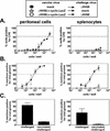
The fundamental question of whether a primed immune system is capable of preventing latent gammaherpesvirus infection remains unanswered. Recent studies showing that vaccination can reduce acute replication and short-term latency but cannot alter long-term latency further call into question the possibility of achieving sterilizing immunity against gammaherpesviruses. Using the murine gammaherpesvirus 68 (gammaHV68) system, we demonstrate that it is possible to effectively vaccinate against long-term latency. By immunizing mice with a gammaHV68 mutant virus that is deficient in its ability to reactivate from latency, we reduced latent infection of wild-type challenge virus to a level below the limit of detection. Establishment of latency was inhibited by vaccination regardless of whether mice were challenged intraperitoneally or intranasally. Passive transfer of antibody from vaccinated mice could partially reconstitute the effect, demonstrating that antibody is an important component of vaccination. These results demonstrate the potential of a memory immune response against gammaherpesviruses to alter long-term latency and suggest that limiting long-term latent infection in a clinically relevant situation is an attainable goal.
Figures


Similar articles
-
Critical role of CD4 T cells in an antibody-independent mechanism of vaccination against gammaherpesvirus latency.J Virol. 2004 Jul;78(13):6836-45. doi: 10.1128/JVI.78.13.6836-6845.2004. J Virol. 2004. PMID: 15194759 Free PMC article.
-
An optimized CD8+ T-cell response controls productive and latent gammaherpesvirus infection.J Virol. 2005 Feb;79(4):2573-83. doi: 10.1128/JVI.79.4.2573-2583.2005. J Virol. 2005. PMID: 15681457 Free PMC article.
-
Gamma interferon blocks gammaherpesvirus reactivation from latency in a cell type-specific manner.J Virol. 2007 Jun;81(11):6134-40. doi: 10.1128/JVI.00108-07. Epub 2007 Mar 14. J Virol. 2007. PMID: 17360749 Free PMC article. Review.
-
A replication-deficient murine gamma-herpesvirus blocked in late viral gene expression can establish latency and elicit protective cellular immunity.J Immunol. 2007 Dec 15;179(12):8392-402. doi: 10.4049/jimmunol.179.12.8392. J Immunol. 2007. PMID: 18056385
-
Vaccination against murine gamma-herpesvirus infection.Viral Immunol. 2001;14(3):217-26. doi: 10.1089/088282401753266747. Viral Immunol. 2001. PMID: 11572633 Review.
Cited by
-
A gammaherpesvirus 68 gene 50 null mutant establishes long-term latency in the lung but fails to vaccinate against a wild-type virus challenge.J Virol. 2006 Feb;80(3):1592-8. doi: 10.1128/JVI.80.3.1592-1598.2006. J Virol. 2006. PMID: 16415035 Free PMC article.
-
Antibody-independent control of gamma-herpesvirus latency via B cell induction of anti-viral T cell responses.PLoS Pathog. 2006 Jun;2(6):e58. doi: 10.1371/journal.ppat.0020058. Epub 2006 Jun 23. PLoS Pathog. 2006. PMID: 16789842 Free PMC article.
-
A Gammaherpesvirus MicroRNA Targets EWSR1 (Ewing Sarcoma Breakpoint Region 1) In Vivo To Promote Latent Infection of Germinal Center B Cells.mBio. 2019 Jul 30;10(4):e00996-19. doi: 10.1128/mBio.00996-19. mBio. 2019. PMID: 31363027 Free PMC article.
-
CD4 memory T cells divide poorly in response to antigen because of their cytokine profile.Proc Natl Acad Sci U S A. 2008 Sep 23;105(38):14521-6. doi: 10.1073/pnas.0807449105. Epub 2008 Sep 11. Proc Natl Acad Sci U S A. 2008. PMID: 18787120 Free PMC article.
-
Critical role of CD4 T cells in an antibody-independent mechanism of vaccination against gammaherpesvirus latency.J Virol. 2004 Jul;78(13):6836-45. doi: 10.1128/JVI.78.13.6836-6845.2004. J Virol. 2004. PMID: 15194759 Free PMC article.
References
-
- Asher, L. V., M. A. Walz, and A. L. Notkins. 1978. Effect of immunization on the development of latent ganglionic infection in mice challenged intravaginally with herpes simplex virus types 1 and 2. Am. J. Obstet. Gynecol. 131:788-791. - PubMed
-
- Babcock, G. J., D. Hochberg, and D. A. Thorley-Lawson. 2000. The expression pattern of Epstein-Barr virus latent genes in vivo is dependent upon the differentiation stage of the infected B cell. Immunity 13:497-506. - PubMed
-
- Belz, G. T., P. G. Stevenson, M. R. Castrucci, J. D. Altman, and P. C. Doherty. 2000. Postexposure vaccination massively increases the prevalence of gamma-herpesvirus-specific CD8+ T cells but confers minimal survival advantage on CD4-deficient mice. Proc. Natl. Acad. Sci. USA 97:2725-2730. - PMC - PubMed
-
- Chang, J. Y., E. M. Johnson, Jr., and P. D. Olivo. 1991. A gene delivery/recall system for neurons which utilizes ribonucleotide reductase-negative herpes simplex viruses. Virology 185:437-440. - PubMed
-
- Cremer, K. J., M. Mackett, C. Wohlenberg, A. L. Notkins, and B. Moss. 1985. Vaccinia virus recombinant expressing herpes simplex virus type 1 glycoprotein D prevents latent herpes in mice. Science 228:737-740. - PubMed
Publication types
MeSH terms
Substances
Grants and funding
- R01 CA058524/CA/NCI NIH HHS/United States
- 5T32 CA 09547-14/CA/NCI NIH HHS/United States
- R01 HL060090/HL/NHLBI NIH HHS/United States
- R01 CA043143/CA/NCI NIH HHS/United States
- R01 CA052004/CA/NCI NIH HHS/United States
- CA 96511/CA/NCI NIH HHS/United States
- CA 52004/CA/NCI NIH HHS/United States
- T32 CA009547/CA/NCI NIH HHS/United States
- R01 CA074730/CA/NCI NIH HHS/United States
- CA 74730/CA/NCI NIH HHS/United States
- CA 58524/CA/NCI NIH HHS/United States
- HL 60090/HL/NHLBI NIH HHS/United States
- R01 CA096511/CA/NCI NIH HHS/United States
- CA 43143/CA/NCI NIH HHS/United States
LinkOut - more resources
Full Text Sources
Miscellaneous

