. 2003 Feb;77(4):2675-85.
doi: 10.1128/jvi.77.4.2675-2685.2003.

Interaction of the equine herpesvirus 1 EICP0 protein with the immediate-early (IE) protein, TFIIB, and TBP may mediate the antagonism between the IE and EICP0 proteins

Affiliations

Interaction of the equine herpesvirus 1 EICP0 protein with the immediate-early (IE) protein, TFIIB, and TBP may mediate the antagonism between the IE and EICP0 proteins

Seong K Kim et al. J Virol. 2003 Feb.

Abstract

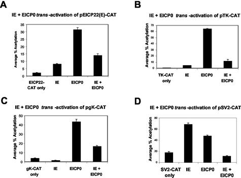
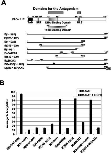
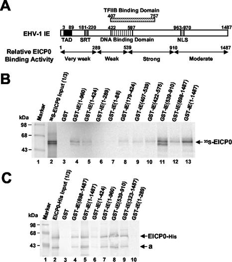
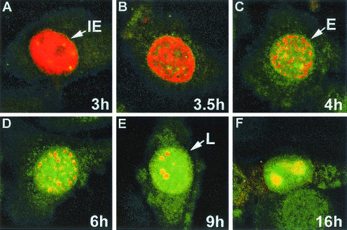
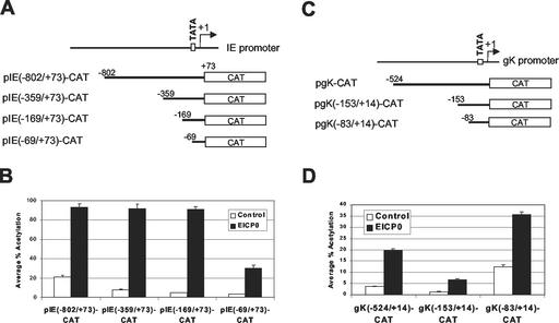
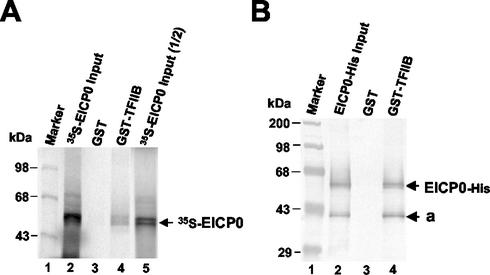
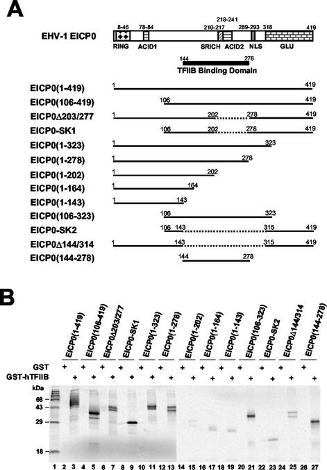
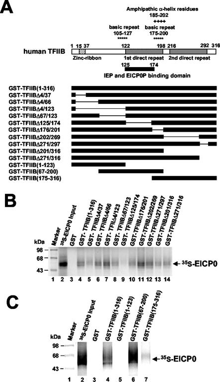
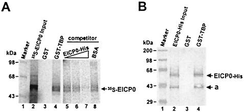
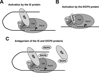
The equine herpesvirus 1 (EHV-1) immediate-early (IE) and EICP0 proteins are potent trans-activators of EHV-1 promoters; however, in transient-transfection assays, the IE protein inhibits the trans-activation function of the EICP0 protein. Assays with IE mutant proteins revealed that its DNA-binding domain, TFIIB-binding domain, and nuclear localization signal may be important for the antagonism between the IE and EICP0 proteins. In vitro interaction assays with the purified IE and EICP0 proteins indicated that these proteins interact directly. At late times postinfection, the IE and EICP0 proteins colocalized in the nuclei of infected equine cells. Transient-transfection assays showed that the EICP0 protein trans-activated EHV-1 promoters harboring only a minimal promoter region (TATA box and cap site), suggesting that the EICP0 protein trans-activates EHV-1 promoters by interactions with general transcription factor(s). In vitro interaction assays revealed that the EICP0 protein interacted directly with the basal transcription factors TFIIB and TBP and that the EICP0 protein (amino acids [aa] 143 to 278) mediated the interaction with aa 125 to 174 of TFIIB. Our unpublished data showed that the IE protein interacts with the same domain (aa 125 to 174) of TFIIB and with TBP. Taken together, these results suggested that interaction of the EICP0 protein with the IE protein, TFIIB, and TBP may mediate the antagonism between the IE and EICP0 proteins.


Figures

FIG. 1.
FIG. 1.
The EHV-1 IE and EICP0 proteins function antagonistically. Transient-transfection assays were performed with the various EHV-1 promoter-CAT reporter plasmids, EICP22 (A), thymidine kinase (TK) (B), gK (C), and SV40 (D) promoter-CAT. Transient-transfection assays were performed as described in Materials and Methods. L-M cells were transfected with 0.5 pmol of the reporter plasmids and 0.3 pmol of effector plasmids (pSVIE and pSVICP0K). Each transfection was performed in triplicate. The data are averages and are representative of several independent experiments. Error bars show standard deviations.
FIG. 2.
FIG. 2.
Domains essential for the antagonism between the IE and EICP0 proteins. (A) Schematic diagram of the deletion mutants of the IE protein. The top diagram represents the 1,487-aa IE protein of EHV-1. TAD, acidic transcriptional activation domain; SRT, serine-rich tract. The numbers refer to the number of amino acids from the N terminus of each protein. The asterisk indicates a point mutation in the DNA-binding domain of the IE protein. (B) CAT assays with the IE deletion mutants. L-M cells were transfected with 2.0 pmol of reporter plasmid (pIR5-CAT) and 0.3 pmol of effector plasmids. Each transfection was performed in triplicate. The data are averages and are representative of several independent experiments. Error bars show standard deviations.
FIG. 3.
FIG. 3.
The EICP0 protein interacts with the IE protein. (A) Relative EICP0 protein-binding activity of the IE protein. The top diagram represents the 1,478-aa IE protein. The numbers refer to the number of amino acids from the N terminus of each protein. (B) GST-pulldown assays. Equal amounts of the IVTT 35S-EICP0 protein were incubated with GST (lane 3) and various GST-IE fusion proteins (lanes 4 to 13) and then precipitated with glutathione-Sepharose 4B beads. The precipitated pellets were electrophoresed through SDS-10% PAGE gels. The bands were quantitated by PhosphorImager analysis (Molecular Dynamics). The numbers on the left represent 14C-methylated protein markers (Pharmacia) in kilodaltons. (C) Equal amounts of purified EICP0-His protein were incubated with GST (lane 3) or various GST-IE fusion proteins (lanes 4 to 10) and then precipitated with glutathione-Sepharose 4B beads. The precipitated pellets were electrophoresed through SDS-10% PAGE gels. Gels were subjected to Western blot analysis. The “a” on the right indicates an isoform of the EICP0 protein. The numbers on the left represent molecular mass standards in kilodaltons.
FIG. 4.
FIG. 4.
Photomicrographs of infected equine ETCC cultures. EHV-1 strain KyA-infected ETCC equine cells were fixed at 3 h (A), 3.5 h (B), 4 h (C), 6 h (D), 9 h (E), and 16 h (F) postinfection. The cells were reacted first with a 1:100 dilution of the A1.4 monoclonal antibody to the IE protein and a 1:200 dilution of a polyclonal antibody to the TrpE-EICP0 protein in PBS-1% BSA for 3 h. After a rinsing, the cells were reacted with a TRITC-conjugated anti-mouse IgG and an fluorescein isothiocyanate-conjugated anti-rabbit IgG for 1 h and examined under a confocal microscope. The IE and EICP0 proteins merged at early and late times of infection. IE, immediate-early pattern (2 to 3 h); E, early pattern (4 to 6 h); L, late pattern (7 to 16 h).
FIG. 5.
FIG. 5.
The EICP0 protein trans-activates the IE and gK promoters by affecting an area proximal to the transcription initiation site. Upstream deletion constructs of the IE and gK promoters were generated and used for CAT assays. (A) Schematic diagram of upstream deletion constructs of the IE promoter. The top diagram represents the EHV-1 IE promoter. (B) Transient-transfection assays with the pIE-CAT reporter plasmids. L-M cells were transfected with 1.4 pmol of reporter plasmid pIE-CAT and 0.3 pmol of effector plasmid pSVICP0K. Each transfection was performed in triplicate. Data are averages and are representative of several independent experiments. Error bars show standard deviations. (C) Schematic diagram of upstream deletion constructs of the gK promoter. The top diagram represents the EHV-1 gK promoter. (D) Transient-transfection assays with the pgK-CAT reporter plasmids. L-M cells were transfected with 1.4 pmol of reporter plasmid pgK-CAT and 0.3 pmol of effector plasmid pSVICP0K. Each transfection was performed in triplicate. The data are averages and are representative of several independent experiments. Error bars show standard deviations.
FIG. 6.
FIG. 6.
GST-pulldown assays with the IVTT and purified EICP0 proteins. (A) The EICP0 protein was in vitro transcribed-translated and radiolabeled with [35S]methionine as described in Materials and Methods. Equal amounts of each radiolabeled species were incubated with GST (lane 2) or GST-TFIIB (lane 3) proteins and then precipitated with glutathione-Sepharose 4B beads. The precipitated pellets were electrophoresed through SDS-10% PAGE gels. The bands were quantitated by PhosphorImager analysis. The numbers on the left represent 14C-methylated protein markers in kDa. (B) The purified EICP0 protein was preincubated with the GST (lane 2) or GST-TFIIB (lane 3) proteins and then precipitated with glutathione-Sepharose 4B beads. The precipitated pellets were electrophoresed through SDS-10% PAGE gels. Gels were subjected to Western blot analysis. The numbers on the left represent molecular mass standards (Gibco-BRL) in kilodaltons.
FIG. 7.
FIG. 7.
GST-pulldown assays with various deletion mutants of the EICP0 protein. (A) Schematic diagram and relative TFIIB-binding activities of the deletion mutants of the EICP0 protein. The top diagram represents the 419-aa EICP0 protein of EHV-1. RING, Ring finger domain; ACID1 and ACID2, acidic regions 1 and 2; SRICH, serine-rich region; GLU, glutamine-rich region. The numbers refer to the number of amino acids from the N terminus of each protein. (B) Equal amounts of each radiolabeled species were incubated with GST (lanes 2, 4, 6, 8, 10, 12, 14, 16, 18, 20, 22, 24, and 26) or GST-TFIIB (lanes 3, 5, 7, 9, 11, 13, 15, 17, 19, 21, 23, 25, and 27) proteins and then precipitated with glutathione-Sepharose 4B beads. The precipitated pellets were electrophoresed through SDS-10% PAGE gels. The bands were quantitated by PhosphorImager analysis. The numbers on the left represent 14C-methylated protein markers in kilodaltons. +, Present.
FIG. 8.
FIG. 8.
The EICP0 protein interacts with human TFIIB. (A) Schematic diagram of the deletion mutants of human TFIIB. The top diagram represents the 316-aa human TFIIB. The numbers refer to the number of amino acids from the N terminus of each protein. (B) Equal amounts of the IVTT 35S-EICP0 protein were incubated with the GST (lane 3) and GST-TFIIB (lanes 4 to 14) proteins and precipitated with glutathione-Sepharose 4B beads. The precipitated pellets were electrophoresed through SDS-10% PAGE gels. The bands were quantitated by PhosphorImager analysis. (C) Equal amounts of the IVTT 35S-EICP0 protein were incubated with GST (lane 3), GST-TFIIB(1-316) (lane 4), GST-TFIIB(1-123) (lane 5), GST-TFIIB(67-200) (lane 6), or GST-TFIIB(175-316) (lane 7) proteins and then precipitated with glutathione-Sepharose 4B beads. The precipitated pellets were electrophoresed through SDS-10% PAGE gels. The bands were quantitated by PhosphorImager analysis. The numbers on the left represent 14C-methylated protein markers in kilodaltons.
FIG. 9.
FIG. 9.
GST-pulldown assays with the IVTT and purified EICP0 proteins. (A) Equal amounts of the IVTT 35S-EICP0 protein were incubated with GST (lane 2) or GST-TBP (lane 3) proteins and then precipitated with glutathione-Sepharose 4B beads. For competition assays, the IVTT 35S-EICP0 protein was incubated with the GST-TBP (lanes 5 to 8) proteins in the presence of specific competitor (purified EICP0-His) or nonspecific competitor (BSA) and then precipitated with glutathione-Sepharose 4B beads. The precipitated pellets were electrophoresed through SDS-10% PAGE gels. The numbers on the left represent 14C-methylated protein markers in kilodaltons. (B) The purified EICP0-His protein was incubated with GST (lane 3) or GST-TFIIB (lane 4) proteins and precipitated with glutathione-Sepharose 4B beads. The precipitated pellets were electrophoresed through SDS-10% PAGE gels. Gels were subjected to Western blot analysis. The numbers on the left represent molecular mass standards in kilodaltons.
FIG. 10.
FIG. 10.
Models of how the IE and EICP0 proteins may activate transcription. (A) The IE protein binds to the IE protein-binding consensus 5′-ATCGT-3′ sequence of an EHV-1 promoter and then interacts with the basal transcription factors TFIIB and TBP to trans-activate EHV-1 promoters. (B) The EICP0 protein directly interacts with TFIIB and TBP to trans-activate EHV-1 promoters. (C) The mechanism of the antagonism observed between the IE and EICP0 proteins. Two possibilities were proposed according to our data. First, the IE and EICP0 proteins antagonize by an interaction between the two proteins. Second, the IE and EICP0 proteins antagonize by competing for binding to TFIIB and TBP. The arrows indicate the transcription initiation sites.

Similar articles

Cited by

References

    1. Bowles, D. E., S. K. Kim, and D. J. O'Callaghan. 2000. Characterization of the trans-activation properties of equine herpesvirus 1 EICP0 protein. J. Virol. 74:1200-1208. - PMC - PubMed
    1. Bowles, D. E., V. R. Holden, Y. Zhao, and D. J. O'Callaghan. 1997. The ICP0 protein of equine herpesvirus 1 is an early protein that independently transactivates expression of all classes of viral promoters. J. Virol. 71:4904-4914. - PMC - PubMed
    1. Bruatowski, S. 1994. The basics of basal transcription by RNA polymerase II. Cell 77:1-3. - PubMed
    1. Bruni, R., B. Fineschi, O. Ogle, and B. Roizman. 1999. A novel cellular protein, p60, interacting with both herpes simplex virus 1 regulatory proteins ICP22 and ICP0 is modified in a cell-type-specific manner and is recruited to the nucleus after infection. J. Virol. 73:3810-3817. - PMC - PubMed
    1. Caughman, G. B., J. Staczek, and D. J. O'Callaghan. 1985. Equine herpesvirus type 1 infected cell polypeptides: evidence for immediate-early/early/late regulation of viral gene expression. Virology 145:49-61. - PubMed

Publication types

MeSH terms

LinkOut - more resources