. 2003 Feb;77(4):2686-95.
doi: 10.1128/jvi.77.4.2686-2695.2003.

Herpes simplex virus gE/gI expressed in epithelial cells interferes with cell-to-cell spread

Affiliations

Herpes simplex virus gE/gI expressed in epithelial cells interferes with cell-to-cell spread

Wendy J Collins et al. J Virol. 2003 Feb.

Abstract

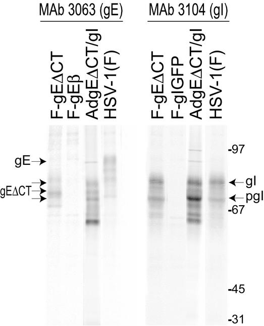
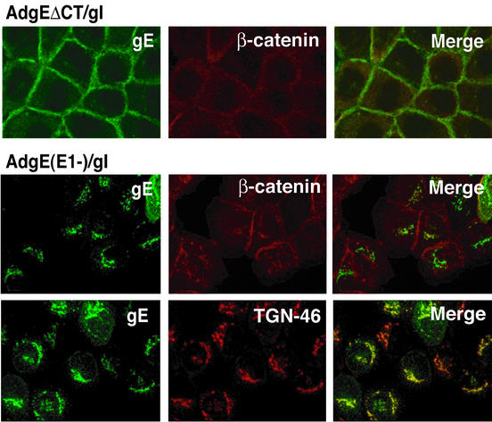
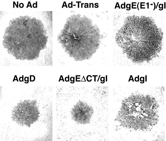
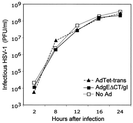
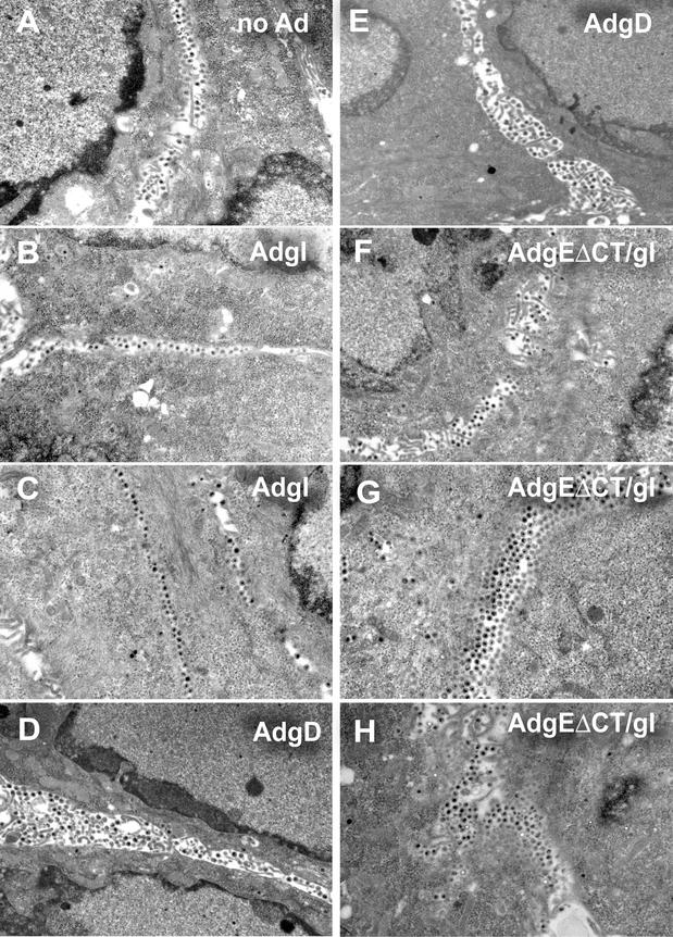
The herpes simplex virus (HSV) glycoprotein heterodimer gE/gI plays an important role in virus cell-to-cell spread in epithelial and neuronal tissues. In an analogous fashion, gE/gI promotes virus spread between certain cell types in culture, e.g., keratinocytes and epithelial cells, cells that are polarized or that form extensive cell junctions. One mechanism by which gE/gI facilitates cell-to-cell spread involves selective sorting of nascent virions to cell junctions, a process that requires the cytoplasmic domain of gE. However, the large extracellular domains of gE/gI also appear to be involved in cell-to-cell spread. Here, we show that coexpression of a truncated form of gE and gI in a human keratinocyte line, HaCaT cells, decreased the spread of HSV between cells. This truncated gE/gI was found extensively at cell junctions. Expression of wild-type gE/gI that accumulates at intracellular sites, in the trans-Golgi network, did not reduce cell-to-cell spread. There was no obvious reduction in production of infectious HSV in cells expressing gE/gI, and virus particles accumulated at cell junctions, not at intracellular sites. Expression of HSV gD, which is known to bind virus receptors, also blocked cell-to-cell spread. Therefore, like gD, gE/gI appears to be able to interact with cellular components of cell junctions, gE/gI receptors which can promote HSV cell-to-cell spread.


Figures

FIG. 1.
FIG. 1.
Expression of gE/gI in cells infected with recombinant Ad vectors and HSV. HaCaT cells were infected with AdgEΔCT and AdtetgI with 100 PFU/cell for 22 h or were infected with wild-type HSV-1 strain F, HSV gE-null mutant F-gEβ, HSV gE mutant F-gEΔCT (lacking the gE CT domain), or HSV gI-null mutant F-gIGFP for 5 h. The cells were radiolabeled with [35S]methionine-cysteine for 3 h, cell extracts were produced, and gE/gI complexes were immunoprecipitated with MAb 3063, which recognizes gE in gE/gI complexes, or MAb 3104, which recognizes gI in gE/gI complexes. Samples were subjected to electrophoresis in polyacrylamide gels, and radiolabeled proteins were identified by using a PhosphorImager. The positions of gE (mature gE expressed by wild-type HSV-1), gEΔCT/gI (expressed by F-gEΔCT and by coinfection with AdgEΔCT and AdtetgI), gI (mature gI), and pgI (immature gI) and molecular mass markers of 97, 67, 45, and 31 kDa are indicated.
FIG. 2.
FIG. 2.
Confocal immunofluorescence microscopy of cells expressing gEΔCT/gI. HaCaT cells were infected with either AdgEΔCT (150 PFU/cell), AdtetgI (150 PFU/cell) and AdTet-trans (80 PFU/cell), or AdgE(E1−) and AdgI(E1−) with 150 PFU of each per cell. After 20 to 24 h the cells were fixed, permeabilized, and stained for gE with MAb 3114 and simultaneously with anti-β-catenin or anti-TGN46 antibodies.
FIG. 3.
FIG. 3.
HSV plaques produced on cells expressing gEΔCT/gI. HaCaT cells were left uninfected (No Ad) or infected with AdTet-trans (Ad-Trans); with AdgE(E1−) and AdgI(E1−) with 150 PFU of each per cell [AdgE(E1−)/gI]; with AdgD with 300 PFU/cell; with AdgEΔCT (150 PFU/cell), AdtetgI (150 PFU/cell), and AdTet-trans with 80 PFU/cell (AdgEΔCT/gI); or with AdtetgI (AdgI) with 300 PFU/cell for 12 h. The cells were then infected with wild-type HSV-1 F, with ∼100 PFU/dish, and after 2 to 2.5 days the cells were fixed and stained with anti-HSV polyclonal antibodies and peroxidase-conjugated secondary antibodies.
FIG. 4.
FIG. 4.
Replication of HSV-1 on cells expressing gEΔCT/gI. HaCaT cells were left uninfected (No Ad) or infected with AdTet-trans (300 PFU/cell) or AdgEΔCT and AdtetgI (150 PFU of each per cell) for 12 h. The cells were then infected with HSV-1 (10 PFU/cell). Cells and cell culture supernatants were collected at various times and sonicated, and infectious HSV was assayed by plaque assays.
FIG. 5.
FIG. 5.
Electron microscopic analyses of cells expressing AdgEΔCT and then infected with HSV. HaCaT cells were left uninfected (no Ad) (A) or infected with AdtetgI (300 PFU/cell; designated AdgI) (B and C), AdgD (300 PFU/cell) (D and E), or AdgEΔCT and AdtetgI (150 PFU of each per cell) and AdTet-trans (80 PFU/cell) (F to H), in each case for 12 h. The cells were then infected with HSV-1 with 3 PFU/cell (A, B, D, and F) or with 0.3 PFU/cell (C, E, G, and H), and at 17 h the cells were washed, fixed with 2.5% glutaraldehyde, stained, sectioned, and viewed by electron microscopy.

Similar articles

Cited by

References

    1. Balan, P., N. Davis-Poynter, S. Bell, H. Atkinson, H. Browne, and T. Minson. 1994. An analysis of the in vitro and in vivo phenotypes of mutants of herpes simplex virus type 1 lacking glycoproteins gG, gE, gI or the putative gJ. J. Gen. Virol. 75:1245-1258. - PubMed
    1. Brack, A. R., B. G. Klupp, H. Granzow, R. Tirabassi, L. W. Enquist, and T. C. Mettenleiter. 2000. Role of the cytoplasmic tail of pseudorabies virus glycoprotein E in virion formation. J. Virol. 74:4004-4016. - PMC - PubMed
    1. Brunetti, C. R., K. S. Dingwell, C. Wale, F. L. Graham, and D. C. Johnson. 1998. Herpes simplex virus gD and virions accumulate in endosomes by mannose 6-phosphate-dependent and -independent mechanisms. J. Virol. 72:3330-3339. - PMC - PubMed
    1. Campadelli-Fiume, G., M. Arsenakis, F. Farabegoli, and B. Roizman. 1988. Entry of herpes simplex virus 1 in BJ cells that constitutively express viral glycoprotein D is by endocytosis and results in degradation of the virus. J. Virol. 62:159-167. - PMC - PubMed
    1. Campadelli-Fiume, G., S. Qi, E. Avitabile, L. Foa-Tomasi, R. Brandimarti, and B. Roizman. 1990. Glycoprotein D of herpes simplex virus encodes a domain which precludes penetration of cells expressing the glycoprotein by superinfecting herpes simplex virus. J. Virol. 64:6070-6079. - PMC - PubMed

Publication types

Substances

LinkOut - more resources