

doi: 10.1128/jvi.77.4.2779-2783.2003.
The latency-associated nuclear antigen of Kaposi's sarcoma-associated herpesvirus permits replication of terminal repeat-containing plasmids
Affiliations
- PMID: 12552022
- PMCID: PMC141125
- DOI: 10.1128/jvi.77.4.2779-2783.2003
Item in Clipboard
The latency-associated nuclear antigen of Kaposi's sarcoma-associated herpesvirus permits replication of terminal repeat-containing plasmids
J Virol.
2003 Feb.
Erratum in
- J Virol. 2003 Apr;77(7):4470
Abstract
The latency-associated nuclear antigen (LANA) of Kaposi's sarcoma-associated herpesvirus can associate with mitotic chromosomes and promote latent episome maintenance and segregation. Here we report that LANA also mediates the replication of plasmid DNAs bearing viral terminal repeats. The predicted secondary structure of LANA's C terminus reveals striking similarity to the known structure of the DNA-binding domain of Epstein-Barr virus EBNA1, despite the absence of primary sequence homology between these proteins, suggesting conservation of the key mechanistic features of latent gammaherpesvirus DNA replication.
Figures
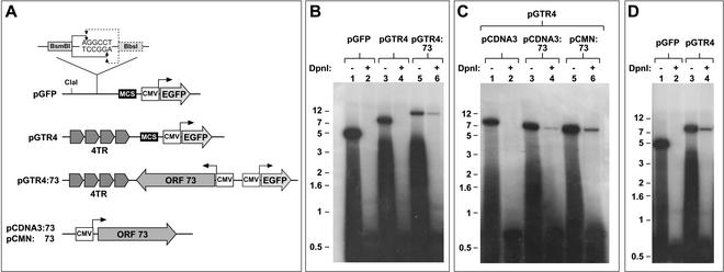
Replication of TR-containing plasmids in B cells. (A) Functional elements of reporter and expression constructs. The reporter constructs used in this study are based on the vector backbone pGFP. The vector contains a GFP expression cassette driven by the CMV promoter and a multiple-cloning site (MCS) upstream of the CMV promoter. The linker inserted into the BsaI site of pGFP and the cleavage sites of BsmBI and BbsI are shown enlarged. Cleavage with either enzyme generates identical NotI-compatible overhangs. These sites together with the indicated ClaI site were used to assemble the four head-to-tail-oriented TR units of pGTR4 (see text for details). The reporter construct pGTR4:73 contains the CMV-driven ORF73 expression cassette of pCDNA3:73 inserted into the MCS of pGTR4. The LANA expression constructs pCDNA3:73 and pCMN:73 were used to provide LANA in trans. EGFP, enhanced GFP. (B to D) DpnI resistance assays. The indicated constructs were introduced into either BJAB cells (B and C) or the stable LANA-expressing cell line BJAB:73 (D) as described in the text. Episomal plasmids were recovered 72 h later by Hirt extraction and subjected to digestion with either XhoI (which cuts once in the reporters) (−) or XhoI and DpnI (+). Standards were analyzed by Southern blotting, and reporter constructs were detected with a radioactively marked GFP probe. The positions of size markers (in kilobase pairs) are shown. Quantitation of linearized versus DpnI-digested plasmids was carried out with a phosphorimager (Molecular Dynamics Storm860 and ImageQuant software). A relatively large proportion of cells undergo cell death following the electroporation procedure; the smears below the linearized bands result from degraded DNA released from dead cells as well as degraded, adherent extracellular plasmid DNA and thus were not included in the quantitation.
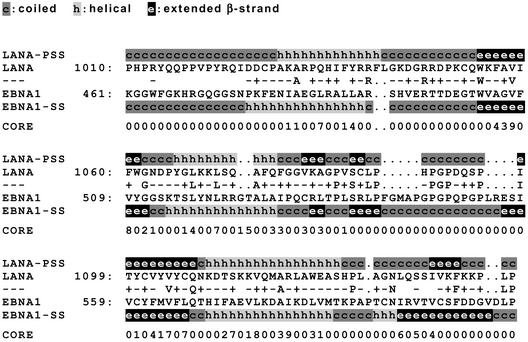
3D-PSSM alignment of EBNA1 and LANA. The carboxy-terminal domain of LANA (aa 932 to 1162; GenBank accession no. AAC57158) was used to search the 3D-PSSM database (program version 2.6.0; Imperial College of Science, Technology and Medicine [http://www.sbg.bio.ic.ac.uk/∼3dpssm/ ]). Shown is a modified version of the alignment of the relevant regions in LANA (aa 1010 to 1144) and EBNA1 (aa 461 to 607) generated by 3D-PSSM. The primary sequences of LANA and EBNA1 (accession no. NP_039875) are given. Dots, gaps introduced into the aligned sequences. Identities and positive (+) or negative (−) scores for aligned residues as assigned by 3D-PSSM are shown between the primary sequence data for LANA and EBNA1. The secondary structure of LANA (LANA-PSS) as predicted by the 3D-PSSM algorithm and the known secondary structure of EBNA1 (EBNA1-SS) are shown above or below the primary sequences of LANA and EBNA1, respectively. The CORE values shown below the aligned sequences provide an index for the contribution of residues to hydrophobic interactions (on a scale from 0 to 9) within the resolved EBNA1 structure. High values indicate buried residues important for the core structure, whereas low values indicate residues on the surface of the protein.
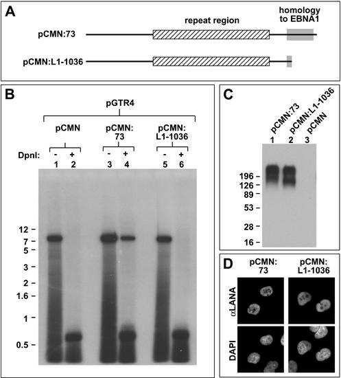
The carboxy-terminal region of LANA is required for replication activity. (A) Cartoon showing the proteins encoded by the expression vectors pCMN:73 (full-length LANA) and pCMN:L1-1036 (lacking the carboxy-terminal 126 aa of LANA). Mutant L1-1036 is truncated within the region showing structural homology to EBNA1. All amino acid positions conform to the LANA sequence with GenBank accession no. AAC57158. (B) DpnI resistance assays. Expression construct pCMN:73 or pCMN:L1-1036 or the empty expression vector pCMN was transfected into BJAB cells together with the reporter construct pGTR4. Episomes were recovered 72 h later by Hirt extraction and analyzed as described for Fig. 1. (C) Western blot analysis of LANA expression. Aliquots of the transfected BJAB cells (4 × 10 6 cells) described above were harvested 48 h posttransfection, and extracts containing 40 μg of total protein were analyzed by Western blotting using a polyclonal antiserum against LANA (18). The positions of size markers (in kilodaltons) are shown. Note that LANA shows multiple bands and runs higher than expected from its calculated molecular mass (135 kDa). The protein shows similar behavior in KSHV-positive BCBL1 cells (data not shown). (D) Immunofluorescence analysis of endothelial SLK cells transfected with the expression construct pCMN:73 (left) or pCMN:L1-1036 (right). LANA was detected with polyclonal antibodies, and DNA was stained with DAPI (4′,6′-diamidino-2-phenylindole).
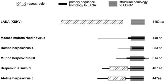
ORF73-encoded proteins of rhadinoviruses: homology to LANA and EBNA1. Shown is a graphical representation of the results presented in Table 1. Hatched boxes, repeat regions within LANA and ORF73-encoded proteins of other rhadinoviruses. Primary amino acid homology of the ORF73-encoded proteins to LANA was evaluated by BLASTP pairwise sequence alignment. Black bars, regions showing significant homology to LANA; grey boxes, regions which show predicted structural homology to EBNA1, as judged by 3D-PSSM analysis (see Table 1 for details). The ORF73-encoded proteins of alcelaphine herpesvirus 2 and ovine herpesvirus 2 are not depicted since they do not show homology to either LANA or EBNA1.
Similar articles
-
The latency-associated nuclear antigen of Kaposi's sarcoma-associated herpesvirus supports latent DNA replication in dividing cells.J Virol. 2002 Nov;76(22):11677-87. doi: 10.1128/jvi.76.22.11677-11687.2002. J Virol. 2002. PMID: 12388727 Free PMC article.
-
Kaposi's Sarcoma-Associated Herpesvirus LANA-Adjacent Regions with Distinct Functions in Episome Segregation or Maintenance.J Virol. 2019 Mar 5;93(6):e02158-18. doi: 10.1128/JVI.02158-18. Print 2019 Mar 15. J Virol. 2019. PMID: 30626680 Free PMC article.
-
Mutational analysis of the latency-associated nuclear antigen DNA-binding domain of Kaposi's sarcoma-associated herpesvirus reveals structural conservation among gammaherpesvirus origin-binding proteins.J Gen Virol. 2010 Sep;91(Pt 9):2203-15. doi: 10.1099/vir.0.020958-0. Epub 2010 May 19. J Gen Virol. 2010. PMID: 20484563 Free PMC article.
-
The latency-associated nuclear antigen, a multifunctional protein central to Kaposi's sarcoma-associated herpesvirus latency.Future Microbiol. 2011 Dec;6(12):1399-413. doi: 10.2217/fmb.11.137. Future Microbiol. 2011. PMID: 22122438 Free PMC article. Review.
-
Kaposi's Sarcoma-Associated Herpesvirus Latency-Associated Nuclear Antigen: Replicating and Shielding Viral DNA during Viral Persistence.J Virol. 2017 Jun 26;91(14):e01083-16. doi: 10.1128/JVI.01083-16. Print 2017 Jul 15. J Virol. 2017. PMID: 28446671 Free PMC article. Review.
Cited by
-
Mechanism of angiopoietin-1 upregulation in Kaposi's sarcoma-associated herpesvirus-infected PEL cell lines.J Virol. 2015 May;89(9):4786-97. doi: 10.1128/JVI.03144-14. Epub 2015 Jan 28. J Virol. 2015. PMID: 25631079 Free PMC article.
-
A short sequence immediately upstream of the internal repeat elements is critical for KSHV LANA mediated DNA replication and impacts episome persistence.Virology. 2014 Jan 5;448:344-55. doi: 10.1016/j.virol.2013.10.026. Epub 2013 Nov 12. Virology. 2014. PMID: 24314665 Free PMC article.
-
Identification of properties of the Kaposi's sarcoma-associated herpesvirus latent origin of replication that are essential for the efficient establishment and maintenance of intact plasmids.J Virol. 2014 Aug;88(15):8490-503. doi: 10.1128/JVI.00742-14. Epub 2014 May 14. J Virol. 2014. PMID: 24829342 Free PMC article.
-
Maintenance of large numbers of virus genomes in human cytomegalovirus-infected T98G glioblastoma cells.J Virol. 2014 Apr;88(7):3861-73. doi: 10.1128/JVI.01166-13. Epub 2014 Jan 22. J Virol. 2014. PMID: 24453365 Free PMC article.
-
Kaposi's sarcoma-associated herpesvirus LANA protein downregulates nuclear glycogen synthase kinase 3 activity and consequently blocks differentiation.J Virol. 2007 May;81(9):4722-31. doi: 10.1128/JVI.02548-06. Epub 2007 Feb 21. J Virol. 2007. PMID: 17314169 Free PMC article.
References
-
- Arad, U. 1998. Modified Hirt procedure for rapid purification of extrachromosomal DNA from mammalian cells. BioTechniques 24:760-762. - PubMed
-
- Ballestas, M. E., P. A. Chatis, and K. M. Kaye. 1999. Efficient persistence of extrachromosomal KSHV DNA mediated by latency-associated nuclear antigen. Science 284:641-644. - PubMed
-
- Bochkarev, A., J. A. Barwell, R. A. Pfuetzner, E. Bochkareva, L. Frappier, and A. M. Edwards. 1996. Crystal structure of the DNA-binding domain of the Epstein-Barr virus origin-binding protein, EBNA1, bound to DNA. Cell 84:791-800. - PubMed
-
- Bochkarev, A., J. A. Barwell, R. A. Pfuetzner, W. Furey, Jr., A. M. Edwards, and L. Frappier. 1995. Crystal structure of the DNA-binding domain of the Epstein-Barr virus origin-binding protein EBNA 1. Cell 83:39-46. - PubMed
MeSH terms
Substances
LinkOut - more resources
Full Text Sources
Other Literature Sources
