

doi: 10.1128/jvi.77.4.2784-2788.2003.
Molecular chaperone GRP78/BiP interacts with the large surface protein of hepatitis B virus in vitro and in vivo
Affiliations
- PMID: 12552023
- PMCID: PMC141094
- DOI: 10.1128/jvi.77.4.2784-2788.2003
Item in Clipboard
Molecular chaperone GRP78/BiP interacts with the large surface protein of hepatitis B virus in vitro and in vivo
J Virol.
2003 Feb.
Abstract
The proper folding and assembly of viral envelope proteins are mediated by host chaperones. In this study, we demonstrated that an endoplasmic reticulum luminal chaperone GRP78/BiP bound specifically to the pre-S1 domain of the L protein in vitro and in vivo where complete viral particles were secreted, suggesting that GRP78/BiP plays an essential role in the proper folding of the L protein and/or assembly of viral envelope proteins.
Figures
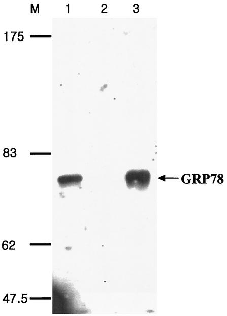
Purification and Western blot analysis of GRP78/BiP. Nonbiotinylated HepG2 lysates (lane 1), proteins bound to a GST-Sepharose column (lane 2), and proteins bound to a GST-pre-S1-Sepharose column (lane 3) were subjected to Western blotting using goat anti-BiP and HRP-conjugated anti-goat IgG antibodies. Molecular size markers (in kilodaltons) (lane M) are shown on the left. GRP78/BiP is indicated with an arrow.
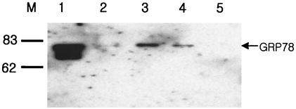
GRP78/BiP binding is ATP dependent. Biotinylated HepG2 lysates were incubated with GST and further incubated with glutathione-Sepharose beads. The unbound proteins (precleared lysate; lane 1) were incubated with GST-pre-S1 fusion protein that bound to glutathione-Sepharose beads. After the beads were extensively washed with lysis buffer, the bound proteins were eluted with PBS (lane 2), ATP elution buffer (lane 3), and ATP elution buffer again (lane 4), in that order, before they were eluted by boiling in the protein sample buffer (lane 5). All eluates were divided into two parts and subjected to SDS-PAGE and Western blotting using goat anti-BiP and HRP-conjugated anti-goat IgG antibodies. Molecular size markers (in kilodaltons) (lane M) are shown on the left.
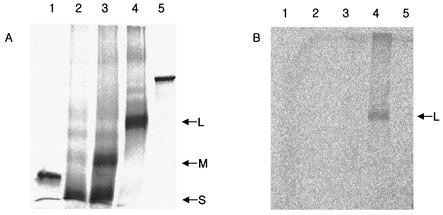
GRP78/BiP binds to the L protein in vitro. The S (lane 2), M (lane 3), and L (lane 4) proteins of the HBV envelope, along with GST (lane 1) and luciferase (lane 5) as negative controls, were synthesized in vitro using a TnT quick-coupled transcription-translation system and canine microsomes. Equal amounts of proteins were subjected to SDS-PAGE either directly (A) or after coimmunoprecipitation with anti-BiP antibody (B).
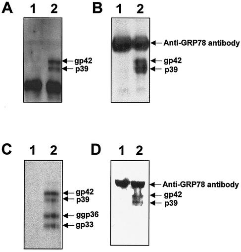
GRP78/BiP binds to the L protein in vivo. (A) The COS7 cell lysates, transfected with pCDNA3 as a control vector (lane 1) or the L protein expression plasmid, pCDNAL (lane 2), were subjected to Western blotting with the humanized version of anti-pre-S1 antibody. (B) The COS7 cell lysates, transfected with pCDNA3 (lane 1) or pCDNAL (lane 2), were immunoprecipitated with anti-BiP antibody. The immunoprecipitates were subjected to Western blot analysis using the humanized version of anti-pre-S1 antibody and HRP-conjugated anti-human IgG antibody. (C) The HepG2 cell lysates, transfected with mock DNA (lane 1) or pHBV5.2 (lane 2), were subjected to Western blotting with anti-pre-S2 antibody. (D) The HepG2 cell lysates, transfected with mock DNA (lane 1) or pHBV5.2 (lane 2), were immunoprecipitated with anti-BiP antibody, and the immunoprecipitates were subjected to Western blot analysis using anti-pre-S2 monoclonal antibody and HRP-conjugated anti-murine IgG antibody. The L and M proteins and the heavy chain of anti-BiP antibody that was reacted with HRP-conjugated anti-murine IgG antibody or anti-human IgG antibody are indicated. Molecular size markers (in kilodaltons) (lane M) are shown on the right.
References
-
- Brodsky, J. L., and R. Schekman. 1994. Heat shock cognate proteins and polypeptide translocation across the endoplasmic reticulum membrane, p. 85-109. In R. I. Marimoto, A. Tissieres, and C. Georgopoulos (ed.), The biology of heat shock proteins and molecular chaperones. Cold Spring Harbor Laboratory Press, Plainview, N.Y.
Publication types
MeSH terms
Substances
LinkOut - more resources
Full Text Sources
Molecular Biology Databases
Miscellaneous
