. 2003 Feb 4;100(3):1004-9.
doi: 10.1073/pnas.242735899. Epub 2003 Jan 27.

Molecular features of adult mouse small intestinal epithelial progenitors

Affiliations

Molecular features of adult mouse small intestinal epithelial progenitors

Thaddeus S Stappenbeck et al. Proc Natl Acad Sci U S A. .

Abstract

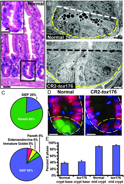
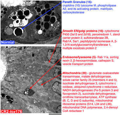
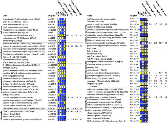
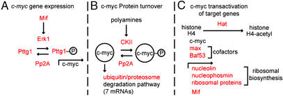
The adult mouse small intestinal epithelium undergoes perpetual regeneration, fueled by a population of multipotential stem cells and oligopotential daughters located at the base of crypts of Lieberkühn. Although the morphologic features of small intestinal epithelial progenitors (SiEPs) are known, their molecular features are poorly defined. Previous impediments to purification and molecular characterization of SiEPs include lack of ex vivo clonigenic assays and the difficulty of physically retrieving them from their niche where they are interspersed between their numerous differentiated Paneth cell daughters. To overcome these obstacles, we used germ-free transgenic mice lacking Paneth cells to obtain a consolidated population of SiEPs with normal proliferative activity. These cells were harvested by laser capture microdissection. Functional genomics analysis identified 163 transcripts enriched in SiEPs compared with Paneth cell-dominated normal crypt base epithelium. The dataset was validated by (i) correlation with the organellar composition of SiEPs versus Paneth cells, (ii) similarities to databases generated from recent mouse hematopoietic and neural stem cell genome anatomy projects, and (iii) laser capture microdissectionreal-time quantitative RT-PCR studies of progenitor cell-containing populations retrieved from the small intestines, colons, and stomachs of conventionally raised mice. The SiEP profile has prominent representation of genes involved in c-myc signaling and in the processing, localization, and translation of mRNAs. This dataset, together with our recent analysis of gene expression in the gastric stem cell niche, discloses a set of molecular features shared by adult mouse gut epithelial progenitors.


Figures

Figure 1
Figure 1
The fractional representation of SiEPs is markedly increased in the crypt bases of Paneth cell-ablated CR2-tox176 mice. (A) Hematoxylin and eosin-stained section of the distal jejunum of a normal GF mouse. (Inset) An enlarged view of the boxed crypt. The crypt base is populated with Paneth cells that contain distinctive eosinophilic secretory granules (e.g., arrow). (B) EM images of distal jejunal crypt base epithelium from GF normal and transgenic CR2-tox176 mice. Paneth cells located at the normal crypt base (below white dashed line) contain characteristic electron-dense cytoplasmic granules. The CR2-tox176 crypt base is populated with poorly differentiated SiEPs. (C) Quantitative EM analysis of distal jejunal crypt base epithelial populations in normal (Upper) and CR2-tox176 (Lower) animals. (D) S-phase cells in GF normal and CR2-tox176 crypts. Mice were injected with BrdUrd 1 h before death. Sections of distal jejunum were stained with goat anti-BrdUrd, Alexa Fluor 594-labeled donkey anti-goat Ig (red), FITC-conjugated U. europeaus agglutinin-1 (to mark Paneth cells as green), and bis-benzimide (blue nuclear stain). The outer margin of each crypt is demarcated by a white dashed line and the base is outlined by yellow dashed lines. (E) Quantifying S-phase cells. Mean values ± SEM are plotted. See text for details. (Scale bars: A and D = 25 μm; B = 5 μm.)
Figure 2
Figure 2
Correlations between cytoplasmic organelle composition and mRNAs represented in SiEP and Paneth cell datasets. (Upper) Transmission EM of Paneth cells located at the base of normal GF crypts. Gene products that are enriched in Paneth cells and are affiliated with their distinctive electron dense secretory granules are listed. The number of genes per category is indicated in parentheses. (Lower) EM of SiEPs. mRNAs encoding proteins associated with organelles that are well represented in these progenitors are listed. (Scale bars: 5 μm.)
Figure 3
Figure 3
Representation of components of the SiEP database in mouse NSC and HSC databases. Gene abbreviations are in parentheses. HSC1/NSC1 datasets are from ref. . HSC2/NSC2 are from ref. . A yellow box indicates that the identical mRNA is present in the SiEP and HSC or NSC databases. A blue box indicates that one or more transcripts encoding functionally similar members of the gene family are expressed. A box with blue and yellow sectors means that the identical transcript plus one or more related family members are present. A white box indicates that the identical or similar mRNA is absent. See Table 2 for additional details. aAverage fold difference, defined by qRT-PCR, in progenitor versus differentiated epithelial populations harvested by LCM from the distal jejunum (SI), descending colon, and stomachs of GF or conventionally raised (CONV) normal adult male FVB/N mice.
Figure 4
Figure 4
Overview of SiEP-associated factors that affect c-myc signaling. Members of the SiEP dataset are indicated in red. See text for additional details.

Similar articles

Cited by

References

    1. Cheng H, Leblond C P. Am J Anat. 1974;141:461–479. - PubMed
    1. Bjerknes M, Cheng H. Gastroenterology. 1999;116:7–14. - PubMed
    1. Ouellette A J. Am J Physiol. 1999;277:G257–G261. - PubMed
    1. Cheng H. Am J Anat. 1974;141:521–535. - PubMed
    1. Bjerknes M, Cheng H. Am J Anat. 1981;160:51–63. - PubMed

Publication types