. 2003 Feb 4;100(3):1034-9.
doi: 10.1073/pnas.0237312100. Epub 2003 Jan 27.

Involvement of bone morphogenetic protein 4 (BMP-4) in pituitary prolactinoma pathogenesis through a Smad/estrogen receptor crosstalk

Affiliations

Involvement of bone morphogenetic protein 4 (BMP-4) in pituitary prolactinoma pathogenesis through a Smad/estrogen receptor crosstalk

Marcelo Paez-Pereda et al. Proc Natl Acad Sci U S A. .

Abstract

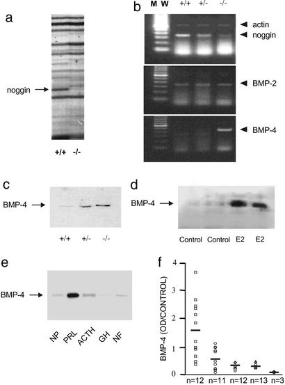
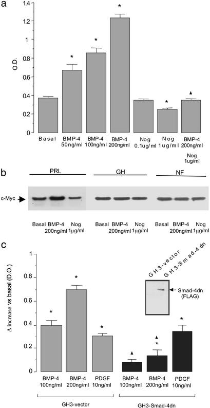
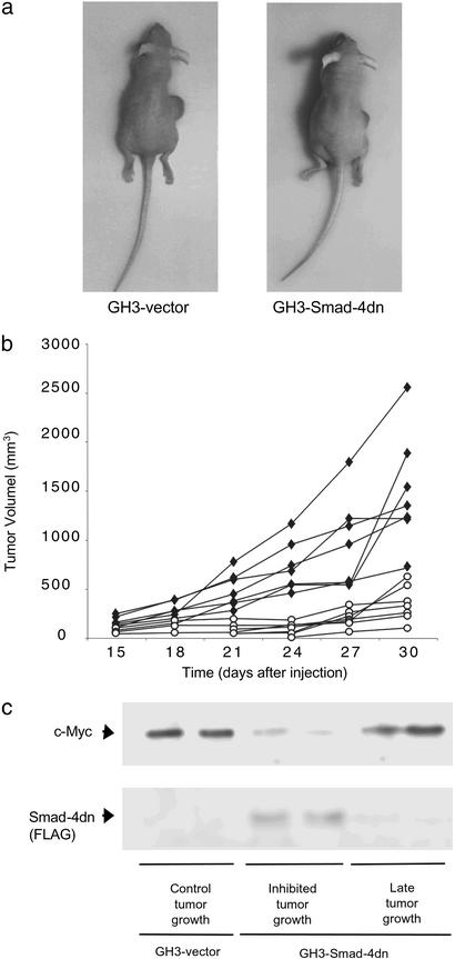
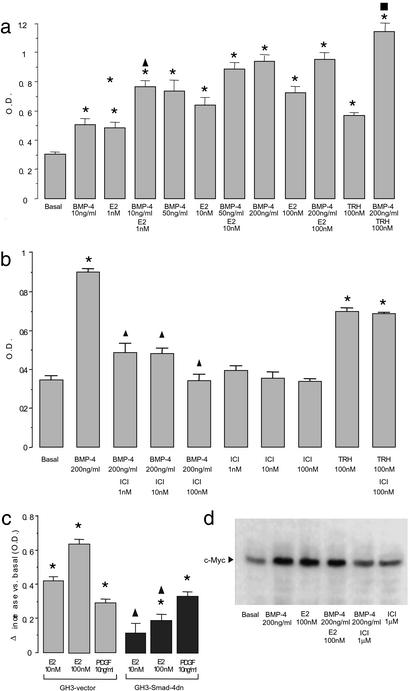
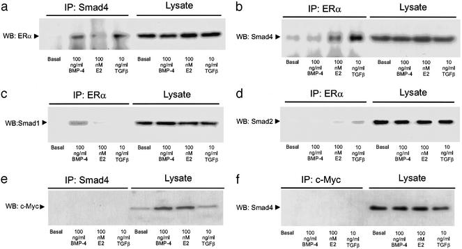
Pituitary tumor development involves clonal expansion stimulated by hormones and growth factorscytokines. Using mRNA differential display, we found that the bone morphogenetic protein (BMP) inhibitor noggin is down-regulated in prolactinomas from dopamine D2-receptor-deficient mice. BMP-4 is overexpressed in prolactinomas taken from dopamine D2-receptor-deficient female mice, but expression of the highly homologous BMP-2 does not differ in normal pituitary tissue and prolactinomas. BMP-4 is overexpressed in other prolactinoma models, including estradiol-induced rat prolactinomas and human prolactinomas, compared with normal tissue and other pituitary adenoma types (Western blot analysis of 48 tumors). BMP-4 stimulates, and noggin blocks, cell proliferation and the expression of c-Myc in human prolactinomas, whereas BMP-4 has no action in other human pituitary tumors. GH3 cells stably transfected with a dominant negative of Smad4 (Smad4dn; a BMP signal cotransducer) or noggin have reduced tumorigenicity in nude mice. Tumor growth recovered in vivo when the Smad4dn expression was lost, proving that BMP-4Smad4 are involved in tumor development in vivo. BMP-4 and estrogens act through overlapping intracellular signaling mechanisms on GH3 cell proliferation and c-myc expression: they had additive effects at low concentrations but not at saturating doses, and their action was inhibited by blocking either pathway with the reciprocal antagonist (i.e., BMP-4 with ICI 182780 or 17beta-estradiol with Smad4dn). Furthermore, coimmunoprecipitation studies demonstrate that under BMP-4 stimulation Smad4 and Smad1 physically interact with the estrogen receptor. This previously undescribed prolactinoma pathogenesis mechanism may participate in tumorigenicity in other cells where estrogens and the type beta transforming growth factor family have important roles.


Figures

Figure 1
Figure 1
Differential expression of the BMP-4 system in prolactinomas. (a) mRNA differential display of pituitaries from D2R+/+ (control) mice and D2R−/− mice with prolactinoma. Arrow indicates the band identified as noggin. These results represent four independent amplifications. (b) Comparative RT-PCR was performed to detect noggin, BMP-2, and BMP-4 in RNA extracted from pituitaries taken from D2R+/+, D2R+/−, or D2R−/− mice. Actin amplification was performed in the same reaction tubes under suboptimal conditions as an internal standard. These results represent three independent RT-PCR reactions. (c) Differential expression of BMP-4 protein was confirmed by Western blot analysis in protein extracts from normal pituitaries taken from D2R+/+ (control) mice and from D2R+/− mice without tumors, and from prolactinomas taken from D2R−/− mice. Equal loading was assessed by β-actin detection. These results represent two independent experiments with similar results. (d) BMP-4 was measured by Western blot analysis in pituitaries from four female Sprague–Dawley rats treated with vehicle (control; plasma PRL, 38.65 ± 0.45 ng/ml) or 200 mg/ml 17β-estradiol (E2; plasma PRL, 446.5 ± 42.5 ng/ml) as detailed in Methods. Equal loading was assessed by β-actin detection. (e) BMP-4 was examined by Western blot analysis in 51 protein homogenates, each obtained from one of 51 individual samples (distributed as shown in f) from normal human pituitary tissue (NP; n = 3); human prolactinomas (PRL; n = 12); human corticotropin (ACTH)-secreting tumors (ACTH; n = 11); human growth hormone (GH)-secreting tumors (GH; n = 12); or clinically inactive pituitary tumors (NF; n = 13); one representative example of each case is shown. Equal loading was assessed by β-actin detection. (f) BMP-4 detections by Western blot from the 51 samples used in e were analyzed by densitometry and normalized by using β-actin values as loading control. ●, normal human pituitary tissue; □, human prolactinomas; ○, human ACTH-secreting tumors; ⋄, human GH-secreting tumors; ▵, clinically inactive pituitary tumors. In many cases, symbols corresponding to individual tumors overlap. BMP-4 levels in human prolactinomas are significantly different (P < 0.01) with respect to all other groups, which show no statistical difference among them (ANOVA with Scheffé's test).
Figure 2
Figure 2
BMP-4 stimulates the proliferation of a lactotroph tumor cell line. (a) GH3 cells were treated with BMP-4, noggin (Nog), or their combination for 72 h. Cell proliferation was measured by WST-1 assay. Results represent the mean ± SE of quadruplicates from four independent experiments. *, P < 0.01 with respect to basal values; ▴, P < 0.01 with respect to 200 ng/ml BMP-4 stimulation (ANOVA with Scheffé's test). (b) Tumor explants were treated with BMP-4 or noggin (Nog) for 90 min, and c-Myc expression was analyzed by Western blot analysis as a parameter related to cell proliferation in different types of pituitary tumors (n = 9). PRL, human prolactinoma; GH, human GH-secreting pituitary tumor; NF, clinically inactive pituitary tumor. Equal loading was assessed by β-actin detection. (c) Cell proliferation under BMP-4 treatment was compared between GH3 cells stably transfected with an empty vector and GH3 cells with Smad4dn. Cells were treated for 72 h, and proliferation was measured by WST-1 assay. PDGF stimulation of GH3 cell proliferation was used as a positive control unrelated to BMP signaling. Bars represent the mean ± SE of the differences between the treated and the corresponding basal values of quadruplicates from three independent experiments. *, P < 0.01 with respect to the corresponding basal values (GH3–vector, 0.305 ± 0.015; GH3–Smad4dn, 0.365 ± 0.020); ▴, P < 0.01 comparing GH3–vector and GH3–Smad4dn cells under the same treatment (ANOVA with Scheffé's test). (Inset) Smad4dn (FLAG) expression was checked by Western blot analysis against a FLAG epitope contained in the GH3 cells with Smad4dn, as described in Methods.
Figure 3
Figure 3
Smad4dn inhibits tumor growth in vivo. (a) Nude mice were injected s.c. with GH3 cells stably transfected with an empty vector (GH3–vector) or with a vector expressing Smad4dn (GH3–Smad4dn). At 30 days after injection, large tumors formed by GH3–vector cells were observed and compared with the smaller tumors in animals injected with GH3–Smad4dn cells. (b) Tumors of animals injected as indicated in a were detected 15 days after injection, and growth was monitored for a total of 30 days. ♦, GH3–vector; ○, GH3–Smad4dn. These results represent two independent experiments in which three and four animals, respectively, were injected with each cell line. (c) c-Myc and Smad4dn (FLAG) expression was analyzed by Western blot in tumor samples from nude mice injected with GH3 cells stably transfected with an empty vector (control tumor growth) or Smad4dn-expressing GH3 cells (inhibited tumor growth) after 30 days. Two additional tumor samples that escaped from the initial Smad4dn inhibition and presented a late increase in tumor size (late tumor growth) were analyzed for c-Myc and Smad4dn (FLAG) expression by Western blot. Equal loading was assessed by β-actin detection.
Figure 4
Figure 4
Crosstalk between BMP-4 and estrogen signaling. (a) GH3 cells were treated with BMP-4, 17β-estradiol (E2), or their combination as indicated. After 72 h, cell proliferation was measured by WST-1 assay. As a control, cells were treated with TRH in combination with BMP-4, which, at the same saturating dose at which it did not interact with estrogen, produced a greater effect than did BMP-4 or TRH individually. *, P < 0.01 compared with basal values; ▴, P < 0.01 compared with 10 ng/ml BMP-4 or 1 nM 17β-estradiol individually; ■, P < 0.01compared with TRH or 200 ng/ml BMP-4 individually (ANOVA with Scheffé's test). (b) GH3 cells were treated with BMP-4, ICI 182780 (ICI), or their combination as indicated. After 72 h, cell proliferation was measured by WST-1 assay. The effect of TRH (used as a control) was not inhibited, indicating the specificity of the effect of ICI 182780. Similar results were obtained with tamoxifen. Bars represent the mean ± SE of quadruplicates from three independent experiments. *, P < 0.01compared with basal values; ▴, P < 0.01 compared with 200 ng/ml BMP-4 (ANOVA with Scheffé's test). (c) 17β-estradiol stimulation of cell proliferation was compared between GH3–vector and GH3–Smad4dn cells. Cells were treated with 17β-estradiol for 72 h, and cell proliferation was measured by WST-1 assay. PDGF stimulation was performed as a positive control of similar responsiveness between cell lines. Bars represent the mean ± SE of the differences between the treated and the corresponding basal values of quadruplicates from three independent experiments. *, P < 0.01 compared with the corresponding basal values (GH3–vector, 0.335 ± 0.011; GH3–Smad4dn, 0.395 ± 0.020); ▴, P < 0.01 between the two cell lines under the same treatment (ANOVA with Scheffé's test). (d) GH3 cells were treated with BMP-4, 17β-estradiol, ICI 182780, or their combination as indicated. After a 1-h treatment, cells were lysed, and the protein extracts were analyzed by Western blot for c-Myc as described in Methods. Equal loading was assessed by β-actin detection. One representative of three independent experiments with similar results is shown.
Figure 5
Figure 5
Smad and ER physical association. GH3 cells were treated with BMP-4, 17β-estradiol (E2), or TGFβ for 1 h. Cell lysates were immunoprecipitated with Protein A Sepharose in combination with the following primary antibodies: (a and e) anti-Smad4, (b–d) anti-ERα, and (f) anti-c-Myc. The immunoprecipitated fractions and the whole lysates were analyzed by Western blot as described in Methods, by using the following antibodies: (a) anti-ERα, (b and f) anti-Smad4, (c) anti-Smad1, (d) anti-Smad2, and (e) anti-c-Myc, which was used as an unrelated control. One representative of four independent experiments with similar results is shown. Similar results were obtained by using anti-Smad4 conjugated with agarose.

References

    1. Stefaneanu L, Kovacs K, Lloyd R V, Scheithauer B W, Young W F, Jr, Sano T, Jin L. Virchows Arch B Cell Pathol Incl Mol Pathol. 1992;62:291–296. - PubMed
    1. Holmgren U, Bergstrand G, Hagenfeldt K, Werner S. Acta Endocrinol. 1986;111:452–459. - PubMed
    1. Garcia M M, Kapcala L P. J Endocrinol Invest. 1995;18:450–455. - PubMed
    1. Stefaneanu L, Kovacs K, Horvath E, Lloyd R V, Buchfelder M, Fahlbusch R, Smyth H. J Clin Endocrinol Metab. 1994;78:83–88. - PubMed
    1. Lee E J, Duan W R, Jakacka M, Gehm B D, Jameson J L. Endocrinology. 2001;142:3756–3763. - PubMed

Publication types

MeSH terms