. 2003 Feb 4;100(3):1334-9.
doi: 10.1073/pnas.0335785100. Epub 2003 Jan 27.

Eye opening induces a rapid dendritic localization of PSD-95 in central visual neurons

Affiliations

Eye opening induces a rapid dendritic localization of PSD-95 in central visual neurons

Akira Yoshii et al. Proc Natl Acad Sci U S A. .

Abstract

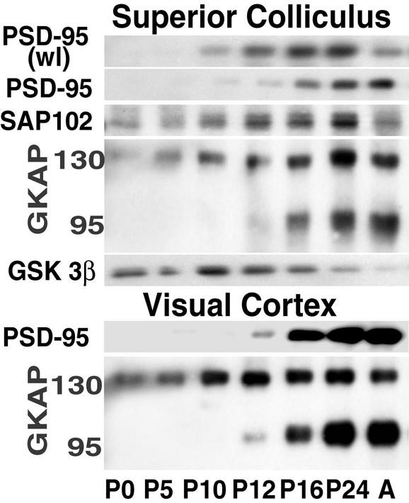
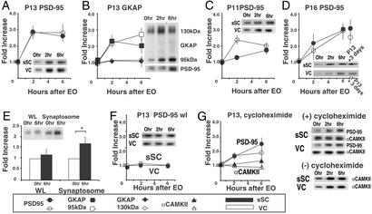
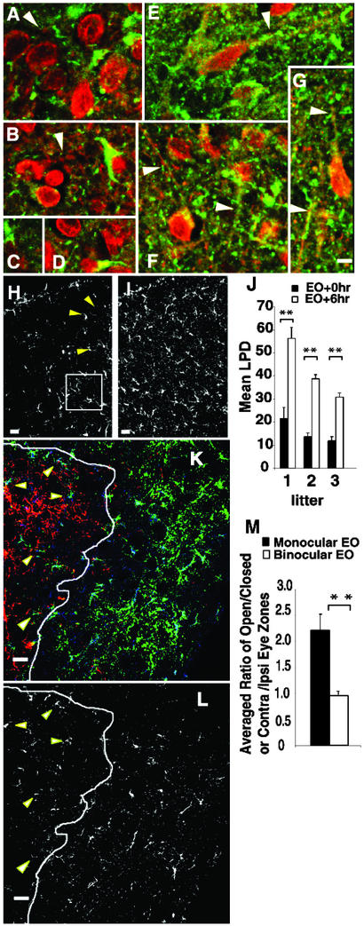
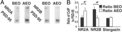
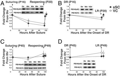
The membrane-associated guanylate kinase PSD-95 scaffolds N-methyl-d-aspartate receptors to cytoplasmic signaling molecules, and associates with other glutamate receptors at central synapses. However, regulation of PSD-95 in vivo is poorly understood. We provide evidence of an activity-dependent redistribution of PSD-95 to dendrites in central visual neurons that is tied to eye opening. Six hours after eye opening, increased dendritic PSD-95 coimmunoprecipitates with the same proportions of stargazin, increased proportions of the N-methyl-d-aspartate receptor subunit NR2A, and decreased proportions of NR2B. Sustained high levels of PSD-95 in dendrites are dependent on continued pattern vision in juvenile but not mature animals, suggesting that the stabilization of PSD-95 at synapses may be involved in the control of developmental plasticity.


Figures

Figure 1
Figure 1
PSD-95 and GKAP95 increase in synaptoneurosomes from sSC and VC in the interval surrounding EO. PSD-95 increases gradually in wl of the sSC from P10 through P24 with some decrease in adulthood. Synaptoneurosomal PSD-95 increased abruptly between P12 and P16. In contrast, SAP102, examined on the same blots, is present in the sSC synaptoneurosome fractions from P0 and its level increases gradually with a drop in the adult (A). The two families of GKAP splice variants also increase in synaptoneurosome fractions from the sSC. The 95-kDa isoforms follow PSD-95 levels, whereas 130-kDa variants follow SAP102 levels. The same changes are observed in protein from VC synaptoneurosomes.
Figure 2
Figure 2
Rapid increases in PSD-95 synaptoneurosome are tied to EO, not age, and the increases appear to be caused by redistribution, not translation. The density of each band at 2 and 6 h is shown as a ratio relative to band density from littermates killed at the same age without EO (0 h). (A) In both sSC and VC increased PSD-95 synaptoneurosome levels increase at 2 and 6 h after EO when controlled EO is performed on P13, the normal day of EO (95% confidence intervals at 6 h, sSC: 2.59 ± 0.63, VC: 2.80 ± 1.02). (B) Levels of GKAP95 increase after EO on P13 with the same timing as PSD-95, whereas levels of GKAP130 on the same gels remain constant (GKAP 95, 95% confidence intervals at 6 h, sSC: 1.83 ± 0.70, VC: 2.76 ± 0.77; GKAP130, 95% confidence intervals at 6 h, sSC: 1.09 ± 0.49, VC: 1.19 ± 0.21). (C and D) Levels of PSD-95 in synaptoneurosomes from sSC and VC also increase 2 and 6 h after EO at P11 (95% confidence intervals at 6 h, sSC: 1.74 ± 0.48, VC: 2.02 ± 0.34) and P16 (95% confidence intervals at 6 h, sSC: 3.04 ± 0.71, VC: 2.96 ± 0.98). In D, the last data point represents the level of PSD-95 in littermates that opened their eyes at the normal time (P13) and were killed 3 days later. (E) Synaptosome fractions also show increases of PSD-95 6 h after EO (95% confidence intervals at 6 h, wl: 1.14 ± 0.15, synaptosome: 1.54 ± 0.27). (F) No change in PSD-95 level is observed 2 or 6 h after EO in wl from sSC and VC of P13 animals (95% confidence intervals at 6 h, sSC: 0.98 ± 0.36, VC: 1.02 ± 0.36). (G) Cycloheximide does not block the PSD-95 protein increase in synaptoneurosomes (95% confidence intervals at 6 h, sSC: 2.45 ± 0.83, VC: 1.76 ± 0.68), whereas it does block α-CAMKII synthesis (confidence intervals at 6 h, sSC: 1.21 ± 0.31, VC: 0.90 ± 0.153), which normally increases significantly because of local synaptic synthesis after EO. Representative gels are shown at left. (Quantitation of α-CAMKII increase in untreated animals not shown.) All error bars indicate SEM.
Figure 3
Figure 3
PSD-95 becomes localized along sSC dendrites after EO. (AD and H) Before EO on P13, confocal reconstructions of sSC coronal sections show PSD-95 immunoreactivity (green) as large concentrations in the soma and proximal dendrites identified with anti-MAP2 (red). (A and B) Arrows indicate segments of MAP2-labeled dendrites running longitudinally within the con- focal reconstructions. (H) Arrowheads show PSD-95 immunoreactivity in perinuclear cytoplasm. (EG and J) Equivalent reconstructions from littermates killed 6 h after EO at P13 show punctate label distributed along longitudinally viewed dendrites (arrows) as well as many small puncta in the neuropil probably reflecting label in dendrites cut in cross-section. This punctate labeling pattern is characteristic of PSD-95 staining at synapses. (I) In the lower power views from another set of littermates, the PSD-95 change with EO is seen as an increase in small puncta dispersed throughout the neuropil. (J) The density of pixels with any PSD-95 immunoreactivity (LPD) was increased nearly 2-fold in reconstructions at EO + 6 h (I) compared with littermates with unopened eyes (EO + 0 h, H). All differences were significant (paired sample Student's t test; **, P < 0.01). Error bars indicate SEM. (K) Monocular EO at P13 induces more dendritic PSD-95 in the open eye as compared with the closed eye termination zone of the dLGN. Open eye input is green, closed eye input is red, and PSD-95 immunoreactivity is blue. The margin between the zones of each eye is shown as a white line. These zones of segregation are not absolute. (L) PSD-95 label alone. Arrowheads point to five open eye axons within the closed eye zone that are juxtaposed to heavy PSD-95 immunoreactivity. (M) Average of the PSD-95 LPD ratios between open and closed eye or contra- and ipsilateral eye termination zones. A significant increase (paired t test; **, P < 0.01) in the open eye area in animals with monocular EO, suggesting more dispersion of PSD-95 in the open eye zone, despite the fact that PSD-95 immunoreactivity adjacent to open eye axons in the closed eye zone is not taken into account in this assay. No differences in PSD-95 LPD are observed between the two zones in animals with binocular EO (paired t test, P = 0.2). Error bars indicate SEM. (All scale bars = 10 μm.)
Figure 4
Figure 4
EO induces rapid synaptosome increases in PSD-95 bound NR2A subunits and decreases in PSD-95 bound NR2B subunits. (A) Different amounts of NR2 subunits coimmunoprecipitate with the PSD-95 antibody before (BEO) and 6 h after EO (AEO) at P13. (B) Quantification of changes in the proportion of NR2A (n = 4 blots) and NR2B (n = 5 blots) subunits coimmunoprecipitating with PSD-95 AEO as compared with BEO (paired Student t test; *, P < 0.05). Identical results were obtained with protein from VC (data not shown). Data were normalized against optical density measurements for PSD-95-immunoreactive bands detected on the same gels. The proportion of stargazin immunoprecipitating with PSD-95 was unchanged (n = 4, paired Student t test, P = 0.274). Error bars indicate SEM.
Figure 5
Figure 5
Synaptoneurosomal PSD-95 levels decrease reversibly with deprivation of pattern vision in developing but not mature rats. (A) Four days of eye closure, starting at P16 after 3 days of pattern vision, decreases synaptoneurosome PSD-95 (95% confidence intervals at 96 h, sSC: 0.25 ± 0.13, VC: 0.38 ± 0.20). Levels increase rapidly at 2 and 6 h after eye reopening. (B) Four days of DR caused a significant decrease in the synaptoneurosome level of PSD-95 (95% confidence intervals at 96 h, sSC: 0.81 ± 0.17, VC: 0.68 ± 0.11) but the decrease was smaller than that seen with eyelid suturing (LR, light rearing). A separate series of blots were performed on wl from similarly treated animals. No significant differences in PSD-95 protein levels were observed at any interval (data not shown). (C and D) The same deprivation and EO protocols do not change levels of dendritic PSD-95 in young adult rats (95% confidence intervals at 96 h eye suture, sSC: 1.08 ± 0.10, VC: 1.28 ± 0.35; 95% confidence intervals at 96 h dark-rearing, sSC: 1.08 ± 0.12, VC: 1.39 ± 0.48). All error bars indicate SEM.

Similar articles

Cited by

References

    1. Kornau H C, Schenker L T, Kennedy M B, Seeburg P H. Science. 1995;269:1737–1740. - PubMed
    1. Garcia E P, Mehta S, Blair L A, Wells D G, Shang J, Fukushima T, Fallon J R, Garner C C, Marshall J. Neuron. 1998;21:727–739. - PubMed
    1. Chen L, Chetkovich D M, Petralia R S, Sweeney N T, Kawasaki Y, Wenthold R J, Bredt D S, Nicoll R A. Nature. 2000;408:936–943. - PubMed
    1. Husi H, Ward M A, Choudhary J S, Blackstock W P, Grant S G. Nat Neurosci. 2000;3:661–669. - PubMed
    1. Kennedy M B. Science. 2000;290:750–754. - PubMed

Publication types

MeSH terms

Substances

LinkOut - more resources