. 2003 Jan 28:3:1.
doi: 10.1186/1472-6807-3-1. Epub 2003 Jan 28.

Evolutionary connection between the catalytic subunits of DNA-dependent RNA polymerases and eukaryotic RNA-dependent RNA polymerases and the origin of RNA polymerases

Affiliations

Evolutionary connection between the catalytic subunits of DNA-dependent RNA polymerases and eukaryotic RNA-dependent RNA polymerases and the origin of RNA polymerases

Lakshminarayan M Iyer et al. BMC Struct Biol. .

Abstract

Background: The eukaryotic RNA-dependent RNA polymerase (RDRP) is involved in the amplification of regulatory microRNAs during post-transcriptional gene silencing. This enzyme is highly conserved in most eukaryotes but is missing in archaea and bacteria. No evolutionary relationship between RDRP and other polymerases has been reported so far, hence the origin of this eukaryote-specific polymerase remains a mystery.

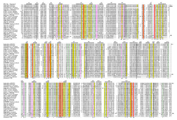
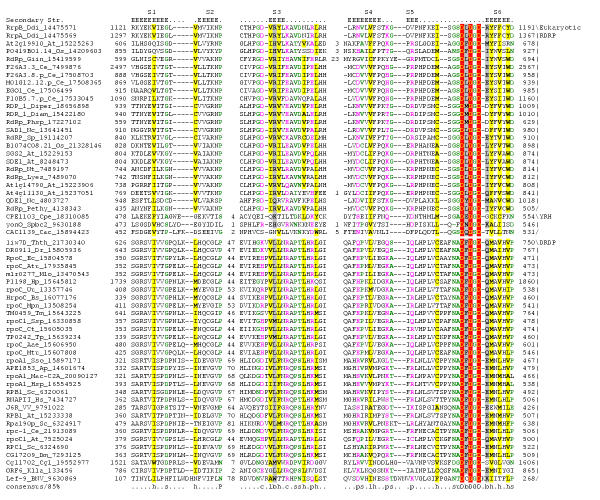
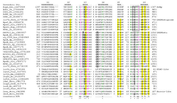
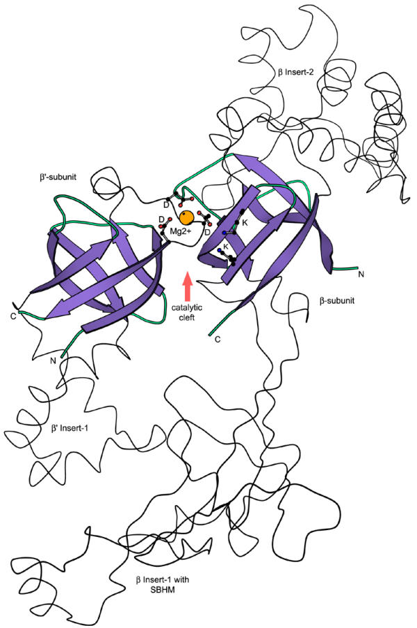
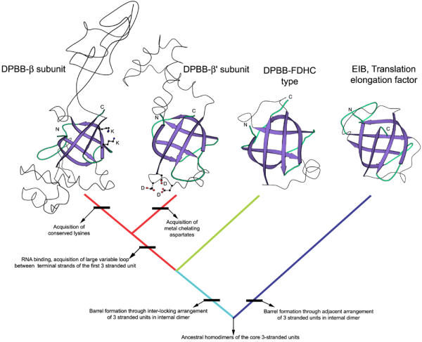
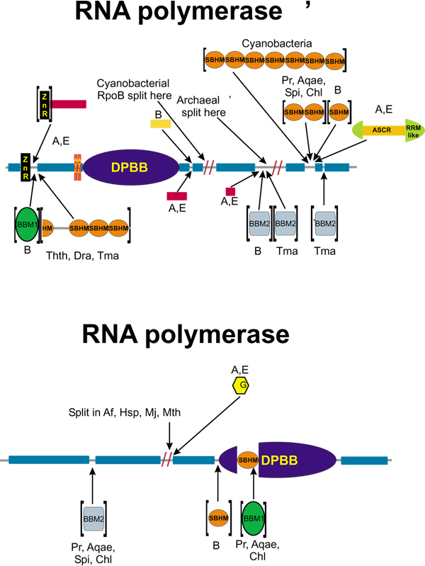
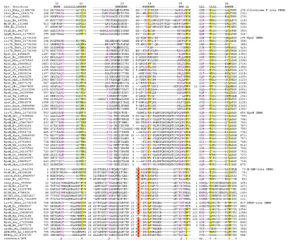
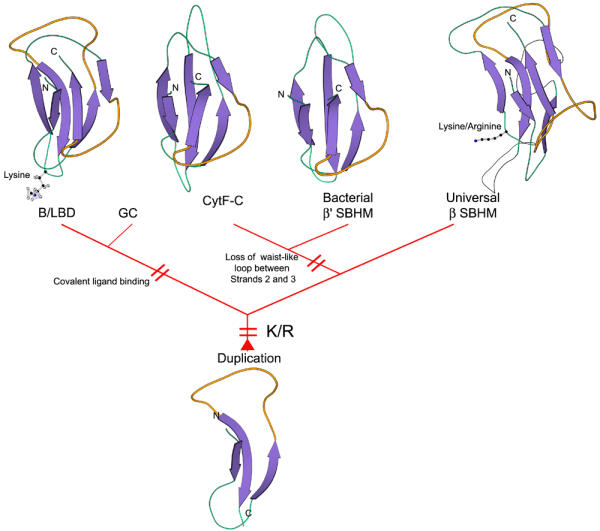
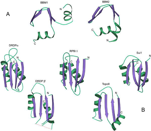
Results: Using extensive sequence profile searches, we identified bacteriophage homologs of the eukaryotic RDRP. The comparison of the eukaryotic RDRP and their homologs from bacteriophages led to the delineation of the conserved portion of these enzymes, which is predicted to harbor the catalytic site. Further, detailed sequence comparison, aided by examination of the crystal structure of the DNA-dependent RNA polymerase (DDRP), showed that the RDRP and the beta' subunit of DDRP (and its orthologs in archaea and eukaryotes) contain a conserved double-psi beta-barrel (DPBB) domain. This DPBB domain contains the signature motif DbDGD (b is a bulky residue), which is conserved in all RDRPs and DDRPs and contributes to catalysis via a coordinated divalent cation. Apart from the DPBB domain, no similarity was detected between RDRP and DDRP, which leaves open two scenarios for the origin of RDRP: i) RDRP evolved at the onset of the evolution of eukaryotes via a duplication of the DDRP beta' subunit followed by dramatic divergence that obliterated the sequence similarity outside the core catalytic domain and ii) the primordial RDRP, which consisted primarily of the DPBB domain, evolved from a common ancestor with the DDRP at a very early stage of evolution, during the RNA world era. The latter hypothesis implies that RDRP had been subsequently eliminated from cellular life forms and might have been reintroduced into the eukaryotic genomes through a bacteriophage. Sequence and structure analysis of the DDRP led to further insights into the evolution of RNA polymerases. In addition to the beta' subunit, beta subunit of DDRP also contains a DPBB domain, which is, however, distorted by large inserts and does not harbor a counterpart of the DbDGD motif. The DPBB domains of the two DDRP subunits together form the catalytic cleft, with the domain from the beta' subunit supplying the metal-coordinating DbDGD motif and the one from the beta subunit providing two lysine residues involved in catalysis. Given that the two DPBB domains of DDRP contribute completely different sets of active residues to the catalytic center, it is hypothesized that the ultimate ancestor of RNA polymerases functioned as a homodimer of a generic, RNA-binding DPBB domain. This ancestral protein probably did not have catalytic activity and served as a cofactor for a ribozyme RNA polymerase. Subsequent evolution of DDRP and RDRP involved accretion of distinct sets of additional domains. In the DDRPs, these included a RNA-binding Zn-ribbon, an AT-hook-like module and a sandwich-barrel hybrid motif (SBHM) domain. Further, lineage-specific accretion of SBHM domains and other, DDRP-specific domains is observed in bacterial DDRPs. In contrast, the orthologs of the beta' subunit in archaea and eukaryotes contains a four-stranded alpha + beta domain that is shared with the alpha-subunit of bacterial DDRP, eukaryotic DDRP subunit RBP11, translation factor eIF1 and type II topoisomerases. The additional domains of the RDRPs remain to be characterized.

Conclusions: Eukaryotic RNA-dependent RNA polymerases share the catalytic double-psi beta-barrel domain, containing a signature metal-coordinating motif, with the universally conserved beta' subunit of DNA-dependent RNA polymerases. Beyond this core catalytic domain, the two classes of RNA polymerases do not have common domains, suggesting early divergence from a common ancestor, with subsequent independent domain accretion. The beta-subunit of DDRP contains another, highly diverged DPBB domain. The presence of two distinct DPBB domains in two subunits of DDRP is compatible with the hypothesis that the ith the hypothesis that the ultimate ancestor of RNA polymerases was a RNA-binding DPBB domain that had no catalytic activity but rather functioned as a homodimeric cofactor for a ribozyme polymerase.


Figures

Figure 1
Figure 1
Multiple sequenced alignment of the conserved RDRP module from eukaryotic RNA-dependent RNA polymerases and their bacteriophage homologs. The sequences are denoted by gene names, abbreviated species names and Gene Identification (GI) numbers from the GenBank database. Species name abbreviations: At: Arabidopsis thaliana, Cac: Clostridium acetobutylicum, Ce: Caenorhabditis elegans, Cpe: Clostridium perfringens, Ddi: Dictyostelium discoideum, Diam: Diaporthe ambigua, Diper: Diaporthe perjuncta, Giin: Giardia intestinalis, Lyes: Lycopersicon esculentum, Nc: Neurospora crassa, Nt: Nicotiana tabacum, Os: Oryza sativa, Pethy: Petunia hybrida, Phsp: Phomopsis sp., Sp: Schizosaccharomyces pombe, Spbc2: Bacteriophage Spβc2. The positions of the first and the last residue of the aligned region in the corresponding protein are indicated before and after each sequence, respectively. The numbers between aligned blocks represent poorly conserved inserts that are not shown. The coloring is based on the 95% consensus shown underneath the alignment; h indicates hydrophobic residues (ACFILMVWY), a indicates aromatic residues (FYW), l indicates aliphatic residues (ILVA), p indicates polar residues (STEDKRNQHC), c indicates charged residues (DEKR), bi indicates bulky residue (ILFYWMKREQ), s indicates small residues (AGSVCDN). The predicted secondary structure elements are shown below the alignment; H indicates α-helix and E indicate extended conformation (β-strand). The predicted helices are marked H1-16 and the predicted strands S1-20. The signature motif DbDGD, which is predicted to form part of the catalytic site, is shown in reverse shading. The three sequences at the bottom are those of bacteriophage homologs of the RDRPs (the YRH proteins); the remaining are RDRP sequences.
Figure 2
Figure 2
Multiple alignment of the double-psi β-barrel (DPBB) domains from the β' subunit of DNA-dependent RNA polymerases with the predicted DPBB domains of RNA-dependent RNA polymerases. The conventions for naming sequences and coloring conserved residues are as described in the legend to Figure 1. The shared secondary structure elements are shown above the alignment with E denote the β-strand (extended) conformation. Non-conserved regions are depicted as numbers with inserts. The species abbreviations are as in figure Fig. 1. The species abbreviations that are not listed in the legend to Figure 1 are: Thth: Thermus thermophilus, Dr: Deinococcus radiodurans, Ec: Escherischia coli, Atu: Agrobacterium tumefaciens, Mlo: Mesorhizobium loti, Hp: Helicobacter pylori, Uu:Ureaplasma ureolyticum, Bs: Bacillus subtilis, Tm: Thermotoga maritima, Ssp: Synechocystis species, Aae: Aquifex aeolicus, Ct Chlamydia trachomatis, Mpn Mycoplasma pneumoniae, Mtu: Mycobacterium tuberculosis, Tp: Treponema pallidum, Sso: Sulfolbus solfataricus, Ap: Aeropyrum pernix, Mac: Methanosarcina acetivorans, Hsp: Halobacterium species, Sc: Saccharomyces cerevisiae, Hs: Homo sapiens, VV: Vaccinia virus, BNV: Bombyx mori Nuclear Polyhedrosis virus, Cgl: Corynebacterium glutamicum, Klla: Kluyveromyces lactis.
Figure 3
Figure 3
A structure-based multiple alignment of the DPBB domains from the β and β' subunits of DDRPs with a selection of other structurally characterized DPBB domains and the predicted DPBB domains of RDRP. The alignments were generated by structural superposition of representatives of the DPBB domains followed by addition of sequence neighbors. The predicted DPBB domain of the RDRPs was included on the basis of the alignment with the DDRP β' subunit's DPBB domain. The functionally important lysines in the β subunits and the metal-coordinating aspartates in the β' subunits are boxed. The conserved positions shared by the RDRPs with the structurally characterized DPBBs are indicated below the alignment by asterisks. The other conventions and abbreviations are as in the legends to Fig. 1 and Fig. 2.
Figure 4
Figure 4
Structure of the catalytic cleft of DDRP formed by interacting DPBB domains of the β and β' subunits. The metal-coordinating DbDGD motif of the β' subunit and the functionally important lysines projecting into the catalytic cleft of the β subunit are shown in ball and stick representation. The two double psi-barrels are juxtaposed in an asymmetric head to tail configuration.
Figure 5
Figure 5
A hypothetical scheme of evolution of two types of 6-stranded β-barrels from 3-stranded units. The scheme was derived as the most parsimonious explanation for the phyletic patterns and structural peculiarities of each lineage of 6-stranded barrels. The emergence of particular properties or characters characteristic of a given clade is indicated by horizontal bars.
Figure 6
Figure 6
Domain architectures of the β and β' subunits of DDRP. Domain designations: DPBB, double-psi β-barrel, SBHM, sandwich-barrel hybrid motif, ZnR, Zn-ribbon, ATL, AT-hook like, BBM, β, β'-(specific) module. βG is a domain containing a minimal version of the β grasp fold. Other designations: A, archaea, B, bacteria, E, eukaryota, Pr, Proteobacteria, Aqae, Aquifex, Spi, spirochetes, Chl, Chlamydia, Tth, Thermus thermophilus, Dra, Deinococcus radiodurans, Tma, Thermotoga maritima; Af, Archaeoglobus fulgidus, Hsp, Halobacterium sp., Mj, Methanocaldococcus jannaschii, Mth, Methanothermobacter thermoautotrophicus. Other globular regions that are conserved between the cellular DDRPs are shown by blue rectangles and non-conserved regions are shown as gray lines. Globular regions conserved in the archaeo-eukaryotic lineage are shown by red rectangles, whereas those conserved in all bacteria are shown by yellow rectangles. The proteins are not shown to scale. Splits indicated by arrows and slashes are instances when different portions of the respective subunits are encoded in separate genes.
Figure 7
Figure 7
A structure-based multiple alignment of the sandwich-barrel hybrid motif (SBHM) domains from β and β' subunits of DDRPs and other proteins. The alignments were generated by structural superposition of representatives of the SBHM domains followed by addition of sequences neighbors of the representative structures. The conserved basic residue that forms a covalent linkage with the organic radical in biotin/lipoate-binding domain-type SBHM is shown in reverse shading. The individual sequence families of SBHMs are indicated to the right of the alignment. The waist-like loops are marked as 'L' in the secondary structure shown above the alignment. Species abbreviations are as in Fig. 1 and Fig. 2 Additional species abbreviations not given above are: Ana, Anabaena sp., Azvi, Azotobacter vinelandii, Bb, Borrelia burgdorferi, Brra, Brassica rapa, Ccr,Caulobacter crescentus, Cj, Campylobacter jejuni, Cpn, Chlamydophila pneumoniae, Haeso, Haemophilus somnus, Mge, Mycoplasma genitalium, Mj, Methanocaldococcus jannaschii, Nm, Neisseria meningitidis, Pab, Pyrococcus abyssi, PhLa, Phormidium laminosum, Pisa, Pisum sativum, Pmar, Prochlorococcus marinus, Prfr, Propionibacterium freudenreichii
Figure 8
Figure 8
An evolutionary scenario for the SBHM domains from the DDRP subunits and other proteins. The scenario for the derivation of various versions of the SHBM fold from the simple, ancestral 3-stranded unit was inferred from phyletic patterns, structural features and the internal duplication. The conserved basic residue present in the biotin/lipoate-binding domain-type SBHMs and the β subunit of the DDRPs is shows as a ball-and-stick model. The emergence of different lineage-specific specializations is indicated to the side of each clade.
Figure 9
Figure 9
Structures of other conserved modules of DDRPs. (A) The core BBMC module of the BBM1 and BBM2 domains (B) Different versions of the α-subunit-core related (ASCR) domain ASCR domains from the bacterial DDRP α subunit, RBP11 (the eukaryotic ortholog of α), the β' subunit of the archaeo-eukaryotic DDRPs, topoisomerase II/gyrase globular domain 3, and the eukaryotic translation initiation factor 1 (Sui1) are shown. Inserts or regions of poor X-ray diffraction are shown with dotted lines. The ASCR domains from the archaeo-eukaryotic β' subunits are most similar to those from the α-subunits.

Similar articles

Cited by

References

    1. Anantharaman V, Koonin EV, Aravind L. Comparative genomics and evolution of proteins involved in RNA metabolism. Nucleic Acids Res. 2002;30:1427–1464. doi: 10.1093/nar/30.7.1427. - DOI - PMC - PubMed
    1. Nelson DL, Cox MM. InLehninger Principles of Biochemistry. Worth Publishers Inc. 2000.
    1. Goodman MF, Tippin B. The expanding polymerase universe. Nat Rev Mol Cell Biol. 2000;1:101–109. doi: 10.1038/35040051. - DOI - PubMed
    1. Burgers PM, Koonin EV, Bruford E, Blanco L, Burtis KC, Christman MF, Copeland WC, Friedberg EC, Hanaoka F, Hinkle DC, et al. Eukaryotic DNA polymerases: proposal for a revised nomenclature. J Biol Chem. 2001;276:43487–43490. doi: 10.1074/jbc.R100056200. - DOI - PubMed
    1. Cheetham GM, Steitz TA. Insights into transcription: structure and function of single-subunit DNA-dependent RNA polymerases. Curr Opin Struct Biol. 2000;10:117–123. doi: 10.1016/S0959-440X(99)00058-5. - DOI - PubMed

MeSH terms

Substances