Review
. 2003 Feb 3;22(3):355-61.
doi: 10.1093/emboj/cdg044.

From Alzheimer to Huntington: why is a structural understanding so difficult?

Affiliations
Review

From Alzheimer to Huntington: why is a structural understanding so difficult?

Piero Andrea Temussi et al. EMBO J. .

Abstract

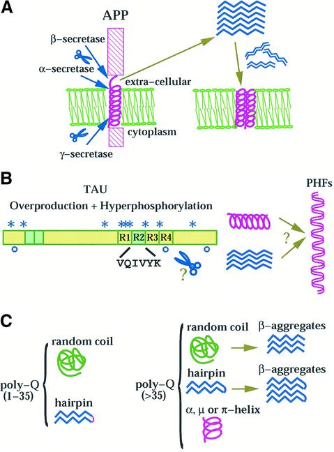
An increasing family of neurodegenerative disorders such as Alzheimer's, Parkinson's and Huntington's diseases, prion encephalopathies and cystic fibrosis is associated with aggregation of misfolded polypeptide chains which are toxic to the cell. Knowledge of the three-dimensional structure of the proteins implicated is essential for understanding why and how endogenous proteins may adopt a non-native fold. Yet, structural work has been hampered by the difficulty of handling proteins insoluble or prone to aggregation, and at the same time that is why it is interesting to study these molecules. In this review, we compare the structural knowledge accumulated for two paradigmatic misfolding disorders, Alzheimer's disease (AD) and the family of poly-glutamine diseases (poly-Q) and discuss some of the hypotheses suggested for explaining aggregate formation. While a common mechanism between these pathologies remains to be proven, a direct comparison may help in designing new strategies for approaching their study.


Figures

None
Fig. 1. Hypothetical mechanisms of conformational transitions for Aβ peptides (A), tau (B) and poly-Q containing proteins (C). (A) The peptides are proteolytically cleaved from APP, precipitate as β-structures in equilibrium with oligomers which may eventually redissolve into the membrane. The lipid double layer is shown schematically in green. (B) A schematic picture of the architecture of tau. The four repeats are indicated with R1–R4. Regions differentially spliced are coloured in green. The sites identified as calpain and caspase-3 cleavage are indicated with stars and open circles respectively (Canu et al., 1998). The proteolytic products may assemble or nucleate aggregate formation (PHFs). (C) Summary of the current models of conformational transitions involving poly-Q tracts both in the pathological and non-pathological range. For a more detailed account see Masino and Pastore (2002).
None
Fig. 2. Comparison of current models of amyloid fibres (A), tangles (B) and poly-Q aggregates (C). (A) The amyloid protofilament of 2.5–3.5 nm diameter would originate from β-strands running perpendicular to the fibre axes, although it is still disputed whether they run parallel or anti-parallel (Serpell, 2000). Several protofilaments are thought to further assemble to form mature fibrils. (B) Although no details are available yet about the molecular arrangement of tau in tangles, ample evidence of their morphology suggests an arrangement of paired helical filaments (Moreno-Herrero et al., 2001). (C) Model of water-filled nanotube in which the poly-Q forms a helical fibre with 20 residues per turn (Perutz et al., 2002). The main chains are shown in purple. The side chains (shown in green) protrude inside and outside the cylinder surface, according to β-sheet periodicity. Hydrogen bonds are formed both between the main chains and the side chains and would stabilize the structure.

References

    1. Balbach J.J., Petkova,A.T., Oyler,N.A., Antzutkin,O.N., Gordon,D.J., Meredith,S.C. and Tycko,R. (2002) Supramolecular structure in full-length Alzheimer’s β-amyloid fibrils: evidence for a parallel β-sheet organization from solid-state nuclear magnetic resonance. Biophys. J., 83, 1205–1216. - PMC - PubMed
    1. Brandt R., Leger,J. and Lee,G. (1995) Interaction of tau with the neural plasma membrane mediated by tau’s amino-terminal projection domain. J. Cell Biol., 13, 1327–1340. - PMC - PubMed
    1. Canu N. et al. (1998) Tau cleavage and dephosphorylation in cerebellar granule neurons undergoing apoptosis. J. Neurosci., 18, 7061–7074. - PMC - PubMed
    1. Coles M., Bicknell,W., Watson,A.A., Fairlie,D.P. and Craik,D.J. (1998) Solution structure of amyloid β-peptide(1–40) in a water-micelle environment. Is the membrane-spanning domain where we think it is? Biochemistry, 37, 11064–11077. - PubMed
    1. Crescenzi O., Tomaselli,S., Guerrini,R., Salvadori,S., D’Ursi,A., Temussi,P.A. and Picone,D. (2002) Solution structure of the Alzheimer amyloid β-peptide (1–42) in an apolar microenvironment. Eur. J. Biochem., 269, 5642–5648. - PubMed