. 2003 Feb 3;22(3):395-403.
doi: 10.1093/emboj/cdg035.

C-terminal domains implicated in the functional surface expression of potassium channels

Affiliations

C-terminal domains implicated in the functional surface expression of potassium channels

Marc Jenke et al. EMBO J. .

Abstract

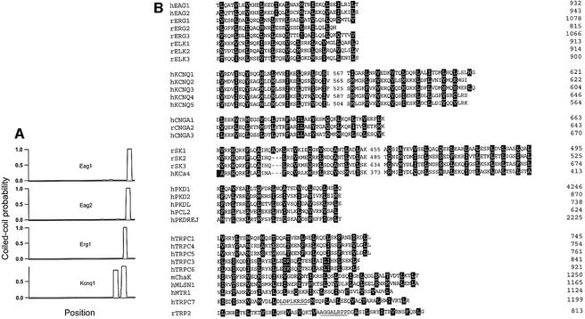
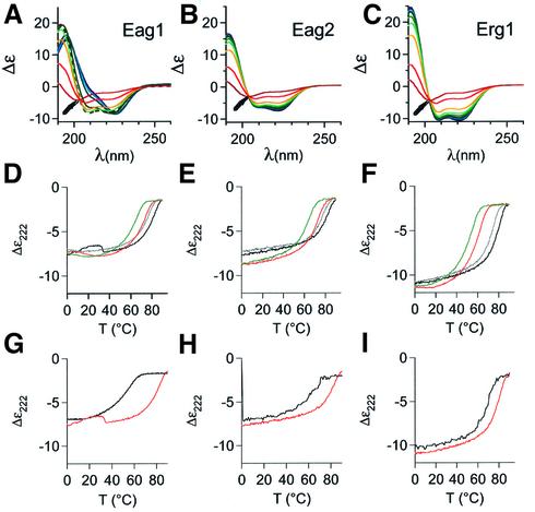
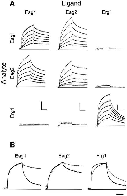
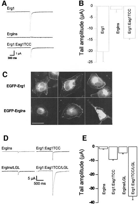
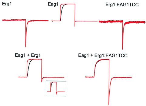
A short C-terminal domain is required for correct tetrameric assembly in some potassium channels. Here, we show that this domain forms a coiled coil that determines not only the stability but also the selectivity of the multimerization. Synthetic peptides comprising the sequence of this domain in Eag1 and other channels are able to form highly stable tetrameric coiled coils and display selective heteromultimeric interactions. We show that loss of function caused by disruption of this domain in Herg1 can be rescued by introducing the equivalent domain from Eag1, and that this chimeric protein can form heteromultimers with Eag1 while wild-type Erg1 cannot. Additionally, a short endoplasmic reticulum retention sequence closely preceding the coiled coil plays a crucial role for surface expression. Both domains appear to co-operate to form fully functional channels on the cell surface and are a frequent finding in ion channels. Many pathological phenotypes may be attributed to mutations affecting one or both domains.


Figures

None
Fig. 1. (A) Coiled-coil probability for several 6TM1P ion channels. (B) Alignments of the amino acid sequences of TCC domains from different gene families. The domains corresponding to the KCNQ channels as well as the SK calcium channels, TRP2 and TRPC7 are bipartite [see (A) and text]. The first letter of the abbreviated names indicates the species [human (h), rat (r), or mouse (m)] and numbering refers to the position of the last depicted residue. Heptad positions a and d are emphasized (black boxes). Putative non-helical stretches are underlined.
None
Fig. 2. Stoichiometry of TCC peptides. SEC performed under native conditions. The figures show normalized optical density (nOD) versus retention volume (Vret) of (A) TCCEag1 (4591 Da, solid curve) and TCCEag1L20Y (broken curve), (B) TCCEag2 (4462 Da, solid curve) and TCCEag2L13Y (broken curve), and (C) TCCErg1 (4467 Da, solid curve) and TCCErg1L20Y (broken curve). Elution volumes corresponding to 43, 19.2, 13.7 and 6.5 kDa standards are marked by arrowheads. The insets to (A) and (C) show the determination of the apparent molecular weight under native conditions using MALLS. The concentration determined (left axis, solid curve) and the evaluated molecular weight (right axis, symbols) are plotted against the retention volume (Vret).
None
Fig. 3. Secondary structure and thermal stability of TCC peptides determined by CD spectroscopy. The temperature-dependent molar ellipticity (Δε) in the far UV from 190 to 260 nm of (A) TCCEag1, (B) TCCEag2 and (C) TCCErg1 is shown in 10°C increments from 0°C (dark blue) to 90°C (red). Note the nearly isodichroic point around 204 nm at all temperatures in the CD spectra of (B) TCCEag2 and (B) TCCERG1, as well as in (A) TCCEag1 at temperatures above 50°C (arrows; the spectrum corresponding to 50°C is represented by a broken curve). All peptides have a similar CD spectrum at 90°C, to which no defined protein structure can be assigned. (DF) Molar ellipticities at 222 nm (Δε) of the thermal denaturation from 0 to 90°C (black) and the subsequent renaturation from 90 to 0°C (grey) in PBS and in 3.5 M guanidinium hydrochloride (red, green). (GI) Molar ellipticities at 222 nm (Δε) of the thermal denaturation from 0 to 90°C of the mutant peptides (black) (G) TCCEagL20Y, (H) TCCEag2L13Y and (I) TCCErg1L20Y compared with those of the wild-type peptides from (D–F) (red).
None
Fig. 4. (A) Kinetics of TCC peptide homo- and heteromeric assembly. Peptides were immobilized on the sensor surface (ligand, columns) and homomeric and heteromeric interactions were studied by surface plasmon resonance. Peptides in solution (analyte, rows) were brought into contact with the ligands for 480 s (association) and dissociation was monitored for 600 s (dots) at 1 Hz. When the injection of peptide at 3 µM yielded more than 5 RU, the interaction was studied in triplicate at decreasing concentrations of 3 µM, 1 µM, 300 nM, 100 nM, 30 nM and 10 nM, leading to decreasing responses. Fits are superimposed as solid lines. Scale bars: 200 s; 10 RU. (B) Kinetics of the heteromeric assembly of TCCEag1L20Y, TCCEag2L13Y and TCCErg1L20Y (as analytes) with the corresponding wild-type peptides (grey, as ligands). The corresponding homomeric interaction between wild-type peptides is shown in black. Scale bar: 200 s.
None
Fig. 5. Characterization of TCCErg1Ins peptide. (A) SEC under native conditions. Arrowheads mark the elution volumes of standard proteins. (B) Secondary structure measured by CD spectroscopy. The spectrum is not compatible with a helical structure. (C) Kinetics of TCCErgIns peptide assembly both in heteromeric interaction and with TCCErg1. Scale bars: 10 RU, 200 s.
None
Fig. 6. (A) Disruption of the TCC domain in Erg1 by a frame shift (InsG3107) renders non-functional channels that are rescued by insertion of the Eag1TCC domain. Upper trace, Erg1 wild type; middle trace, ErgIns; lower trace, chimera ErgIns:Eag1TCC. (B) Mean tail current amplitudes in Erg1 (N = 8), ErgIns (N = 21) and ErgIns: Eag1TCC (N = 20) (Mean ± SEM). (C) EGFP fluorescence distribution of chimerical proteins EGFP-Erg1wt (upper row) and EGFP-ErgIns (lower row). Both show similar distribution with a significant portion of the fluorescence located at the plasma membrane. Scale bar: 25 µm. (D)  Representative traces showing that LGL mutation gives rise to much larger currents in both the ErgIns and the ErgIns: Eag1TCC background. (E) Mean tail current amplitudes in ErgIns (N = 21), ErgIns/LGL (N = 22), ErgIns:Eag1TCC (N = 22) and ErgIns: Eag1TCC/LGL (N = 19). In these experiments, external potassium was 30 mM (mean ± SEM).
None
Fig. 7. Heteromeric interaction between Eag1 and Erg1:Eag1TCC. Currents were obtained in oocytes expressing wild-type Erg1, wild-type Eag1 or the chimera Erg1:Eag1TCC alone (upper traces), or co-expressing Eag1 + Erg1 or Eag1 + Erg1:Eag1TCC (lower traces). Current traces were obtained using a protocol designed to highlight both Eag1 and HERG properties which consisted of a long (5 s) preconditioning pulse to –140  mV (black traces) or –60 mV (red traces) followed by a depolarizing pulse to +60 mV for 1 s and a depolarization to –120 mV lasting for 1.5 s, and are shown after normalization to the maximal current value. Note the strong voltage dependence of the activation in Eag1 which is abolished in oocytes injected with Eag1+Erg1:Eag1TCC.

Similar articles

Cited by

References

    1. Berggrun A. and Sauer,R.T. (2001) Contributions of distinct quaternary contacts to cooperative operator binding by Mnt repressor. Proc. Natl Acad. Sci. USA, 98, 2301–2305. - PMC - PubMed
    1. Biervert C., Schroeder,B.C., Kubisch,C., Berkovic,S.F., Propping,P., Jentsch,T.J. and Steinlein,O.K. (1998) A potassium channel mutation in neonatal human epilepsy. Science, 279, 403–406. - PubMed
    1. Bohm G., Muhr,R. and Jaenicke,R. (1992) Quantitative analysis of protein far UV circular dichroism spectra by neural networks. Protein Eng., 5, 191–195. - PubMed
    1. Brüggemann A., Pardo,L.A., Stühmer,W. and Pongs,O. (1993) Ether-à-go-go encodes a voltage-gated channel permeable to K+ and Ca2+ and modulated by cAMP. Nature 365, 445–448. - PubMed
    1. Cabezon E., Arechaga,I., Butler,P.J.G. and Walker,J.E. (2000) Dimerization of bovine F-1-ATPase by binding the inhibitor protein, IF1. J. Biol. Chem., 275, 28353–28355. - PubMed

Publication types

MeSH terms