. 2003 Feb 3;22(3):404-17.
doi: 10.1093/emboj/cdg045.

Scrapie-like prion protein accumulates in aggresomes of cyclosporin A-treated cells

Affiliations

Scrapie-like prion protein accumulates in aggresomes of cyclosporin A-treated cells

Ehud Cohen et al. EMBO J. .

Abstract

Prion diseases are infectious, sporadic and inherited fatal neurodegenerations that are propagated by an abnormal refolding of the cellular prion protein PrP(C). Which chaperones assist the normal folding of PrP(C) is unknown. The linkage of familial Gerstmann- Sträussler-Scheinker (GSS) syndrome with proline substitutions in PrP raised the prospect that peptidylprolyl cis-trans isomerases (PPIases) may play a role in normal PrP metabolism. Here we used cyclo sporin A (CsA), an immunosuppressant, to inhibit the cyclophilin family of PPIases in cultured cells. CsA-treated cells accumulated proteasome-resistant, 'prion-like' PrP species, which deposited in long-lived aggresomes. PrP aggresomes also formed with disease-linked proline mutants when proteasomes were inhibited. These results suggest mechanisms whereby abnormally folded cytosolic PrP may in some cases participate in the development of spontaneous and inherited prion diseases.


Figures

None
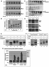
Fig. 1. Protease-resistant and detergent-insoluble PrP species accumulate in CsA-treated cells (western blots). N2a-M cells were incubated with CsA as detailed below, and then lysed either in Triton-doc (A, C and D) or NOG (B) lysis buffers. Post nuclear supernatants (PNS) were then subjected to proteolysis or sedimentation as detailed below, and the PrP species were analyzed in western blots developed with the PrP mAb 3F4. (A) Cells were incubated with CsA at the indicated concentration for 24 h, and the lysates were analyzed with (+PK) or without (–PK) prior stringent proteolysis (20 µg/ml proteinase K, 30 min, 37°C). A major 26 kDa PrP species accumulated at high CsA concentrations (–PK, left panel, arrow). This band probably represents full-length, unglycosylated PrP. Proteolysis generated a major protease-resistant core of 19 kDa (+PK, right panel, arrowhead). (B) Cells were incubated for 36 h with or without 20 µg/ml CsA, as indicated. PNS were made 1% with Sarkosyl and centrifuged through 10–60% sucrose gradients containing 1% Sarkosyl. The CsA-induced 26 kDa species sedimented to the bottom of the gradient (arrow). (C) Cells were incubated with 15 µg/ml CsA for the indicated time. PNS were made 1% with Sarkosyl and separated into high-speed supernatants (not shown) and pellets. Densitometry of the 26 kDa band shows first a slight decrease and then a significant increase in the sedimenting PrP species (four independent experiments). (D) Cells were incubated for 6 h with the indicated CsA concentrations, and high-speed pellets were prepared as in (C). The insoluble PrP species increased until the CsA concentration reached 60 µg/ml, and then leveled off.
None
Fig. 2. CsA-induced PrP: kinetics of accumulation, proteasome resistance and comparison with PrPSc. (A–C) N2a-M cells were incubated with 150 µM ALLN, 25 µg/ml CsA, or both inhibitors (as indicated), for the indicated time. Sarkosyl-insoluble PrP species were then analyzed in western blots, which were first developed with 3F4 (A) and then reprobed with a ubiquitin (ub) mAb (C). Densitometry of the 26 kDa in (A) is shown in (B) (four independent experiments). (A and B) In contrast to CsA, ALLN caused an immediate increase in insoluble 26 kDa PrP. (C) ALLN, but not CsA, induced the accumulation of insoluble poly-ub conjugates (presumably proteasome bound). Since CsA did not inhibit the ubiquitylation machinery (ALLN + CsA), the lack of poly-ub conjugates with CsA alone indicates that proteasomes were not inhibited by this drug. (D) N2a-M cells were treated for 24 h with ALLN (75 µM), CsA (25 µg/ml) or left untreated (cont). PNS were separated into high-speed pellets (upper panel) and supernatants (data not shown) or subjected to stringent proteolysis (middle and bottom panels). The ALLN- and CsA-induced 26 and 22 kDa PrP bands co-migrated with the equivalent species in untreated cells. The 19 kDa protease-resistant cores elicited by CsA and ALLN also co-migrated, both when probed with 3F4 or with the C-terminal PrP antiserum R009. (E) N2a-M cells were either treated for 24 h with 25 µg/ml CsA or left untreated. Sarkosyl-insoluble fractions (lanes 3 and 6) were prepared by ultracentrifugation as described above. Lanes 1, 2, 4 and 5: total cell membranes were prepared from parallel cultures and extracted with sodium carbonate. Three-quarters of each membrane preparation was enzymatically deglycosylated with PNGase F (lanes 2 and 5), while the remaining membranes were analyzed without further treatment (lanes 1 and 4). That the Sarkosyl-insoluble 26 kDa bands strictly co-migrate with the deglycosylated membranal PrP confirms the ER origin of the former PrP species. (F) Comparison with PrPSc. The proteinase K-resistant core (20 µg/ml proteinase K, 30 min, 37°C) of CsA-treated (25 µg/ml CsA, 24 h) N2a-M cells (lanes 1 and 3) is smaller than that of the prion isoform PrPSc in untreated ScN2a-M cells (lanes 2 and 5). In ScN2a-M cells treated with CsA (lane 4), these two PK-resistant cores appeared as a doublet. (G) Histogram: denaturation-dependent immunoassay. Untreated, ALLN- (75 µM, 24 h) and CsA- (25 µg/ml, 24 h) treated N2a-M cells, untreated ScN2a-M cells and an uninfected hamster brain were lysed and spotted on nitrocellulose strips that were incubated with the indicated concentration of GdnSCN prior to development with 3F4. The ALLN and CsA samples, as well as the ScN2a-M lysates, were subjected to proteolysis (20 µg/ml, 37°C, 30 min) prior to blotting. The ALLN and CsA samples resembled the scrapie samples in that denaturation increased their immunoreactivity, in contrast to the PrPC present in the untreated N2a-M and the control brain.
None
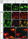
Fig. 3. PrP accumulates in vimentin-surrounded aggresomes in CHO-M cells treated with CsA but not with ALLN. (A–C) CHO-M cells were incubated for 24 h either with 20 µg/ml CsA (B) or with 75 µM ALLN (C), or left untreated (A). Focal PrP deposits were seen in ∼10% of the CsA-treated cells (B). In contrast, in ALLN-treated cells, PrP accumulated diffusely throughout the cytosol (C). (D–K) To further characterize the PrP-containing deposits, CHO-M cells were incubated for 48 h without (DF) or with (GK) 30 µg/ml CsA. The cells were labeled using the RO73 PrP antiserum (green) and with an anti-vimentin mAb (red). In the CsA-treated cells, the PrP foci were surrounded by collapsed vimentin fibers. A higher magnification (J and K) confirmed that vimentin cages enclose the PrP deposits (see arrowheads in K).
None
Fig. 4. The PrP aggresomes co-localize with γ-tubulin and 20S proteasomes but not with ubiquitin; they are inhibited by nocodazole and contain protease-resistant PrP. (A and B) CHO-M cells were incubated for 24 h either with or without 30 µg/ml CsA, as indicated, and examined by immunofluorescent confocal microscopy. PrP was detected using either RO73 (A, green channel) or 3F4 (B, red) and additional Abs as indicated. PrP aggresomes co-localized with the centrosomal marker γ-tubulin (A) and with 20S proteasomes (B) but not with ub (A), and they were found in the general region of the cells that stained with wheat germ agglutinin (WGA) (B). (C) CHO-M cells were incubated for 24 h either with 10 µg/ml nocodazole, with 30 µg/ml CsA and 10 µg/ml nocodazole or with 30 µg/ml CsA and 75 µM ALLN, and then examined by immunofluorescence for PrP (R073, green) and vimentin (red). Nocodazole prevented the formation of PrP aggresomes by CsA, but multiple PrP aggregates could be seen throughout the cell and especially underneath the cell surface (arrowheads). ALLN and CsA induced diffuse cytosolic accumulation as well as a single juxtanuclear, vimentin-caged deposit of PrP (arrowhead). (D) N2a-M cells treated for 24 h either with 25 µg/ml CsA, or with 25 µg/ml CsA and 10 µg/ml nocodazole, were subjected to proteolysis (20 µg/ml proteinase K, 30 min, 37°C) prior to western blotting with 3F4. Nocodazole did not prevent the formation of PK-resistant PrP, and the formation of aggresomes is thus not a prerequisite for the protease resistance of the CsA-induced PrP species. (E) CHO-M cells were treated with CsA (30 µM, 48 h), and then fixed with formalin and incubated with proteinase K (right panel; 7.5 µg/ml, 30 min, 37°C) prior to immunodetection of PrP with RO73 (red). The PrP immunoreactivity in aggresomes was protease resistant.
None
Fig. 5. PrP aggresomes characterized by thin-section EM. CHO-M cells were incubated with 15 µg/ml CsA for 48 h and examined by transmission EM. (A) Micrograph of a CHO-M cell containing a juxtanuclear aggresome. (B) A higher magnification of (A) shows that the structure impacts on the nucleus and is surrounded by filamentous material. A centriole (Ce) is clearly seen within the aggresome. (C) No similar structures were seen in the centriolar area of untreated cells (n = 100). (DG) CHO-M cells were treated with 15 µg/ml CsA for 48 h and subjected to pre-embedding immunogold staining using RO73 (6 nm gold). Most gold particles were observed in the vicinity of the aggresome, indicating the presence of PrP (D; higher magnification in E). A centriole is clearly seen within the aggresome (D; higher magnification in F). No gold particles were seen around centrioles of untreated cells (G). Nu, nucleus; Ag, aggresome; Fi, filamentous material; Ce, centriole.
None
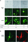
Fig. 6. The double proline PrP mutant (dmPrP) accumulates in aggresomes of ALLN-treated cells. (A) CHO cells expressing P101L-PrP or the double mutant (P101L,P104L) dmPrP, as indicated, were treated either with 30 µg/ml CsA or 15 µM ALLN for 48 h or left untreated and examined by confocal immunofluorescence microscopy using RO73 (green). In untreated cells as well as in cells treated with CsA, both mutants behaved similarly to the wtPrP. In contrast, in ALLN-treated cells, the distribution of the mutants differed significantly from that of wtPrP as: (i) P101L-PrP formed diffuse perinuclear deposits (lower panel), which could sometimes be seen by Nomarski optics as well (upper panel); (ii) in a few cells, dmPrP formed tight juxtanuclear foci, and because these deposits were surrounded by a vimentin cage (B), we identified them as aggresomes.
None
Fig. 7. Possible actions of CsA on PrP metabolism. (A) The influence of CsA on PrP metabolism. (i) In the absence of CsA, ∼90% of nascent PrP molecules in the ER gain their native conformation (stage 1) and are exported to the secretory pathway (stage 2, filled diamonds). ER cyclophilins (presumably cypB) may play a role in normal PrP folding, perhaps through their PPIase activity. PrP chains that fail to fold properly (horizontal triangles) are directed to degradation via the ERAD machinery. In the course of ERAD, these misfolded PrP molecules are dislocated into the cytosol (stage 4) and are degraded by proteasomes (stage 5). Degradation is inhibited by ALLN. (ii) The action of CsA is likely to be manifold and may target several of the normal metabolic steps outlined above. (a) By inhibiting the PPIase activity of cyclophilins, CsA may interfere with stage 1, perhaps by hampering the rescue of a minority of PrP molecules with cis X-Pro peptide bonds (see text and panel B). As a result, specific CsA-induced PrP molecules are formed (filled triangles). The CsA-induced PrP are likely to include non-native X-Pro isomers. (b) CsA could also act on some spontaneously misfolded PrP molecules (horizontal triangles) and convert them into CsA-induced species (stage 3, filled triangles). In either case, the CsA-induced species (filled triangles) are dislocated into the cytosol (stage 4). However, in contrast to the normally misfolded species, they escape proteasomal degradation and accumulate in aggresomes (stage 6). We envisage two mechanisms whereby CsA-induced PrP could avoid proteasomes. First, they might form tight aggregates at the exit of the sec61p dislocon, and these deposits may be unresolvable by the cytosolic chaperone system in the absence of cyclophilin activity. Another possibility is that these species cannot be unfolded to permit proteasomal degradation, perhaps because correcting their aberrant X-Pro bonds requires cytosolic cyclophilins, which are inhibited by CsA. (B) Model for the generation of misfolded PrP by wtPrP and by the P102L and P105L mutants. Upper panel: in all cases the majority of both wild-type and mutant (P102L and P105L) PrP is formed with predominantly trans X-Pro (or trans X-Leu) peptide bonds (which are energetically favored). Lower panel: (i) in untreated cells expressing wtPrP, the small minority of nascent PrP with cis X-Pro bonds is corrected to trans by ER cyclophilins and adopts a native conformation (filled diamonds); (ii) when cells expressing wtPrP are exposed to CsA, the cis-containing PrP molecules fail to be rescued and they misfold and adopt prion-like biochemical properties (panel A, filled triangle); (iii) a very small minority of mutant P→L-PrP molecules is formed with aberrant cis X-Leu bonds (at positions 102 or 105, depending on the mutant). Since cyclophilins fail to correct these molecules, they misfold into prion-like PrP. These molecules are normally degraded, hence ALLN is needed to enable their accumulation in aggresomes. Because spontaneous cis X-Leu bonds (in the mutants) are less probable than their cis X-Pro counterpart (in wtPrP), this model predicts that less prion-like PrP will be generated in (iii) than in (ii).

References

    1. Atkinson K., Biggs,J., Darveniza,P., Boland,J., Concannon,A. and Dodds,A. (1984) Cyclosporin-associated central nervous system toxicity after allogeneic bone marrow transplantation. Transplanta tion, 38, 34–37. - PubMed
    1. Balbach J. and Schmid,F.X. (2000) Proline isomerization and its catalysis in protein folding. In Pain,R.H (ed.), Mechanisms of Protein Folding. Oxford University Press, Oxford, UK, pp. 212–249.
    1. Basler K., Oesch,B., Scott,M., Westaway,D., Walchli,M., Groth,D.F., McKinley,M.P., Prusiner,S.B. and Weissmann,C. (1986) Scrapie and cellular PrP isoforms are encoded by the same chromosomal gene. Cell, 46, 417–428. - PubMed
    1. Bence N.F., Sampat,R.M. and Kopito,R.R. (2001) Impairment of the ubiquitin–proteasome system by protein aggregation. Science, 292, 1552–1555. - PubMed
    1. Bolton D.C., McKinley,M.P. and Prusiner,S.B. (1982) Identification of a protein that purifies with the scrapie prion. Science, 218, 1309–1311. - PubMed

Publication types

MeSH terms