. 2003 Feb 3;22(3):529-36.
doi: 10.1093/emboj/cdg041.

Mapping of the laminin-binding site of the N-terminal agrin domain (NtA)

Affiliations

Mapping of the laminin-binding site of the N-terminal agrin domain (NtA)

Joseph B Mascarenhas et al. EMBO J. .

Abstract

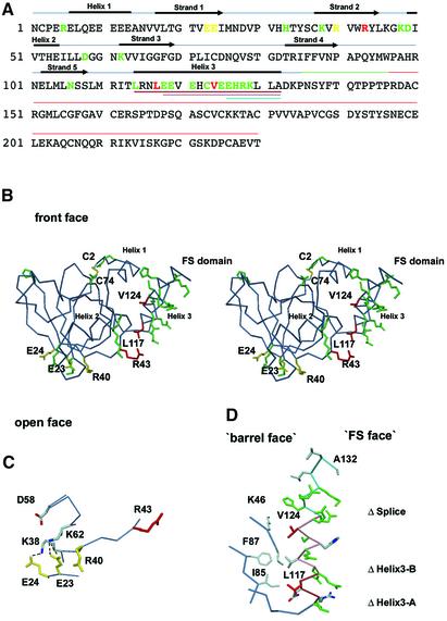
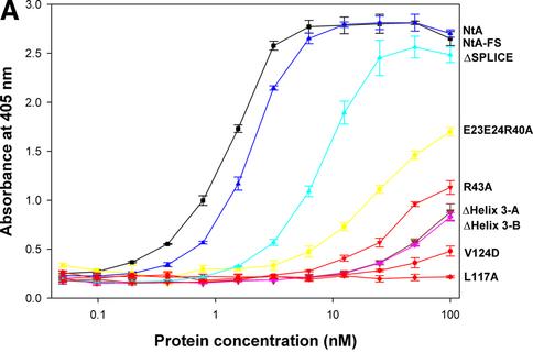
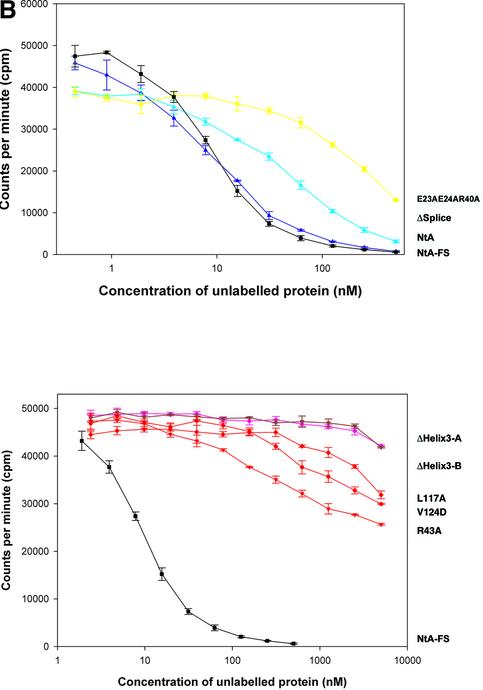
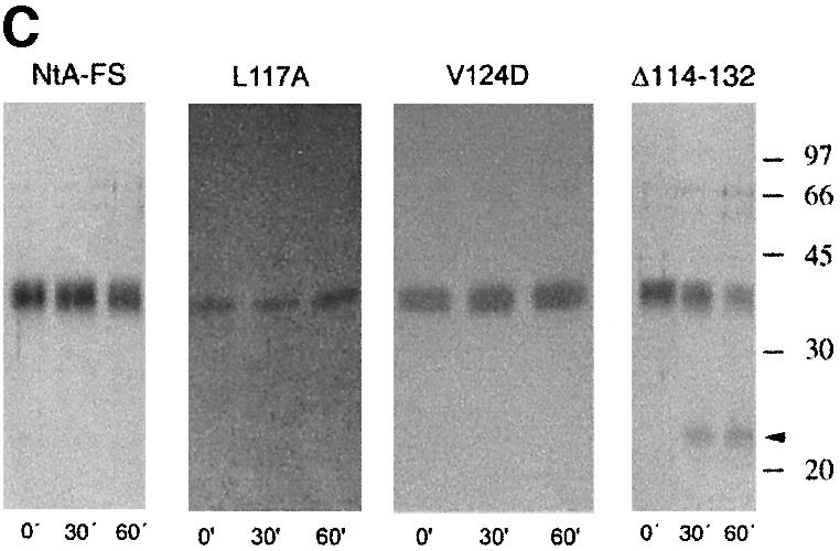
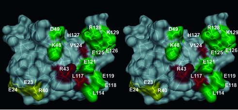
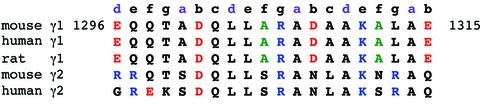
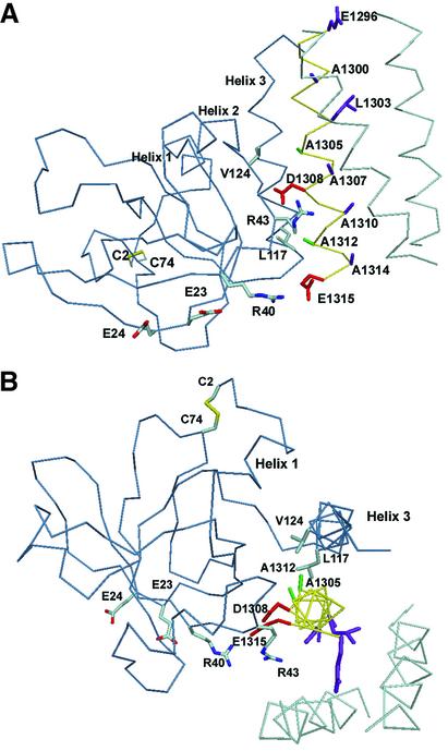
Agrin is a key organizer of acetylcholine receptor (AChR) clustering at the neuromuscular junction. The binding of agrin to laminin is required for its localization to synaptic basal lamina and other basement membranes. The high-affinity interaction with the coiled-coil domain of laminin is mediated by the N-terminal domain of agrin. We have adopted a structurally guided site-directed mutagenesis approach to map the laminin-binding site of NtA. Mutations of L117 and V124 in the C-terminal helix 3 showed that they are crucial for binding. Both residues are located in helix 3 and face the groove between the beta-barrel and the C-terminal helical segment of NtA. Remarkably, the distance between both residues matches a heptad repeat distance of two aliphatic residues which are solvent exposed in the coiled-coil domain of laminin. A lower but significant contribution originates from R43 and a charged cluster (E23, E24 and R40) at the open face of the beta-barrel structure. We propose that surface-exposed, conserved residues of the laminin gamma1 chain interact with NtA via hydrophobic and ionic interactions.


Figures

None
Fig. 1. Mapping of the laminin-binding epitope on NtA. (A) Structure-sequence-alignment of NtA-FS. Domain boundaries for NtA (blue bar), the linker between NtA and FS domain (green bar) and the FS domain (red bar) are highlighted. Secondary structural elements for NtA are indicated above the sequence alignment. β-strands and α-helices are labeled sequentially from strands 1 to 5 and helices 1 to 3. Deletions performed at helix 3 are shown below the sequence and are colored brown for ΔHelix3-A (Δ114–132), pink for ΔHelix3-B (Δ118–132) and cyan for ΔSplice (Δ126–132). Mutated residues are shown in different colors (see also Table I). Mutated residues that cause a significant reduction in laminin-1 binding are in red; those that show a less significant contribution are in yellow and those that cause only a slight or no change in affinity are in green. (B) Stereo Cα trace with all residues mutated shown in the color scheme of (A). Only mutated amino acid residues showing at least a 20-fold decrease in binding are labeled and the very N-terminal disulfide bridge (Cys2–Cys74) is shown in atom color type. (C) Detailed view of the charged cluster at the open face. The triple combination of E23A E24A R40A is shown in yellow. Hydrogen bond distances between E23–K38 and E24–K62 are shown as dotted lines. (D) Detailed view of helix 3 and hydrophobic cluster. Mutated residues are colored as in (A), otherwise in atom color type. The Cα trace of the hydrophobic cluster at the barrel face and the connector between the β-barrel and helix 3 (L109–L114) are shown in steel blue. The Cα trace of deletions of helix 3 is shown according to (A).
None
Fig. 2. Solid-phase assay of NtA or NtA-FS binding to immobilized laminin-1, radioligand competition assay and stability determined by trypsin digestion and CD spectroscopy. (A) Binding of NtA, NtA-FS and mutants within the NtA domain to laminin-1. Soluble ligands were NtA-FS (black squares), NtA (purple triangles), ΔSplice (blue triangles), E23A E24A R40A (yellow squares), R43A (bright red triangles), ΔHelix3-A (dark red triangles), ΔHelix3-B (pink diamonds), L117A (red diamonds) and V124D (red circles). Bars indicate standard deviations of three measurements. (B) Binding of radiolabeled NtA-FS (10 nM) to immobilized laminin-1 was competed with NtA, NtA-FS and mutants of NtA-FS within the NtA domain at different concentrations. Symbols are as in (A). (C) SDS–PAGE analysis after trypsin digestion at different time periods. The native NtA-FS and both single point mutants within helix 3 (L117A and V124D) were stable to trypsin cleavage. Exceptions were the deletion of helix 3, which showed the appearance of an additional band indicated by the arrowhead (only ΔHelix3-A is shown). Molecular weights determined by protein markers are indicated. (D) CD spectra of NtA-FS (squares), ΔHelix3-A (triangles) and ΔHelix3-B (diamonds) recorded in Tris-buffered saline at protein concentrations of 10 µM for the NtA-FS and 6 µM for both the helix 3 deletions.
None
Fig. 3. Surface presentation of the laminin-binding epitope of NtA in stereo view. The protein is shown by van der Waals spheres in a semi-transparent presentation. The molecular surface is shown in gray and all mutated residues are colored according to the scheme in Figure 1. The Cα trace and all marked residues are underlined.
None
Fig. 4. Sequence alignment of different laminin γ chains. The experimentally determined agrin-binding site comprises a 20-residue sequence (Kammerer, 1999). The heptad repeat pattern of residues in a and d position is shown in purple. Charged amino acid residues are in red (negative charge) and blue (positive charge). Both alanine residues (A1305 and A1312), which are flanked by leucines and show an unusual localization at position f (solvent exposed), are drawn in green.
None
Fig. 5. Model of NtA complexed with laminin in two different views. The Cα trace of NtA is shown in blue, laminin in cyan and γ1 chain in yellow. Residues of NtA that mediate the high-affinity binding are labeled and colored according to atom type. The color scheme for atoms of laminin is the same as in Figure 4. Beside electrostatic fixations via R43 (with D1308) and R40 (possible salt bridge with E1315), the equidistant aliphatic residues at helix 3 (L117 and V124 with a Cα–Cα distance of 10.6 Å) and at the solvent-exposed surface of the γ1 chain (A1305 and A1312 separated by 10.7 Å) contribute to the high-affinity binding.

Similar articles

Cited by

References

    1. Beck K., Hunter,I. and Engel,J. (1990) Structure and function of laminin: anatomy of a multidomain glycoprotein. FASEB J., 4, 148–160. - PubMed
    1. Brunger A.T. et al. (1998) Crystallography & NMR system: a new software suite for macromolecular structure determination. Acta Crystallogr. D, 54, 905–921. - PubMed
    1. Burgess R.W., Nguyen,Q.T., Son,Y.J., Lichtman,J.W. and Sanes,J.R. (1999) Alternatively spliced isoforms of nerve- and muscle-derived agrin: their roles at the neuromuscular junction. Neuron, 23, 33–44. - PubMed
    1. Burgess R.W., Skarnes,W.C. and Sanes,J.R. (2000) Agrin isoforms with distinct amino termini: differential expression, localization, and function. J. Cell Biol., 151, 41–52. - PMC - PubMed
    1. Burkin D.J., Kim,J.E., Gu,M. and Kaufman,S.J. (2000) Laminin and α7β1 integrin regulate agrin-induced clustering of acetylcholine receptors. J. Cell Sci., 113, 2877–2886. - PubMed

Publication types

Associated data