. 2003 Feb 3;22(3):548-57.
doi: 10.1093/emboj/cdg062.

Retromer and the sorting nexins Snx4/41/42 mediate distinct retrieval pathways from yeast endosomes

Affiliations

Retromer and the sorting nexins Snx4/41/42 mediate distinct retrieval pathways from yeast endosomes

Ewald H Hettema et al. EMBO J. .

Abstract

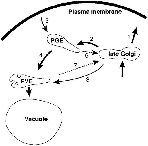
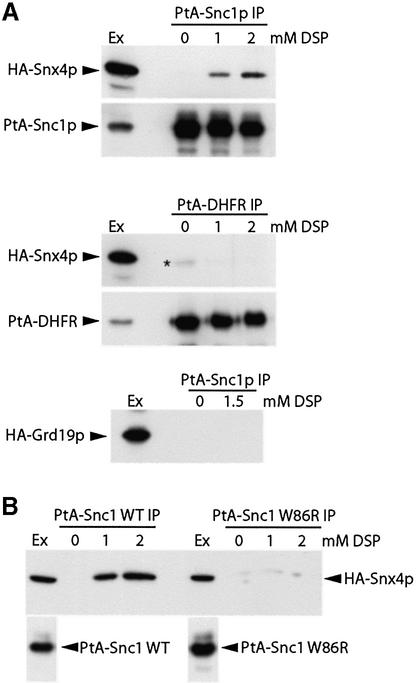
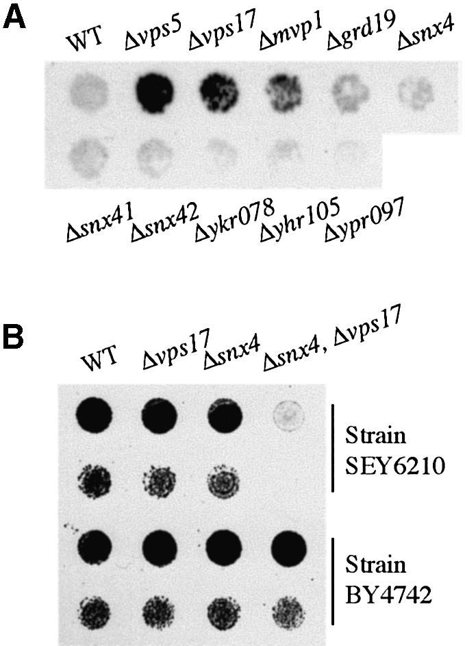
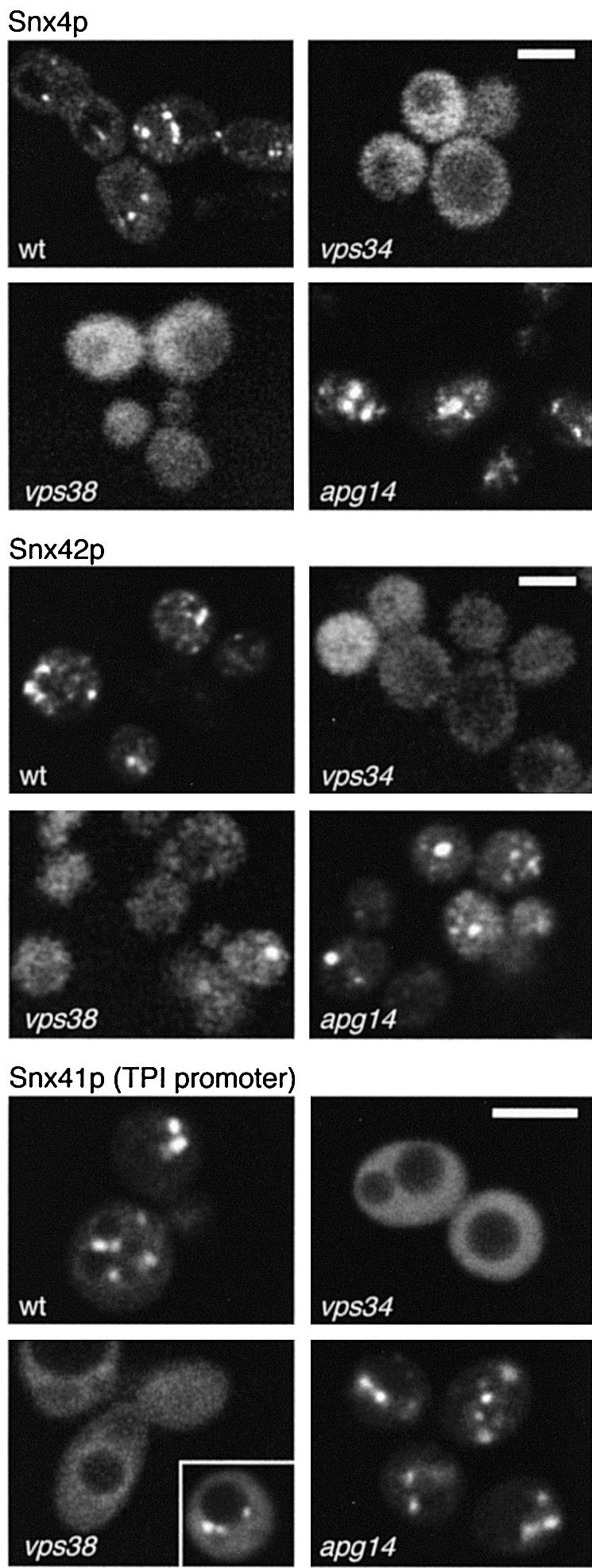
The endocytic pathway in yeast leads to the vacuole, but resident proteins of the late Golgi, and some endocytosed proteins such as the exocytic SNARE Snc1p, are retrieved specifically to the Golgi. Retrieval can occur from both a late pre-vacuolar compartment and early or 'post-Golgi' endosomes. We show that the endosomal SNARE Pep12p, and a mutant version that reaches the cell surface and is endocytosed, are retrieved from pre-vacuolar endosomes. As with Golgi proteins, this requires the sorting nexin Grd19p and components of the retromer coat, supporting the view that endosomal and Golgi residents both cycle continuously between the exocytic and endocytic pathways. In contrast, retrieval of Snc1p from post-Golgi endosomes requires the sorting nexin Snx4p, to which Snc1p can be cross-linked. Snx4p binds to Snx41p/ydr425w and to Snx42p/ydl113c, both of which are also required for efficient Snc1p sorting. Our findings suggest a general role for yeast sorting nexins in protein retrieval, rather than degradation, and indicate that different sorting nexins operate in different classes of endosomes.


Figures

None
Fig. 1. Trafficking pathways discussed herein. From the late Golgi, proteins can pass to the plasma membrane (1) or to post-Golgi endosomes (PGEs) (2) depending on the nature of their TMDs, or via the selective GGA protein-dependent pathway to pre-vacuolar endosomes (PVEs) (3). The FSD motif is required for Pep12p to follow this route. Proteins in PGEs can also pass, without special signals, to PVEs (4) and then to the vacuole. Snc1p is endocytosed to PGEs (5), then retrieved to the Golgi (6) by a mechanism that requires Snx4p. Proteins can also be retrieved from PVEs (7), a step that for Pep12p requires Grd19p. This is also the step mediated by retromer. Other pathways exist but have been omitted for clarity.
None
Fig. 2. Mutants that affect retrieval of Pep12p. (A) GFP–Pep12p bearing the Sso1p TMD and a mutant FSD signal (F20L) is in punctate PGE structures with either the F6L or I71T mutations, but with both it is on vacuolar membranes. (B) Sucrose gradient fractionation of Δpep12 cells expressing FSD Pep12p, with its own TMD, with or without the F6L/I71T mutations. Fraction 1 is the top of the gradient. The positions of PGEs and vacuolar membranes, identified by the presence of Tlg1p and Vam3p, respectively (Black and Pelham, 2000), are indicated. PVEs, defined by Pep12p itself, run in the centre of the gradient. Note that the extracts were pre-cleared by medium-speed centrifugation, resulting in substantial under-representation of vacuoles on the gradients. (C) Quantitation of the indicated proteins in gradient fractions. The left hand panel corresponds to the gradients in (B).
None
Fig. 3. Distribution of Pep12 derivatives in various mutants. (A) GFP–Pep12p is in punctate PGEs in wild-type cells (WT), whether it is delivered there directly from the Golgi (FSD) or via the plasma membrane (FSD, Sso1 TMD), but both constructs are on vacuolar membranes in grd19 cells. (B) Distribution of GFP-tagged FSD Pep12p in deletion mutants of the nine sorting nexin mutants indicated. (C) Immunoblotting of endogenous Pep12p in medium-speed (p13) and high-speed pellet fractions (p100) of the indicated strains. (D) GFP–Pep12p in a vps4 temperature-sensitive mutant grown overnight at 25°C (only vacuoles are visible) or 37°C (pre-vacuolar structures). For the right hand panels, the protein was expressed from the GAL1 promoter for 5 h, expression stopped by shifting to glucose medium for 2 h, then the temperature shifted from 25 to 37°C and cells imaged 2 and 4 h later.
None
Fig. 4. Specific effects of different sorting nexin mutants. (A) The distribution of GFP-tagged Snc1p, Tlg1p and Tlg2p is the same in wild-type (WT) and grd19 cells. (B) GFP–Snc1p is mislocalized to the interior of the vacuole, identified in the upper panels by double labelling with FM4-64, in snx4, snx41 and snx42 cells. Some residual plasma membrane staining is apparent in some cells (arrowheads), and this is true even in an snx4 snx41 snx42 triple mutant (shown at lower magnification to illustrate the range of phenotypes). (C) Profiles of immunoblots of GFP–Snc1p in the indicated strains, detected with anti-GFP antibody. P indicates the phosphorylated forms, and the proteolytic fragment referred to in the text is also indicated. Note the reduction in phosphorylated forms and increased proteolysis of GFP–Snc1p in snx4 cells.
None
Fig. 5. Interactions between nexins. Yeast strains deleted for the appropriate gene or pair of genes were transformed with plasmids expressing the corresponding nexins, one protein A tagged and one HA tagged, as indicated. The protein A-tagged form was isolated, and co-purifying HA-tagged protein detected by immunoblotting (arrowheads). The immunoprecipitates (IP) contain more cell equivalents than the extracts (Ex), but the ratio is the same in each case. The band indicated with an asterisk represents binding of the anti-HA antibody to Snx42p– protein A.
None
Fig. 6. Cross-linking of Snx4p to Snc1p. (A) Yeast strains deleted for snx4 or grd19 were transformed with plasmids expressing HA-tagged Snx4p or Grd19p, together with a plasmid expressing protein A–Snc1p or, as a control, protein A–dihydrofolate reductase. Spheroplasts were treated with the reversible cross-linker DSP prior to lysis, the protein A fusions immunoprecipitated with IgG–Sepharose beads, and protein A-tagged and associated HA-tagged proteins detected by immunoblotting. Results are shown for the cell extracts (Ex) and precipitates (IP). The band indicated by an asterisk corresponds not to HA-Snx4p, but to a small amount of IgG heavy chain eluted from the beads, which weakly cross-reacts with the secondary antibodies used in this experiment. (B) The effects of the W86R mutation in Snc1p on Snx4p binding were tested as in (A), except that in this experiment the bound protein was eluted from the IgG–Sepharose beads by specific proteolytic cleavage between the protein A and the Snc1p.
None
Fig. 7. The Snx4 family of nexins have functions distinct from that of retromer. (A) Detection of carboxypeptidase Y secreted by each sorting nexin mutant. Note that only vps5, vps17 (both encoding retromer subunits) and mvp1 show a detectable phenotype. (B) Synthetic growth defect. The indicated mutants, in two strain backgrounds, were grown to saturation in liquid culture and then spotted at high and low concentrations on plates.
None
Fig. 8. Localization of Snx4p, Snx41p and Snx42p. GFP-tagged versions of the proteins were expressed in the indicated mutants and live cells imaged in a single focal plane. The ‘wt’ cells in each case lacked the endogenous copy of the corresponding gene, but the other strains did not. GFP–Snx4p and GFP–Snx42p were expressed from centromere vectors using the SNX4 promoter; GFP–Snx41p was expressed from the TPI1 promoter, which gives higher levels of fluorescence and hence better quality images. Bars are 3 µm.

References

    1. Abeliovich H., Darsow,T. and Emr,S.D. (1999) Cytoplasm to vacuole trafficking of aminopeptidase I requires a t-SNARE–Sec1p complex composed of Tlg2p and Vps45p. EMBO J., 18, 6005–6016. - PMC - PubMed
    1. Babst M., Sato,T.K., Banta,L.M. and Emr,S.D. (1997) Endosomal transport function in yeast requires a novel AAA-type ATPase, Vps4p. EMBO J., 16, 1820–1831. - PMC - PubMed
    1. Black M.W. and Pelham,H.R. (2000) A selective transport route from Golgi to late endosomes that requires the yeast GGA proteins. J. Cell Biol., 151, 587–600. - PMC - PubMed
    1. Black M.W. and Pelham,H.R. (2001) Membrane traffic: how do GGAs fit in with the adaptors? Curr. Biol., 11, R460–R462. - PubMed
    1. Bravo J. et al. (2001) The crystal structure of the PX domain from p40(phox) bound to phosphatidylinositol 3-phosphate. Mol. Cell, 8, 829–839. - PubMed

Publication types

MeSH terms