. 2003 Feb 3;22(3):588-99.
doi: 10.1093/emboj/cdg052.

Histone H1 enhances synergistic activation of the MMTV promoter in chromatin

Affiliations

Histone H1 enhances synergistic activation of the MMTV promoter in chromatin

Ronald Koop et al. EMBO J. .

Abstract

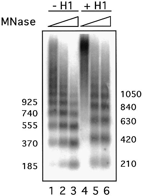
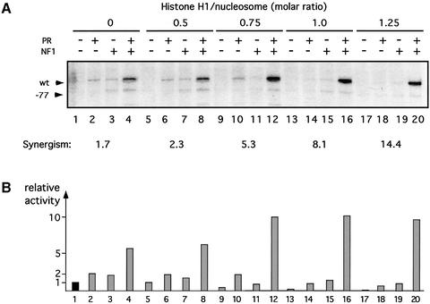
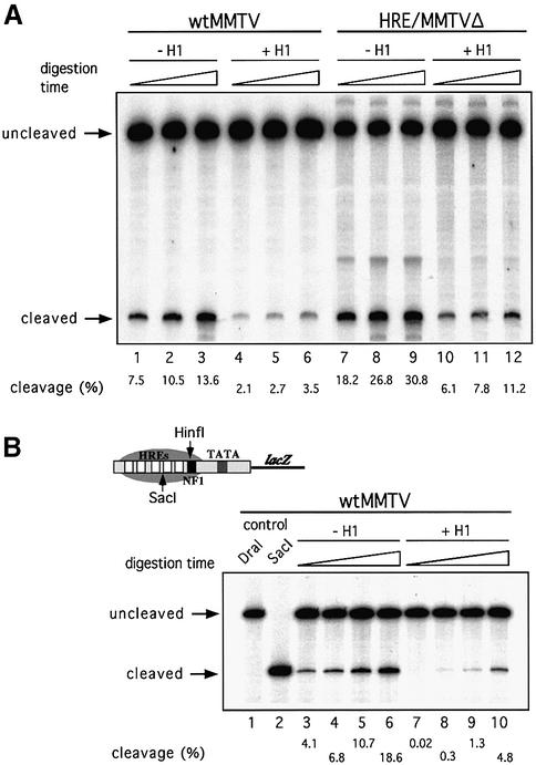
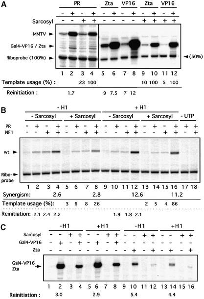
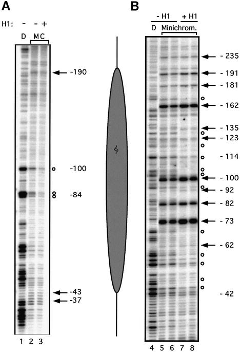
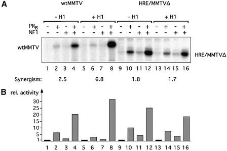
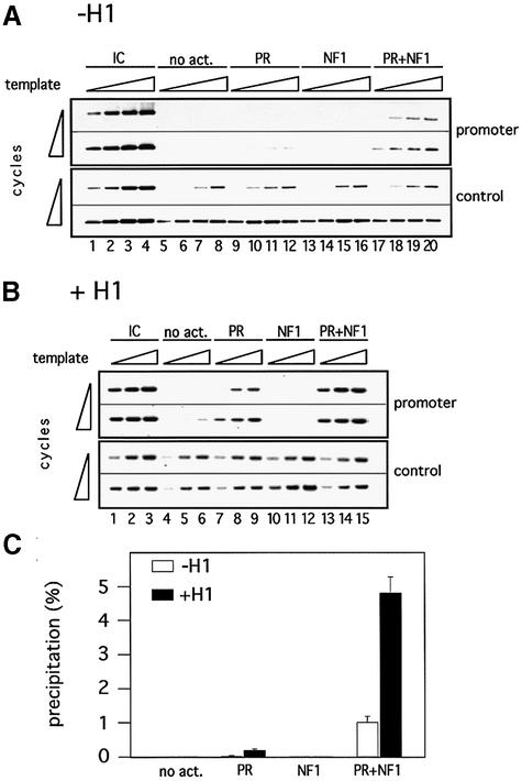
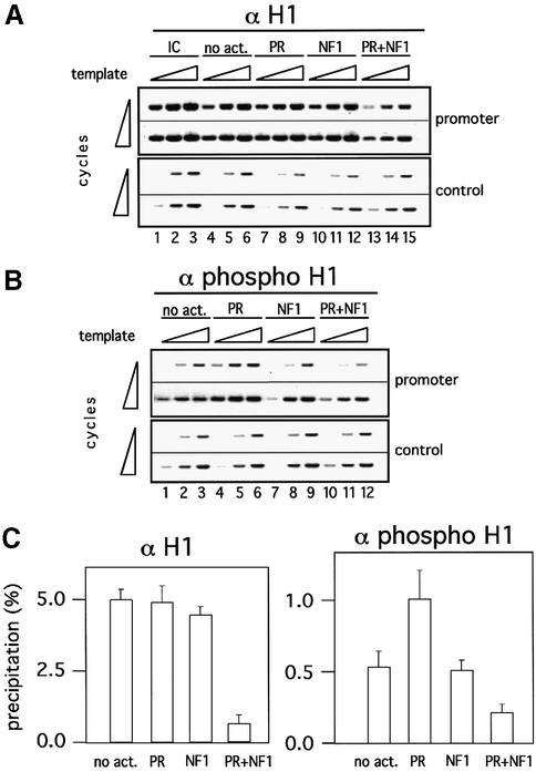
Minichromosomes assembled on the mouse mammary tumor virus (MMTV) promoter in vitro exhibit positioned nucleosomes, one of which covers the binding sites for progesterone receptor (PR) and nuclear factor 1 (NF1). Incorporation of histone H1 into MMTV minichromosomes improves the stability of this nucleosome and decreases basal transcription from the MMTV promoter, as well as its response to either PR or NF1. However, histone H1-containing minichromosomes display better PR binding and support a more efficient synergism between PR and NF1, leading to enhanced transcription initiation. A mutant MMTV promoter lacking positioned nucleosomes does not display enhanced transcriptional synergism in the presence of H1. Binding of PR leads to phosphorylation of H1, which leaves the promoter upon transcription initiation. Thus, H1 assumes a complex and dynamic role in the regulation of the MMTV promoter.


Figures

None
Fig. 1. Incorporation of histone H1 in reconstituted minichromosomes. Effect of H1 on nucleosome spacing. Chromatin assembled with or without histone H1 on pMMTVCAT B-B DNA was digested with micrococcal nuclease (MNase) for increasing times. The resulting DNA fragments were separated by electrophoresis on a 1.3% agarose gel in 1× TBE buffer and stained with ethidium bromide. The numbers on each site indicate the approximate size of the fragments in base pairs as determined by comparison with appropriate size markers.
None
Fig. 2. Influence of histone H1 on transcription from the MMTV promoter. (AIn vitro transcription analysis of MMTV minichromosomes with increasing amounts of histone H1. Chromatin reconstituted with an increasing molar ratio of histone H1 was incubated with purified recombinant PR or/and NF1 as indicated and transcribed in vitro with HeLa nuclear extract. For each template, 25 ng of DNA were used in each reaction. Products were visualized by primer extension analysis and sequencing gel electrophoresis. The positions of the products from the wild-type MMTV promoter (wt) and from a control MMTV promoter lacking the HREs (–77) are indexed on the left. The synergism of PR and NF1 was calculated as: (activity PR + NF1)/(activity PR) + (activity NF1), and is indicated at the bottom. (B) Quantification of the wild-type MMTV transcripts. Transcription signals were quantified in a phosphoimager (Fuji) and are shown in relative units, with the transcription in the absence of H1 and activators (lane 1) set to 1. The numbers on the x-axis refer to the lanes in (A).
None
Fig. 3. Effect of histone H1 on accessibility for restriction enzymes of promoter DNA sequences in chromatin. (A) Cleavage by HinfI of wild-type MMTV (wtMMTV) and mutant MMTV (HRE/MMTVΔ) promoters in minichromosomes assembled in the presence or absence of histone H1. MMTV minichromosomes (200 ng of DNA) were assembled as described in Materials and methods. After assembly, the samples were digested at 26°C with 50 U of HinfI for 2, 4 or 8 min, and the DNA was purified and restricted with DraI (wtMMTV) or PvuII (HRE/MMTVΔ). The digestion products were analyzed by linear PCR. The positions of the HinfI uncleaved and cleaved fragments are indicated. The amount of cleavage as a percentage of total radioactivity is indicated below the lanes. (B) Cleavage by SacI of the wild-type MMTV promoter in minichromosomes assembled in the presence or absence of histone H1. MMTV minichromosomes (200 ng of DNA) were assembled in DREX, digested at 26°C with 50 U of SacI for 1, 2, 4 or 8 min, and the DNA was purified and restricted with DraI. The digestion products were analyzed by linear PCR. The positions of the SacI uncleaved and cleaved fragments are indicated. The amount of cleavage as a percentage of total radioactivity is indicated below the lanes.
None
Fig. 4. Influence of histone H1 on the recruitment of promoters to the active state and on the efficiency of reinitiation. (AIn vitro transcription analysis of free DNA in the presence or absence of sarcosyl. Free reporter DNA (50 ng of template/reaction) for PR (pMMTVCAT B-B), Zta (pZ7E4T CAT) or Gal4-VP16 (pG5E4T CAT) was incubated in the absence of UTP for 30 min with HeLa nuclear extract and purified recombinant PR, Zta or Gal4-Vp16 as indicated. Sarcosyl was added to a final concentration of 0.07% and, after 5 min, transcription was started by addition of UTP. After 30 min, transcription was stopped and a riboprobe was added corresponding to 50 or 100% of the template concentration as indicated. Products were visualized by primer extension analysis and sequencing gel electrophoresis. The positions of the products from the different promoters and the riboprobe are indexed on the left. The template usage was calculated by comparing the signal intensities of the riboprobe with those of the transcription reactions in the presence of 0.07% sarcosyl (single round transcriptions). Comparing the single round transcriptions with the corresponding signals in the absence of sarcosyl (multiple rounds of transcription) yielded the values for the reinitiation rates. (BIn vitro transcription analysis of MMTV minichromosomes in the presence or absence of H1 and sarcosyl. Chromatin reconstituted in the presence or absence of histone H1 (50 ng of template/reaction) was purified via Sephadex G-50 spin columns to remove NTPs. This chromatin showed no transcription signal without addition of UTP (lanes 17 and 18), whereas transcription on non-purified minichromosomes proceeded with internal UTP (data not shown). Purified minichromosomes were incubated in the absence of UTP for 30 min with purified recombinant PR and NF1 as indicated. HeLa nuclear extract was then added for an additional 30 min. Reactions were then processed further with or without sarcosyl addition as in (A). (CIn vitro transcription analysis of pZ7E4T CAT or pG5E4T CAT minichromosomes in the presence or absence of H1 and sarcosyl. Except for the utilization of Gal4-VP16 and Zta instead of PR and NF1, reactions were carried out as in (B).
None
Fig. 5. Structural analysis of the promoter nucleosome in the presence or absence of histone H1. (A) High resolution mapping of the translational positioning. Reconstituted minichromosomes were mildly digested with MNase (20 s at 26°C) and the resulting fragments were amplified by linear PCR. Lane D: MNase digestion pattern on naked DNA. Lanes M and C: minichromosomes reconstituted in the presence or absence of H1 (as indicated above) treated with MNase. Cleavage sites protected in chromatin are indicated by open circles; hypersensitive sites are marked by black arrows. The numbers refer to the distance from the start of transcription. The diagram on the right shows the approximate position of the promoter nucleosome. (B) Rotational phasing. The rotational setting of the promoter nucleosome was determined by DNase I digestion of the minichromosomes followed by linear PCR amplification. Lane D: DNase I digestion pattern on naked DNA. Lanes Minichrom.: minichromosomes (with or without H1 as indicated above) digested for 2 min (lanes 5 and 7) and 4 min (lanes 6 and 8) with DNase I. The alternate enhancements (arrows) and protections (circles) show a periodicity of ∼10 bp. The numbers refer to the distance from the transcription start.
None
Fig. 6. Promoter dependency of the H1 effect on transcription. (AIn vitro transcription analysis of wild-type MMTV and HRE/MMTVΔ minichromosomes reconstituted in the presence or absence of histone H1. Chromatin reconstituted with or without histone H1 was incubated with purified recombinant PR and NF1 as indicated and transcribed in vitro with HeLa nuclear extract (50 ng of DNA template/reaction). Products were visualized by primer extension analysis and sequencing gel electrophoresis. The positions of the products for the wild-type MMTV promoter (wtMMTV) and HRE/MMTVΔ are indicated (note that the HRE/MMTVΔ product is 20 bp shorter). (B) Quantification of the transcription signals. Signals were quantified in a phosphoimager (Fuji) and are shown in relative units with wtMMTV control, no H1 and no activator, set to 1. The synergism of PR and NF1 was calculated as: (activity PR + NF1)/(activity PR) + (activity NF1), and is indicated on the top.
None
Fig. 7. Incorporation of histone H1 improves PR binding to minichromosomes. (A) ChIP of MMTV minichromosomes with α-PR antibody. Minichromosomes without histone H1 were purified by Sephadex G-50 chromatography and incubated with different activators (as indicated on the top) for 30 min at 30°C. ChIPs were performed in the presence of HeLa nuclear extract (120 µl), but without UTP, under conditions of transcription initiation. The upper panel shows the PCR products obtained for the specific region corresponding to the promoter nucleosome (promoter), where different amounts of precipitated material (0.5, 1, 2 and 4 µl) were PCR amplified for 23 and 26 cycles and compared against 10% of the input material (IC). The lower panel displays the results of PCRs run for 32 and 35 cycles for a control distant region of the plasmid (here the precipitates were compared against 0.1% of the input material). (B) ChIP of histone H1-containing MMTV minichromosomes with α-PR antibody. ChIPs were performed as described in (A), except that the minichromosomes were reconstituted with H1, and only 1, 2 and 4 µl of the precipitates were PCR amplified. (C) Quantification of PR binding in ChIP experiments. PR binding is displayed as a percentage of precipitated template with regard to the input material. Two independent experiments with six data points each were evaluated by densitometric measurements of ethidium bromide-stained gels.
None
Fig. 8. Displacement and phosphorylation of histone H1 in the course of transcriptional initiation of the MMTV promoter chromatin. (A) ChIP with α-H1 antibody. ChIPs were performed as described in Figure 7, except that an antibody against histone H1 was used for precipitation. Aliquots of 1, 2 and 4 µl of the precipitates were PCR amplified for 22 and 25 cycles with specific (promoter) and unspecific (control) primers. IC: 10% of input material. (B) ChIP with α-phosphoH1 antibody. ChIPs were performed as described in (A), except that an antibody against phosphorylated histone H1 was used for precipitation, and PCR results for 25 and 28 cycles are shown. (C) Quantification of H1 and phosphorylated H1 binding in ChIP experiments. Factor binding is displayed as a percentage of precipitated template with regard to the input material. The above experiment with six data points for each precipitate was evaluated by densitometric measurement of an ethidium bromide-stained gel.

Similar articles

Cited by

References

    1. Banks G.C., Deterding,L.J., Tomer,K.B. and Archer,T.K. (2001) Hormone-mediated dephosphorylation of specific histone H1 isoforms. J. Biol. Chem., 276, 36467–36473. - PubMed
    1. Bhattacharjee R.N., Banks,G.C., Trotter,K.W., Lee,H.L. and Archer,T.K. (2001) Histone H1 phosphorylation by Cdk2 selectively modulates mouse mammary tumor virus transcription through chromatin remodeling. Mol. Cell. Biol., 21, 5417–5425. - PMC - PubMed
    1. Bonte E. and Becker,P.B. (1999) Preparation of chromatin assembly extracts from preblastoderm Drosophila embryos. Methods Mol. Biol., 119, 187–194. - PubMed
    1. Bouvet P., Dimitrov,S. and Wolffe,A.P. (1994) Specific regulation of Xenopus chromosomal 5S rRNA gene transcription in vivo by histone H1. Genes Dev., 8, 1147–1159. - PubMed
    1. Bresnick E.H., Bustin,M., Marsaud,V., Richard-Foy,H. and Hager,G.L. (1992) The transcriptionally-active MMTV promoter is depeleted of histone H1. Nucleic Acids Res., 20, 273–278. - PMC - PubMed

Publication types

MeSH terms