. 2003 Feb 3;22(3):689-703.
doi: 10.1093/emboj/cdg069.

The proline-rich homeodomain protein, PRH, is a tissue-specific inhibitor of eIF4E-dependent cyclin D1 mRNA transport and growth

Affiliations

The proline-rich homeodomain protein, PRH, is a tissue-specific inhibitor of eIF4E-dependent cyclin D1 mRNA transport and growth

Ivan Topisirovic et al. EMBO J. .

Abstract

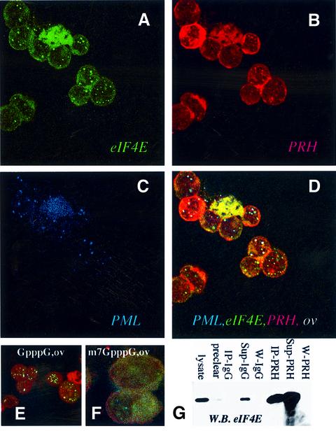
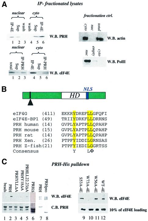
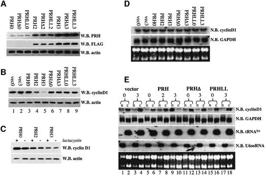
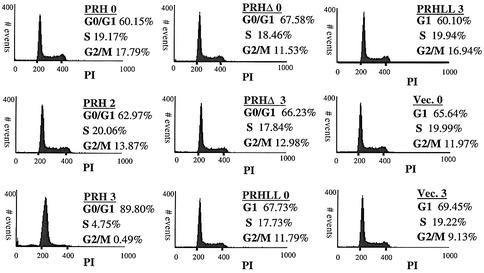
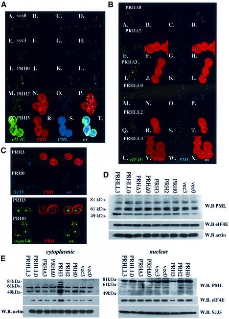
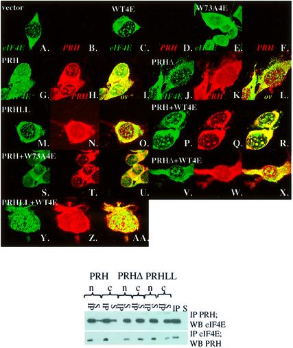
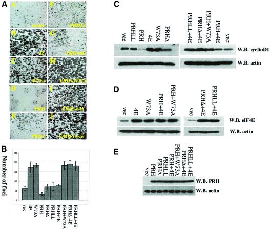
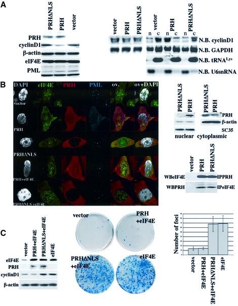
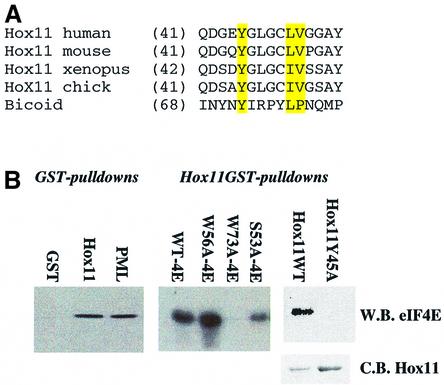
The translation initiation factor eIF4E is involved in the modulation of cellular growth. In the nucleus, where eIF4E is associated with PML nuclear bodies, eIF4E mediates nucleocytoplasmic transport of specific transcripts, and this contributes to its transformation activity. Surprisingly, we found that a trans cription factor, the proline-rich homeodomain protein PRH, is a negative regulator of eIF4E in myeloid cells, interacting with eIF4E through a conserved binding site typically found in translational regulators. Through this interaction, PRH inhibits eIF4E-dependent mRNA transport and subsequent transformation. These activities of PRH are independent of its transcriptional functions. Further, we found that 199 homeodomain proteins contain potential eIF4E-binding sites. Thus, there could be many tissue-specific regulators of eIF4E. These findings provide a model for regulation of a general factor, eIF4E, in tissue- specific contexts, and suggest that its regulation is important in differentiation and development.


Figures

None
Fig. 1. eIF4E, PRH and PML interact in vivo. U937 cells were stained with eIF4E mAb conjugated directly to FITC, in green (A), an affinity-purified PRH polyclonal antibody, in red (B), and PML mAb 5E10, in blue (C). The eIF4E–PRH–PML overlay is shown in white (D). Each channel was recorded independently to avoid cross-talk. The objective is 100× with further 2-fold magnification. Experiments carried out with either mAb eIF4E-conjugated FITC or mAb eIF4E followed by FITC secondary antibody yielded identical results, as observed previously (Cohen et al., 2001). (E and F) K562 cells were triple stained as above and the overlay is shown for cells treated with GpppG or the cap analog, m7GpppG. These confocal micrographs (A–F) represent single optical sections through the plane of the cell. A further 2-fold magnification for (E) and 4-fold for (F). (G) K562 cell lysates were immunoprecipitated with PRH antibody (IP-PRH) or immunoglobulin (IgG), and the resulting western blot (W.B.) was probed as indicated. Sup indicates supernatant after immunoprecipitation; W, final wash of the beads; pre-clear as described previously in Carlile et al. (1998); and lysate, total cell lysate.
None
Fig. 2. PRH and eIF4E interact directly. (A) U937 cells were fraction ated, immunoprecipitated and analyzed by western analysis. IP refers to the immunoprecipitated fraction, Sup to supernatant after IP, and wash to last wash of protein A beads after IP. Fractionation controls are given in the adjacent panels, using actin as a cytoplasmic marker and RNA polymerase II as the nuclear marker. Total indicates total cell lysate; nuc, nuclear fraction; and cyto, cytoplasmic fraction. (B) Schematic of the PRH protein showing the position of the homeodomain (HD) relative to the eIF4E-binding site (indicated by the arrowhead) and the location of the NLS (in blue). The eIF4E-binding site in PRH from several organisms is shown. Accession numbers are as follows: NP-002720 (human), NP-032271 (mouse), NP-077361 (rat), AAB82335 (Xenopus) and NP-571009 (zebrafish). (C) His tag pull-down analysis of the PRH–eIF4E interaction. Wild-type (right panel) or mutant PRH proteins were immobilized on nickel–agarose beads and incubated with wild-type or mutant eIF4E as indicated. Loading of PRH mutants is shown in the bottom left panel. All constructs produced proteins at the expected molecular weight. Faster migrating bands indicate degradation products. eIF4E proteins used in lanes 7 and 8 are slightly degraded; the top band corresponds to full-length protein. Loading of eIF4E mutants is shown by western blot (W.B.) in the lower right panel. Equivalent amounts of eIF4E were used for experiments in the right panel where binding is ∼10% of eIF4E input. C.B. indicates Coomassie Blue-stained SDS–polyacrylamide gel.
None
Fig. 3. PRH suppresses cyclin D1 protein production and cyclin D1 mRNA transport. (A) Induction of PRH, PRHΔ and PRHLL cells is the same in the U937 system as observed by both PRH polyclonal antibody and FLAG tag antibody. PRHLL refers to the PRHLL2324AA mutant and PRHΔ to the deletion mutant. (B) Cyclin D1 levels are suppressed by overexpression of PRH wild-type but not mutant constructs. (C) Addition of 10 µM lactacystin does not reverse the effects of PRH on cyclin D1 protein levels. (D) Northern analysis of RNA isolated from total cell lysates of PRH-overexpressing cells indicates that there are no alterations in the total levels of cyclin D1 mRNA. (E) Fractionation studies in conjunction with northern analysis reveal that PRH overexpression leads to retention of cyclin D1 mRNA in the nucleus. Cells transfected with vector or overexpressing PRH mutants do not have these effects. n indicates the nuclear, and c the cytoplasmic fraction. tRNALys is a marker for the cytoplasmic fraction, and U6snRNA for the nuclear fraction. The numbers 0, 2 and 3 indicate days post-tetracycline withdrawal. W.B., western blot; N.B., northern blot.
None
Fig. 4. PRH overexpression induces G1 arrest in U937 cells. Tet-inducible U937 cells expressing wild-type PRH, PRHΔ and PRHLL2324AA (PRHLL) mutants were stained with propidium iodide (PI), and the DNA content was measured by FACS. Cell phase distributions were determined using the CellQuest software. The numbers 0, 2 and 3 indicate days after tetracycline withdrawal.
None
Fig. 5. PRH overexpression leads to disruption of eIF4E and PML nuclear bodies. (A) Confocal micrographs show that PRH overexpression leads to disruption of nuclear bodies as indicated. Cells are triple stained for eIF4E (green), PRH (red) and PML (blue). FLAG antibody is used to detect PRH, so only exogenous PRH is observed. The numbers 0 and 3 refer to days post-tetracycline withdrawal. (B) Mutant PRH that does not bind to eIF4E does not disrupt eIF4E nuclear bodies. PRHLL refers to the PRHLL2324AA mutation, and PRHΔ the deletion mutant. (C) The subcellular distribution of Sc35 splicing speckles (Sc35), Cajal bodies and nucleoli (nopp140) does not change upon PRH overexpression. Confocal micrographs represent single sections through the plane of the cells. (D) Protein levels in total cell lysates from cells overexpressing PRH, PRHLL, PRHΔ or vector-transfected controls. (E) Fractionation studies reveal that PRH overexpression leads to re-localization of eIF4E and PML to the cytoplasm. W.B. indicates western blot. The PML antibody mAb 5E10 indicates that there is a difference in isoform distribution between the two subcellular compartments, consistent with previous studies (Stuurman et al., 1992; Flenghi et al., 1995). Further, it is possible that some bands represent degradation products rather than different isoforms of PML. Actin is used for the loading control in the cytoplasmic fraction, and Sc35 (splicing speckles) in the nuclear fraction. Note that staining for PRH and eIF4E looks more intense after the bodies are disrupted (A), but by western analysis these levels do not change (D). This is due to differences in antibody availability increasing in the fixed cells after dispersal of the nuclear bodies, whereas this is not an issue on the denaturing gels used for western analysis.
None
Fig. 6. The PRH–eIF4E interaction is independent of PML. Upper panel: PML–/– fibroblasts were stably transfected with eIF4E or PRH constructs as indicated. Confocal micrographs represent single sections through the plane of the cells. Lower panel: PML–/– cells transfected as indicated, immunoprecipitated (IP) with the Xpress antibody (IP PRH) and western blotted for eIF4E (W.B. eIF4E), or vice versa. n, nuclear fraction; c, cytoplasmic fraction; S = supernatant after IP. IP and S in the last two lanes on the right are positive controls for IP, where in the upper panel it is IP eIF4E, and in the lower panel IP PRH, and were probed as indicated.
None
Fig. 7. PRH suppresses oncogenic transformation of eIF4E and expression of cyclin D1. (A) Anchorage-dependent foci formation assays in PML–/– cells. Foci stained with Giemsa formed as a result of different transfections. Cells were transfected as indicated: 4E (wild-type eIF4E), PRH (wild-type PRH), PRHLL (PRHLL2324AA), PRHΔ (PRH ApaI deletion), W73A (eIF4E W73A mutation) and vector. Identically sized areas were taken from representative regions of each Petri dish. The results were quantitated in (B). Foci were counted in five dishes per treatment, and values are ± SD. (C) Cyclin D1 levels. Western blots (W.B.) of the indicated experiments after transfection. (D and E) Western analysis indicated that overexpression of eIF4E and PRH did not alter each other’s levels.
None
Fig. 8. Nuclear localization of PRH is necessary for its function in the suppression of eIF4E-mediated transport and transformation. All experiments were carried out in NIH-3T3 cells. (A) Left panel: western analysis of total cell lysates overexpressing PRH, PRHΔNLS and vector controls. Right panel: northern analysis of total mRNA and (far right) RNA from nuclear (n) and cytoplasmic (c) fractions. (B) Left panel: confocal micrographs of cells transfected as described. Cells were triple stained for eIF4E (green), PRH (red) and PML (Cy5 shown in blue). In addition, DAPI staining is shown in gray. Overlays are as described in Figure 1. The objective is 100× with a further 2-fold magnification. Right panel: western analysis of the nuclear and cytoplasmic fraction where β-actin and Sc35 are markers for the cytoplasmic and nuclear fraction, respectively. Below, co-immunoprecipitation analysis shows that exogenous PRH and PRHΔNLS interact with endogenous eIF4E. Total cell lysates were immunoprecipitated (IP) with eIF4E or PRH antibody. (C) Left panel: western analysis of cells transfected as described. Right panel: anchorage-dependent foci formation assays transfected as described. On the far right, foci were counted in five dishes per treatment and values are ± SD. WB indicates western blot, and N.B. northern blot.
None
Fig. 9. Other homeodomains utilize conserved eIF4E-binding sites to bind eIF4E. (A) The eIF4E-binding site in Hox11 is conserved from chicken to human. Conserved residues are highlighted in yellow. Accession numbers are: XP-046733 (human), P43345 (mouse), AA14453 (Xenopus) and 093366 (chicken). (B) Purified Hox11 binds to purified wild-type eIF4E with the same affinity as PML and PRH (Figure 2), binding ∼10% of eIF4E input. Hox11 and PML RING are immobilized GST fusion proteins, and were incubated with purified wild-type or mutant eIF4E, as indicated. Hox11Y45A mutation does not interact with eIF4E. Loading for Hox11 wild-type and mutants is shown by the Coomassie Blue gel (C.B. Hox11). W.B. indicates western blot.

Similar articles

Cited by

References

    1. Altmann M., Muller,P.P., Pelletier,J., Sonenberg,N. and Trachsel,H. (1989) A mammalian translation initiation factor can substitute for its yeast homologue in vivo. J. Biol. Chem., 264, 12145–12147. - PubMed
    1. Boer J., Bonten-Surtel,J. and Grosveld,G. (1998) Overexpression of the nucleoporin CAN/NUP214 induces growth arrest, nucleocytoplasmic transport defects and apoptosis. Mol. Cell. Biol., 18, 1236–1247. - PMC - PubMed
    1. Boisvert F.M., Hendzel,M.J. and Bazett-Jones,D.P. (2000) Promyelocytic leukemia (PML) nuclear bodies are protein structures that do not accumulate RNA. J. Cell Biol., 148, 283–292. - PMC - PubMed
    1. Borden K.L.B., Boddy,M.N., Lally,J., O’Reilly,N.J., Martin,S., Howe,K., Solomon,E. and Freemont,P.S. (1995) The solution structure of the RING finger domain from the acute promyelocytic leukaemia proto-oncoprotein PML. EMBO J., 14, 1532–1541. - PMC - PubMed
    1. Carlile G.W., Tatton,W.G. and Borden,K.L. (1998) Demonstration of a RNA-dependent nuclear interaction between the promyelocytic leukaemia protein and glyceraldehyde-3-phosphate dehydrogenase. Biochem. J., 335, 691–696. - PMC - PubMed

Publication types

MeSH terms