. 2003 Feb 3;22(3):746-56.
doi: 10.1093/emboj/cdg064.

The alternating ATPase domains of MutS control DNA mismatch repair

Affiliations

The alternating ATPase domains of MutS control DNA mismatch repair

Meindert H Lamers et al. EMBO J. .

Abstract

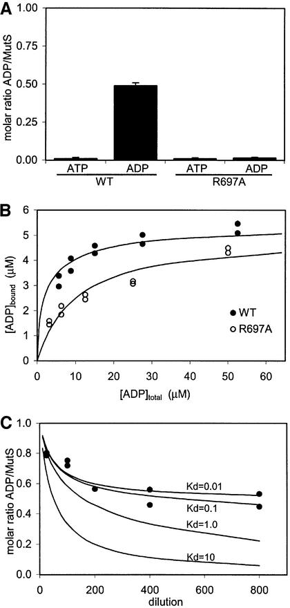
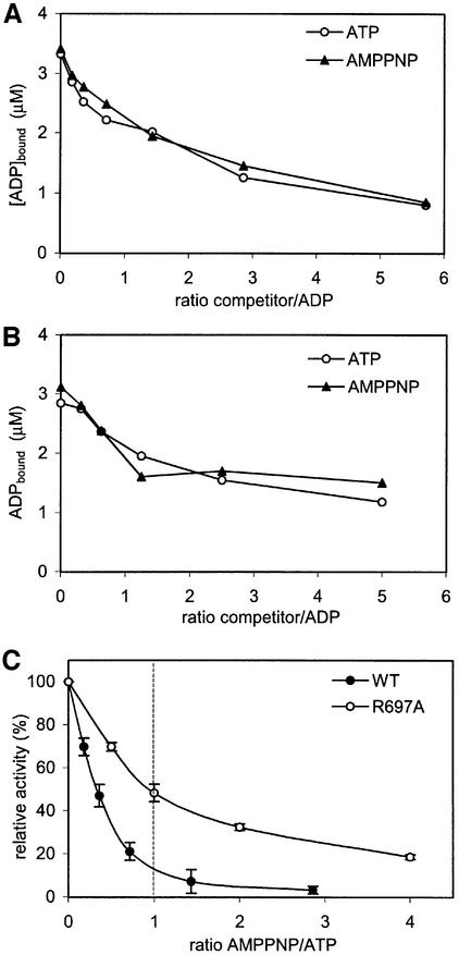
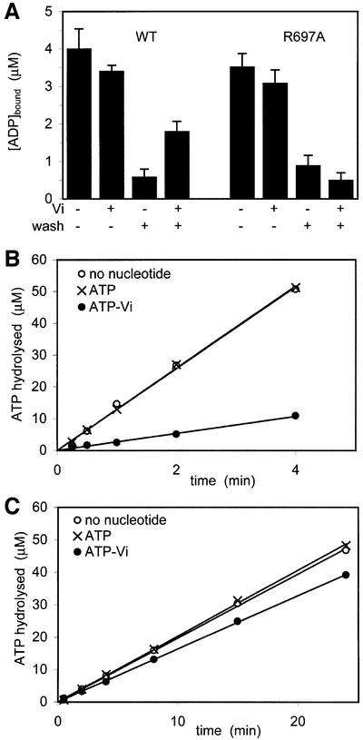
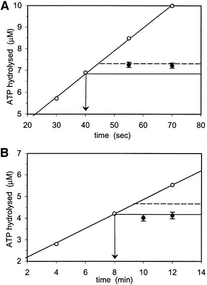
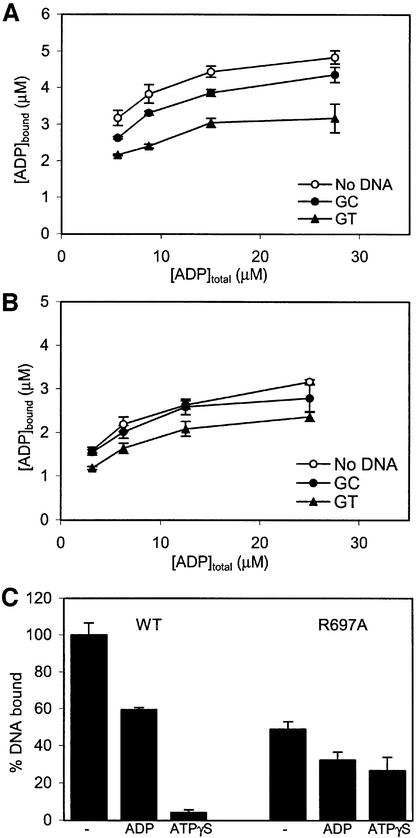
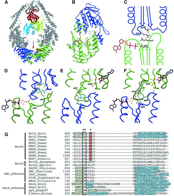
DNA mismatch repair is an essential safeguard of genomic integrity by removing base mispairings that may arise from DNA polymerase errors or from homologous recombination between DNA strands. In Escherichia coli, the MutS enzyme recognizes mismatches and initiates repair. MutS has an intrinsic ATPase activity crucial for its function, but which is poorly understood. We show here that within the MutS homodimer, the two chemically identical ATPase sites have different affinities for ADP, and the two sites alternate in ATP hydrolysis. A single residue, Arg697, located at the interface of the two ATPase domains, controls the asymmetry. When mutated, the asymmetry is lost and mismatch repair in vivo is impaired. We propose that asymmetry of the ATPase domains is an essential feature of mismatch repair that controls the timing of the different steps in the repair cascade.


Figures

None
Fig. 1. The MutS dimer has two non-equivalent ADP-binding sites. (A) Amount of ATP and ADP retained by purified MutS (wild-type and R697A) as measured in a luciferase assay. (B) Filter binding studies on wild-type and R697A MutS (5 µM) with increasing amounts of radiolabelled ADP. (C) Diluting out ADP from wild-type MutS. Black dots show the amount of radiolabelled ADP retained by MutS after dilution in a filter binding assay. Lines indicate theoretical dilution curves of a two-site binding model, with one site having a Kd of 10 µM and the second site as indicated in the graph.
None
Fig. 2. Inhibition of ATPase activity by AMPPNP. Competition of bound radiolabelled ADP with cold ATP or AMPPNP in (A) wild-type MutS and (B) R697A MutS. (C) Relative inhibition of ATPase activity by AMPPNP for wild-type and R697A MutS. The ATP concentration was kept constant, while the AMPPNP concentration was increased. Assay conditions were equal to those in (A) or (B).
None
Fig. 3. Vanadate trapping and ATPase inhibition. (A) MutS (5 µM) was incubated with radiolabelled ATP with or without vanadate (Vi). Samples were spotted directly onto nitrocellulose filters or washed first with a large excess of unlabelled ATP as indicated. (B and C) ATPase inhibition in wild-type and R697A MutS. In conditions identical to (A), MutS was incubated with unlabelled ATP with or without vanadate, after which labelled ATP was added to measure ATPase activity.
None
Fig. 4. Pulse–chase experiments on steady-state ATP hydrolysis for the wild type (A) and R697A MutS (B). During steady-state hydrolysis of radiolabelled ATP (35 µM), ATPase activity was quenched with EDTA at different time points (open circles), or first chased with a large excess (10 mM) of unlabelled ATP (arrow) before EDTA quenching (filled circles).
None
Fig. 5. Communication between ATPase and DNA-binding domains. (A) ADP binding in the absence or presence of homoduplex (GC) or heteroduplex (GT) DNA for wild-type MutS. (B) ADP binding for R697A MutS in the presence of DNA. (C) Relative amounts of DNA bound to MutS in the absence or presence of ADP or ATPγS. DNA bound by wild-type MutS in the absence of nucleotides was set to 100%.
None
Fig. 6. Structural analysis of asymmetry. (A) Structure of the MutS dimer in complex with mismatched DNA. Protein is coloured in grey, with ATPase domains of monomer A and B coloured in green and blue, respectively, and mismatch binding domains of monomer A and B in light green and light blue. DNA and ADP are in red. (B) The two ATPase domains viewed along the arrow in (A). The ADP molecule in monomer A is coloured in brown; the side chains of the two Arg697 (coloured in grey) are located at the centre of the interface of the two ATPase domains. (C) Schematic representation of the asymmetric interactions of the ATPase domains of monomer A (green) and B (blue). (1) ArgB697 hydrogen-bonds to the backbone of GlyA698 in the DE-loop of monomer A, while the reverse contact does not take place. (2) As a result, the DE-loop of monomer A clashes with the P-loop of monomer B, which is thus inhibited from nucleotide binding. (3) Simultaneously, ArgB697 also hydrogen-bonds to and displaces GluB694. (D) Close-up of the nucleotide-binding site of monomer A in green, with the opposing monomer B in blue. (E) Same view as (D), but now viewed from monomer B. (F) Same view as (E), but of R697A MutS. The position of the DE-loop in monomer A and B of wild-type MutS is indicated in grey and black, respectively. In the absence of Arg697, no contacts are made between the two DE-loops, and both monomer A and B now bind ADP-Mg2+. (G) Structure-based sequence alignment of MutS homologues and paralogues, and related ATPases. The conserved Arg697 is coloured in orange, and marked with an asterisk (*). The Walker B motif is boxed, and indicated by (**). Secondary structural elements are indicated by a green arrow (β-strand) or a blue tube (α-helix). PDB accession codes E.coli MutS, 1E3M; Taq MutS, 1EWQ; RAD50, 1F2U; SMC, 1E69; TAP1, 1JJ7; HisP, 1B0U; MalK, 1G29; RecA, 2REB; RepA, 1G8Y; gp4, 1E0J; F1, 1BMF.
None
Fig. 7. The DE-loop in different ATPases showing the different interactions with effectors. The sequences aligned in Figure 6G are coloured in black. (AEscherichia coli MutS dimer binding to mismatch DNA (coloured in dark grey), with the DE-loop contacting the other ATPase domain. (B) Top view from the bovine F1-ATPase, with the DE-loop in contact with the central stalk (γ-subunit) (also coloured in black). (C) Bacteriophage T7 gp4 helicase, and (DE.coli RepA helicase in which the DE-loop is thought to contact a centrally located DNA.

References

    1. Abrahams J.P., Leslie,A.G., Lutter,R. and Walker,J.E. (1994) Structure at 2.8 Å resolution of F1-ATPase from bovine heart mitochondria. Nature, 370, 621–628. - PubMed
    1. Allen D.J., Makhov,A., Grilley,M., Taylor,J., Thresher,R., Modrich,P. and Griffith,J.D. (1997) MutS mediates heteroduplex loop formation by a translocation mechanism. EMBO J., 16, 4467–4476. - PMC - PubMed
    1. Aquilina G. and Bignami,M. (2001) Mismatch repair in correction of replication errors and processing of DNA damage. J. Cell Physiol., 187, 145–154. - PubMed
    1. Bellacosa A. (2001) Functional interactions and signaling properties of mammalian DNA mismatch repair proteins. Cell Death Differ., 8, 1076–1092. - PubMed
    1. Berends M.J. et al. (2002) Molecular and clinical characteristics of MSH6 variants: an analysis of 25 index carriers of a germline variant. Am. J. Hum. Genet., 70, 26–37. - PMC - PubMed

Publication types

MeSH terms