. 2003 Feb;17(2):209-22.
doi: 10.1210/me.2001-0222.

A PIT-1 homeodomain mutant blocks the intranuclear recruitment of the CCAAT/enhancer binding protein alpha required for prolactin gene transcription

Affiliations

A PIT-1 homeodomain mutant blocks the intranuclear recruitment of the CCAAT/enhancer binding protein alpha required for prolactin gene transcription

John F Enwright 3rd et al. Mol Endocrinol. 2003 Feb.

Abstract

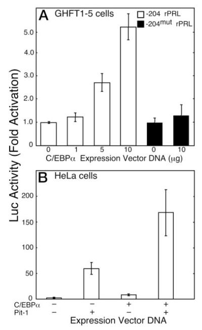
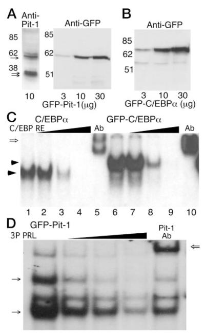
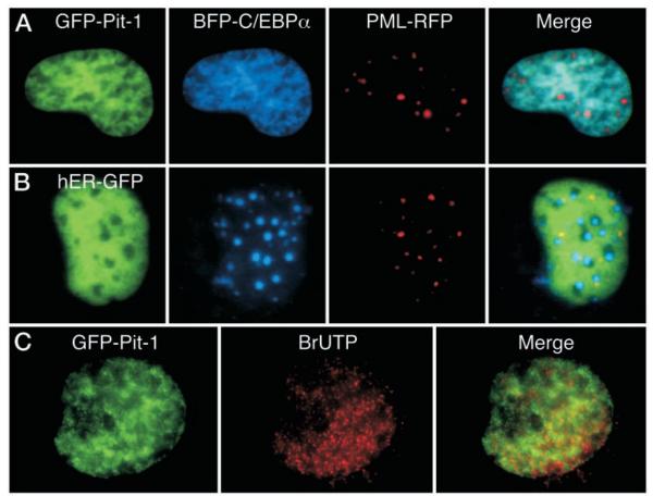
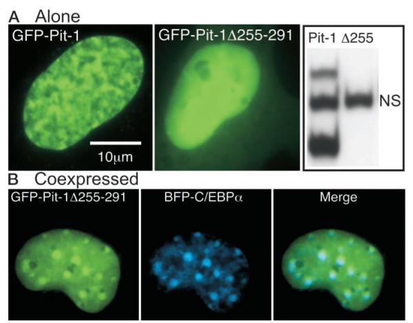
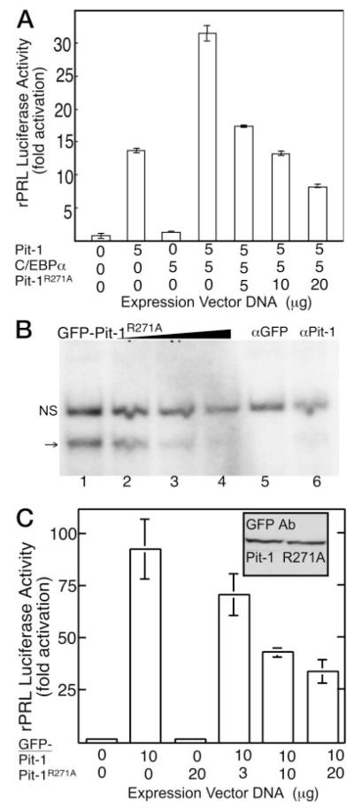
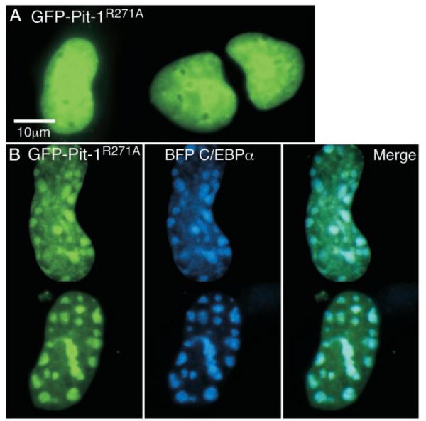
The pituitary-specific homeodomain protein Pit-1 cooperates with other transcription factors, including CCAAT/enhancer binding protein alpha (C/EBPalpha), in the regulation of pituitary lactotrope gene transcription. Here, we correlate cooperative activation of prolactin (PRL) gene transcription by Pit-1 and C/EBPalpha with changes in the subnuclear localization of these factors in living pituitary cells. Transiently expressed C/EBPalpha induced PRL gene transcription in pituitary GHFT1-5 cells, whereas the coexpression of Pit-1 and C/EBPalpha in HeLa cells demonstrated their cooperativity at the PRL promoter. Individually expressed Pit-1 or C/EBPalpha, fused to color variants of fluorescent proteins, occupied different subnuclear compartments in living pituitary cells. When coexpressed, Pit-1 recruited C/EBPalpha from regions of transcriptionally quiescent centromeric heterochromatin to the nuclear regions occupied by Pit-1. The homeodomain region of Pit-1 was necessary for the recruitment of C/EBPalpha. A point mutation in the Pit-1 homeodomain associated with the syndrome of combined pituitary hormone deficiency in humans also failed to recruit C/EBPalpha. This Pit-1 mutant functioned as a dominant inhibitor of PRL gene transcription and, instead of recruiting C/EBPalpha, was itself recruited by C/EBPalpha to centromeric heterochromatin. Together our results suggest that the intranuclear positioning of these factors determines whether they activate or silence PRL promoter activity.


Figures

Fig. 1
Fig. 1
Transcription from the −204 rPRL Promoter Is Induced by C/EBPα A, GHFT1–5 cells were transfected with a plasmid containing either the −204 rPRL promoter (open bars) or −204mut rPRL promoter (black bars) linked to the Luc reporter gene and the indicated amount of C/EBPα expression vector. After 24 h, cell extracts were prepared, and luciferase activity, corrected for total cellular protein, was determined and normalized to the activity of the reporter alone. The error is the SEM from three independent experiments, each done in triplicate. B, HeLa cells were transfected with the −204 rPRL luc reporter gene alone or with expression vectors for either Pit-1 (5 μg), C/EBPα (10 μg), or both. Luciferase activity was corrected for total cellular protein and normalized to the activity of the reporter alone. The error is the SEM from six independent experiments, each done in triplicate.
Fig. 2
Fig. 2
Analysis of the Expression and the DNA Binding Characteristics of GFP-Pit-1 and GFP-C/EBPα A, Western blot analysis of proteins extracted from GHFT1–5 cells transfected with the indicated amount of the GFP-Pit-1 expression plasmid. The transferred proteins were probed with either an antibody against Pit-1 (left panel) or an anti-GFP antibody (right panel). The anti-Pit-1 antibody detected both the doublet for endogenous 31- and 33-kDa Pit-1 proteins (double arrows) and the expressed 60 kDa GFP-Pit-1 (arrow), whereas the GFP antibody detected the 60-kDa GFP-Pit-1. B, Cell extracts from GHFT1–5 cells transfected with the indicated amount of the GFP-C/EBPα expression plasmid were subjected to Western blot analysis using the anti-GFP antibody. The GFP antibody detected the 70-kDa GFP-C/EBPα fusion protein. C, EMSA showing that untagged C/EBPα (lanes 1–5) and GFP-C/ EBPα (lanes 6–10) have similar DNA-binding characteristics. Cell extracts were prepared from GHFT1–5 cells expressing the indicated protein, and samples were incubated with a labeled C/EBPα response element (16) as described in Materials and Methods. After gel electrophoresis a single DNA-protein complex was observed (arrow). Binding specificity was demonstrated by competition with an excess of unlabeled oligonucleotide (lanes 2–4 and 7–9), and the presence of C/EBPα in the complex was verified by a shift in mobility resulting from the addition of an antibody specific for C/EBPα (open arrow, lanes 5 and 10). D, Cell extracts were prepared from 3T3-L1 cells expressing GFP-Pit-1, and samples were incubated with a labeled PRL 3P Pit-1 response element as described in Materials and Methods. GFP-Pit-1 formed two distinct complexes (arrows) with the PRL 3P DNA-element. Probe specificity was demonstrated using 3- to 100-fold excess unlabeled oligonucleotide (wedge) and mobility shift with addition of a Pit-1-specific antibody. Other DNA-protein complexes relate to ETS-protein binding to the 3P DNA element (18).
Fig. 3
Fig. 3
The Expressed GFP-Pit-1 and GFP-C/EBPα Have the Same Intranuclear Distribution as Their Endogenous Counterparts A, Immunohistochemical staining of C/EBPα in mouse 3T3-L1 cells induced to differentiate to adipocytes. Endogenous C/EBPα stained with an anti-C/EBPα antibody and a rhodamine-linked secondary antibody. Overlay of the red C/EBPα fluorescence and blue H33342 fluorescence images appears purple (merge) demonstrating that endogenous, adipocyte C/EBPα was colocalized with the H33342-stained chromatin. In a separate experiment, 3T3-L1 cells were then transfected with the plasmid encoding GFP-C/EBPα, and GFP fluorescence was detected directly by fluorescence microscopy (right panel). B, Immunohistochemical staining of Pit-1 in mouse GHFT1–5 cells. The endogenous Pit-1 was detected with antisera to Pit-1 anda Texas red-linked secondary antibody. Merger of the red Pit-1 fluorescence and blue H33342 fluorescence images show that Pit-1 is not concentrated in regions of heterochromatin preferentially stained by H33342. An image of a GHFT1–5 cell nucleus expressing GFP-Pit-1 is shown for comparison (right panel). GHFT1–5 cells were then transfected with plasmids encoding either GFP-C/EBPα (panel C), GFP-C/EBPΔ244 (panel D), or GFP-Pit1 (panel E) and grown on cover glass in 35-mm culture dishes. After 24 h the living cells were stained for 20 min with the cell-permeable DNA dye H33342. Sequential images were acquired of the GFP fusion protein and the stained DNA in the same focal plane as described in Materials and Methods. The calibration bar indicates 10 μm. The images were merged to show regions of overlap, which appear as cyan color in the merged image. An intensity profile was obtained for both GFP emission (green line) and H33342 fluorescence (blue line) at the position indicted by the line in each merged image, and the results were plotted (right panels).
Fig. 4
Fig. 4
Pit-1 Recruits the Coexpressed C/EBPα GHFT1–5 cells were cotransfected with BFP-C/EBPα, PML-RFP, and either GFP-Pit1 (panel A) or GFP-ER (panel B). Sequential images from the same focal plane were acquired using suitable filters as described in Materials and Methods. The images were merged to show regions of overlap, with blue and green overlap indicated by cyan color, and regions of red and green overlap indicated by yellow color in the merged image. C, GHFT1–5 cells expressing GFP-Pit1 were permeabilized and exposed to BrUTP for 20 min. BrUTP was then immunohistochemically detected in fixed cells with a Texas red-conjugated secondary antibody as described in Materials and Methods. Dual color images were obtained in the same focal plane, and the images were merged to show regions of overlap, which appear yellow in the merged image.
Fig. 5
Fig. 5
The HD of Pit-1 Is Necessary for Specific Subnuclear Interactions with C/EBPα A, Fluorescence images of living GHFT1–5 cells expressing either GFP-Pit-1 (left panel) or GFP-Pit1 lacking the carboxyterminal portion of the HD (GFP-Pit-1Δ255–291, middle panel). Cell extracts were prepared from HeLa cells expressing either GFP-Pit-1 or GFP-Pit-1Δ255–291 and samples were incubated with a labeled PRL 1P Pit-1 response element as described in Materials and Methods. EMSA showed that GFP-Pit-1 (lane 1) bound to the Pit-1 DNA element resolved as two distinct complexes, whereas there was no detectable binding of GFP-Pit1Δ255 (lane 2) to the 1P Pit-1 DNA element (NS, nonspecific, see Fig. 2). B, Dual color fluorescence images of living GHFT1–5 cells coexpressing GFP-Pit1Δ255 and BFP-CEBPα were acquired from the same focal plane. The images were merged to show regions of overlap, which appear as cyan color in the merged image.
Fig. 6
Fig. 6
The Pit-1 Mutant, R271A, Binds Specifically to DNA and Functions as a Dominant Inhibitor of PRL Gene Expression A, HeLa cells were transfected with the rPRL luc reporter and the indicated amounts of the expression plasmids encoding C/EBPα, Pit-1, the Pit-1R271A mutant, or the indicated combination. Luciferase activity was determined after 24 h and was corrected for total cellular protein. The error is the SEM from three independent experiments, each done in triplicate and normalized to reporter alone. B, GFP-Pit1R271A was bound specifically to the PRL promoter 1P DNA element, but formed only a monomeric complex. Probe specificity was demonstrated using 3- to 100-fold excess unlabeled oligonucleotide (wedge, lanes 2–4), and immunoclearing was observed with either a GFP-specific (lane 5) or Pit-1 specific (lane 6) antibody; NS, Nonspecific complex. C, HeLa cells were transfected with the rPRL luc reporter and the indicated amount of GFP-Pit-1 and GFP-Pit1mut. Inset, Western blot demonstrating that the GFP-Pit1 and GFP-Pit-1R271A were expressed at equivalent levels. Error is SEM from three independent experiments, each done in triplicate and normalized to reporter alone.
Fig. 7
Fig. 7
FP-Pit1R271A Fails to Recruit C/EBPα A, GHFT1–5 cells were transfected with the expression plasmid encoding GFP-Pit1mut and grown on cover glasses in 35-mm culture dishes. After 24 h, images were acquired of the GFP-fusion protein. Calibration bar indicates 10 μm. B, GHFT1–5 cells were cotransfected with plasmids encoding BFP-C/EBPα and GFP-Pit-1R271A. Sequential blue and green fluorescent images from the same focal plane were acquired using suitable filters as described in Materials and Methods. The images were merged to show regions of overlap, with blue and green overlap indicated by cyan color.

Similar articles

Cited by

References

    1. FJ Iborra, DA Jackson, PR Cook. The path of transcripts from extra-nucleolar synthetic sites to nuclear pores: transcripts in transit are concentrated in discrete structures containing SR proteins. J Cell Sci. 1996;111:2269–2282. - PubMed
    1. TA Nardozza, MM Quigley, RH Getzenberg. Association of transcription factors with the nuclear matrix. J Cell Biochem. 1996;61:467–477. - PubMed
    1. AI Lamond, WC Earnshaw. Structure and function in the nucleus. Science. 1998;280:547–553. - PubMed
    1. A Pombo, P Cuello, W Schul, JB Yoon, RG Roeder, PR Cook, S Murphy. Regional and temporal specialization in the nucleus: a transcriptionally-active nuclear domain rich in PTF, Oct1 and PIKA antigens associates with specific chromosomes early in the cell cycle. EMBO J. 1998;17:1768–1778. - PMC - PubMed
    1. T Misteli. Protein dynamics: implications for nuclear architecture and gene expression. Science. 2001;291:843–847. - PubMed

Publication types

MeSH terms