. 2003 Jan;9(1):33-43.
doi: 10.1261/rna.2570803.

Identification of the Hfq-binding site on DsrA RNA: Hfq binds without altering DsrA secondary structure

Affiliations

Identification of the Hfq-binding site on DsrA RNA: Hfq binds without altering DsrA secondary structure

Cristin C Brescia et al. RNA. 2003 Jan.

Abstract

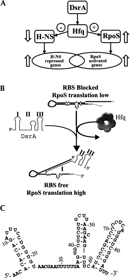
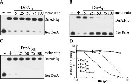
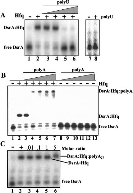
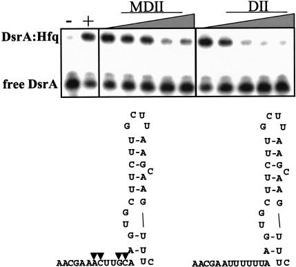
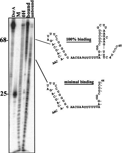
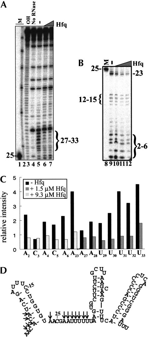
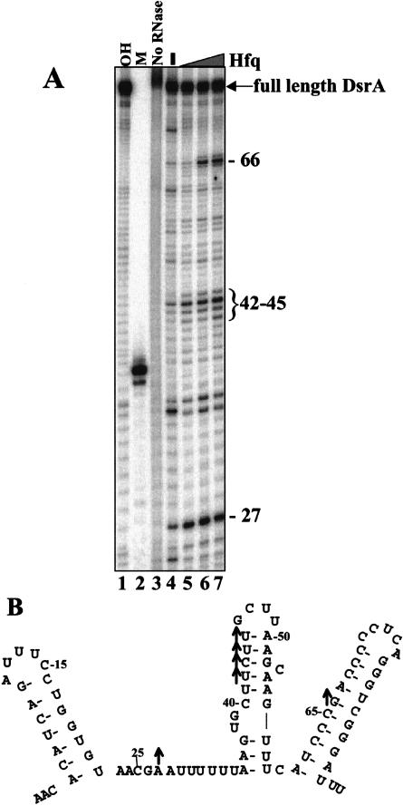
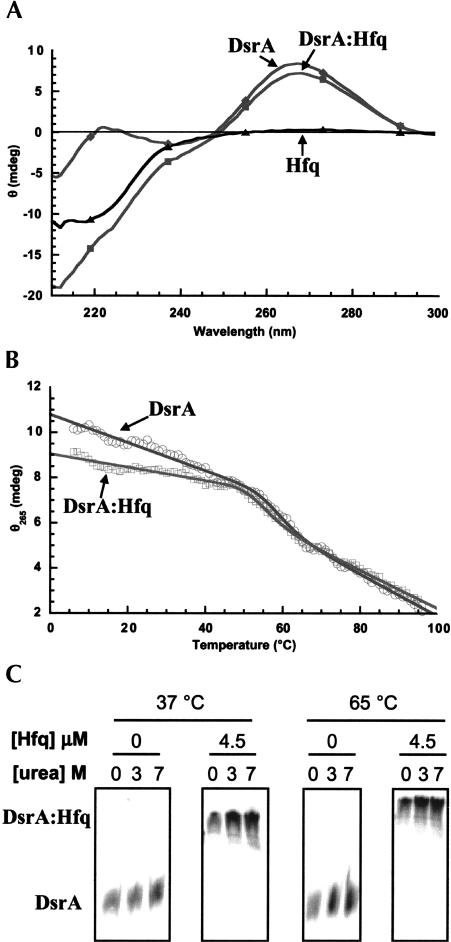
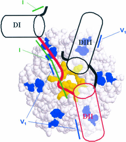
DsrA RNA regulates the translation of two global regulatory proteins in Escherichia coli. DsrA activates the translation of RpoS while repressing the translation of H-NS. The RNA-binding protein Hfq is necessary for DsrA to function in vivo. Although Hfq binds to DsrA in vitro, the role of Hfq in DsrA-mediated regulation is not known. One hypothesis was that Hfq acts as an RNA chaperone by unfolding DsrA, thereby facilitating interactions with target RNAs. To test this hypothesis, we have examined the structure of DsrA bound to Hfq in vitro. Comparison of free DsrA to DsrA bound to Hfq by RNase footprinting, circular dichroism, and thermal melt profiles shows that Hfq does not alter DsrA secondary structures, but might affect its tertiary conformation. We identify the site on DsrA where Hfq binds, which is a structural element in the middle of DsrA. In addition, we show that although long poly(U) RNAs compete with DsrA for binding to Hfq, a short poly(U) stretch present in DsrA is not necessary for Hfq binding. Finally, unlike other RNAs, DsrA binding to Hfq is not competed with by poly(A) RNA. In fact, DsrA:poly(A):Hfq may form a stable ternary complex, raising the possibility that Hfq has multiple RNA-binding sites.


Figures

FIGURE 1.
FIGURE 1.
Overview and model of DsrA/Hfq/RpoS-mediated regulation. (A) Overview of the role of DsrA in H-NS and RpoS-mediated translational regulation. DsrA increases the translation of RpoS mRNA, leading to increased transcription of RpoS-regulated genes. DsrA decreases the translation of H-NS, leading to increased transcription of H-NS-repressed genes (Sledjeski et al. 1996). (B) Model of DsrA/Hfq/RpoS interaction. In this model, Hfq unfolds the RpoS 5′-leader region, allowing DsrA Domain I to base-pair with the RpoS mRNA (Majdalani et al. 1998). This stabilizes an alternative conformer of RpoS mRNA that leads to increased translation by exposing the ribosome-binding site. (C) Sequence and proposed secondary structure of DsrA based on Lease and Belfort (2000).
FIGURE 2.
FIGURE 2.
DsrADII competes with full-length DsrA for binding to Hfq. 5′-end-labeled DsrA was incubated with increasing concentrations of DsrADI, DsrADII, and DsrADIII at 25°C for 5 min, followed by the addition of Hfq and an incubation at 25°C for 5 min. Samples were separated on an 8% native polyacrylamide gel in 1× TBE. (A) DsrADI, (B) DsrADII, and (C) DsrADIII were incubated with full-length 5′-end-labeled DsrA. Samples were incubated without (−) or with (+) 3 μM Hfq. (D) 5′-end-labeled full-length DsrA (+), DsrADI (•), DsrADII (▴), or DsrADIII (▪) were incubated at 25°C for 10 min with the indicated amounts of purified Hfq. The percent of unbound RNA was determined using a phosphorimager (see Materials and Methods) and plotted against Hfq concentration. Only full-length DsrA and DsrADII bound to Hfq.
FIGURE 3.
FIGURE 3.
DsrA binding to Hfq is competed with by poly(U), but not poly(A). 5′-end-labeled DsrA was incubated with increasing concentrations of (A) long polydisperse (200–400 nt) poly(U) (0.5–500 ng, lanes 36); (B) long polydisperse poly(A) (0.5–500 ng, lanes 37, 813); and (C) poly(A)27 (lanes 36) at 25°C for 5 min, followed by the addition of 3 μM Hfq (+) or binding buffer (−) and a second incubation at 25°C for 5 min. To control for RNA, RNA interaction DsrA was incubated (A) with (+, lane 8) or without (−, lane 7) 3.6 μg of polydisperse poly(U); or (B) with (0.5–500 ng, lanes 813) or without (lane 1) polydisperse poly(A). The top complexes in B are trapped in the well.
FIGURE 4.
FIGURE 4.
Modified Domain II (DsrAMDII) competed with full-length DsrA for binding to Hfq. 5′-end-labeled DsrA was incubated with increasing concentrations of DsrAMDII or DsrADII at 25°C for 5 min, followed by the addition of Hfq and an incubation at 25°C for 5 min. The four modified nucleotides are marked by triangles on the structure of DsrAMDII. Samples were incubated without (−) or with (+) 3 μM Hfq. The gels were quantitated with a phosphorimager, and the Kc was calculated for MDII (180 nM) and DII (100 nM).
FIGURE 5.
FIGURE 5.
Minimal binding assay of the DsrA–Hfq complex. 5′-end-labeled DsrA transcript (0.17 pmole) was subjected to limited alkaline hydrolysis to give ∼10% cleavage. The fragments of DsrA were incubated with purified Hfq protein, and bound and unbound fragments were subsequently separated on a native polyacrylamide gel. Bound and unbound DsrA fragments were excised, eluted, and fractionated on a denaturing polyacrylamide gel. Uncleaved DsrA (DsrA), 5′-end-labeled DsrADI (M), DsrA alkaline hydrolysis ladder (OH), DsrA fragments bound to Hfq (bound), and DsrA fragments that are not bound to Hfq (unbound) are represented in the figure. The bands seen in the bound lane at positions 25 and 35 are nonspecific and were likely due to small amounts of RNA degradation during purification of the DsrA bound fraction. These bands were not seen in other experiments
FIGURE 6.
FIGURE 6.
RNase I cleavage of the DsrA–Hfq complex. DsrA in the absence and presence of varying concentrations of Hfq protein was cleaved by RNase I. Samples were analyzed on a denaturing polyacrylamide gel. (A) RNase I cleavage of the DsrA–Hfq complex showing nucleotides 25–85. End-labeled DsrA (16 nM) was mixed with 0 (lane 5), 1.5 μM (lane 6), or 3.1 μM (lane 7) purified Hfq hexamer and incubated at 25°C for 10 min. This was followed by cleavage with 0.05 U of RNase I at 25°C for 2 min. (B) RNase I cleavage of the DsrA–Hfq complex showing nucleotides 2–26. End-labeled DsrA (16 nM) was mixed with 0 (lane 9), 1.5 μM (lane 10), 3.1 μM (lane 11), or 9.3 μM (lane 12) purified Hfq hexamer and treated as above. Markers (M) are 5′-end-labeled DsrADI (lanes 1,8); Decade Marker (Ambion) representing 30, 40, 50, 60, 70, 80, and 90 nt (lane 2); and limited hydrolysis of DsrA (OH, lane 3). (C) Quantitative analysis of protected nucleotides shown in A and B in the absence (lanes 5,9, black bar) or presence (lane 6, dark gray bar) of 1.5 μM Hfq or 9.3 μM Hfq (lane 12, light gray bar). Individual bands were quantitated using a phosphorimager and normalized to the amount of full-length DsrA in the same lane. The data shown are representative of several independent experiments. (D) Structure of DsrA proposed by Lease and Belfort (2000), with arrows showing the nucleotides that are cleaved less by RNase I in the presence of Hfq.
FIGURE 7.
FIGURE 7.
RNase V1 cleavage of the DsrA–Hfq complex. DsrA in the presence or absence of Hfq was cleaved by RNase V1. Samples were fractionated on a denaturing polyacrylamide gel. (A) RNase V1 cleavage of the DsrA–Hfq complex. (Lanes 1,2) Markers: the limited alkaline hydrolysis ladder of DsrA (OH) and 38-nt DsrADII (see Materials and Methods). (Lane 3) 5′-end-labeled DsrA (16 nM) that was not treated with RNase V1. DsrA (16 nM) was treated with RNase V1 with (+) or without (−) purified Hfq protein (3.1 μM). (B) Structure of DsrA proposed by Lease and Belfort (2000), with upward arrows showing the nucleotides that have increased cleavage in the presence of Hfq. The data shown are representative of several independent experiments.
FIGURE 8.
FIGURE 8.
Circular dichroism studies of DsrA and the DsrA:Hfq complexes. (A) Circular dichroism spectra of 0.5 μM DsrA with (▪) and without (♦) 3.0 μM Hfq, in 50 mM cacodylate buffer (pH 6.6), 250 mM NaCl at 37°C. The difference spectrum (▴) corresponds very closely to the spectrum of Hfq alone (data not shown). (B) Trace of circular dichroism intensity at 263 nm as a function of temperature (0°–100°C) for 0.5 μM DsrA and with (□) and without (○) 3.0 μM Hfq in 3 M urea, 50 mM cacodylate buffer (pH 6.6), and 250 mM NaCl. Lines represent fits of the data to a unimolecular two-state folding model. (C) Binding of DsrA to Hfq in the presence of urea and at high temperature. DsrA was incubated with or without 3.0 μM Hfq in 50 mM cacodylate buffer (pH 6.6), 250 mM NaCl, with 0, 3 M, or 7 M urea. Samples were incubated at 37°C or 65°C and run on gels at the same temperature. We observed 100% binding of DsrA to Hfq irrespective of the urea concentration or temperature of incubation.
FIGURE 9.
FIGURE 9.
Schematic model of interaction between DsrA and Hfq. In our present model all three DsrA domains are located on one face of the hexameric complex. The A/U-rich single-stranded region between Domains I and II (red line) might wrap around the inner diameter of the torus by analogy to the interaction between Sm-proteins and short poly(U) substrates (Toro et al. 2001). If both Domains I and II lie on the same face of Hfq, only a portion of the central cavity can be used for interaction with the U-rich region to avoid a steric clash between the 5′- and 3′-extensions from this primary binding site. The additional structural requirements we observe for tight binding of larger RNAs probably result from the need of RNA secondary structure elements to lie down against the rest of the hexamer face of Hfq. Two highly conserved residues (R15 and F41, shown in blue) could assist in this process by providing ionic interactions with the RNA. In our model, the formation of a dodecamer might bring together two RNAs, one bound to either face of the torus, thereby increasing the chances of interactions between the RNAs. Tertiary structure showing the coaxial stacking of Domains II and III of DsrA is purely speculative at this point. The regions of increased RNase V1 nuclease sensitivity (blue lines) or decreased RNase I cleavage (green lines) in the presence of Hfq are indicated. Note that the cleavage of nucleotide A27 by RNase V1 could be interpreted as this region forming a single-stranded helix (Lowman and Draper 1986).

References

    1. Arluison, V., Derreumaux, P., Allemand, F., Folichon, M., Hajnsdorf, E., and Regnier, P. 2002. Structural modelling of the Sm-like Protein Hfq from Escherichia coli. J. Mol. Biol. 320: 705–712. - PubMed
    1. Azam, T.A., Iwata, A., Nishimura, A., Ueda, S., and Ishihama, A. 1999. Growth phase-dependent variation in protein composition of the Escherichia coli nucleoid. J. Bacteriol. 181: 6361–6370. - PMC - PubMed
    1. Bloomfield, V.A., Crothers, D.M., and Tinoco, J.I. 2000. Nucleic acids: Structures, properties and function. University Science Books, Sausalito, CA.
    1. Collins, B.M., Harrop, S.J., Kornfeld, G.D., Dawes, I.W., Curmi, P.M., and Mabbutt, B.C. 2001. Crystal structure of a heptameric Sm-like protein complex from archaea: Implications for the structure and evolution of snRNPs. J. Mol. Biol. 309: 915–923. - PubMed
    1. de Haseth, P.L. and Uhlenbeck, O.C. 1980a. Interaction of Escherichia coli host factor protein with oligoriboadenylates. Biochemistry 19: 6138–6146. - PubMed

Publication types

MeSH terms

LinkOut - more resources