. 2003 Jan;9(1):62-76.
doi: 10.1261/rna.2134303.

A 100-kD complex of two RNA-binding proteins from mitochondria of Leishmania tarentolae catalyzes RNA annealing and interacts with several RNA editing components

Affiliations

A 100-kD complex of two RNA-binding proteins from mitochondria of Leishmania tarentolae catalyzes RNA annealing and interacts with several RNA editing components

Ruslan Aphasizhev et al. RNA. 2003 Jan.

Abstract

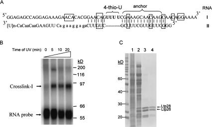
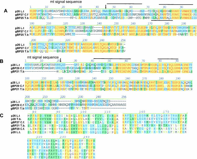
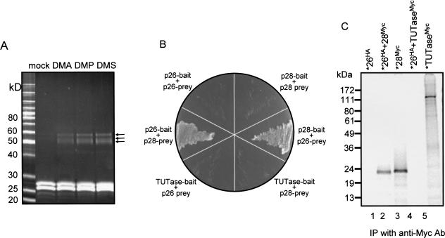
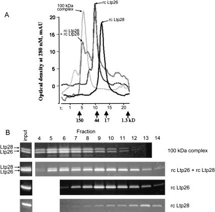
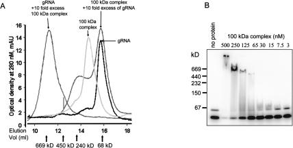
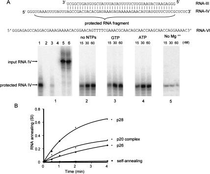
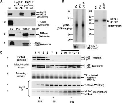
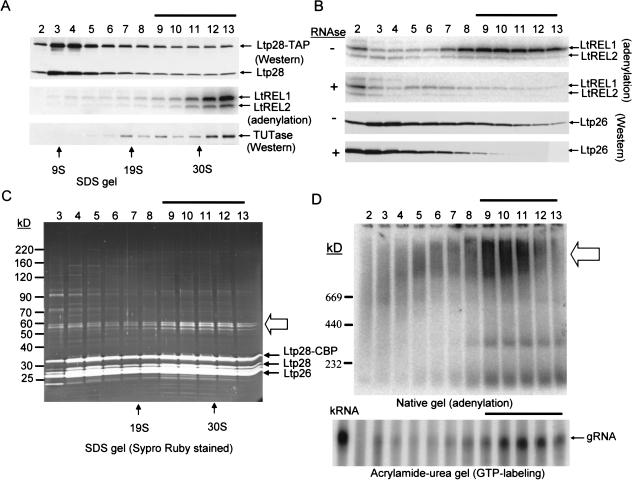
A stable 100-kD complex from mitochondria of Leishmania tarentolae containing two RNA-binding proteins, Ltp26 and Ltp28, was identified by cross-linking to unpaired 4-thiouridine nucleotides in a partially duplex RNA substrate. The genes were cloned and expressed and the complex was reconstituted from recombinant proteins in the absence of RNA or additional factors. The Ltp26 and Ltp28 proteins are homologs of gBP27 and gBP29 from Crithidia fasciculata and gBP25 and gBP21 from Trypanosoma brucei, respectively. The purified Ltp26/Ltp28 complex, the individual recombinant proteins, and the reconstituted complex are each capable of catalyzing the annealing of complementary RNAs, as was previously shown for gBP21 from T. brucei. A high-molecular-weight RNP complex consisting of the Ltp26/Ltp28 complex and several 55-60-kD proteins together with guide RNA could be purified from mitochondrial extract of L. tarentolae transfected with Ltp28-TAP. This complex also interacted in a less stable manner with the RNA ligase-containing L-complex and with the 3' TUTase. The Ltp26/Ltp28 RNP complex is a candidate for catalyzing the annealing of guide RNA and pre-edited mRNA in the initial step of RNA editing.


Figures

FIGURE 1.
FIGURE 1.
Isolation of Ltp26/Ltp28 100-kD complex from L. tarentolae mitochondria. (A) The model mRNA editing substrate contains four 4-thiouridines (in bold) in the first editing site. The anchor region is labeled, and the locations of the original U nucleotides which were mutated to non-U nucleotides outside the editing site are indicated by boxes. In five cases, the complementary nucleotides were also mutated to preserve base pairing. (B) The annealed labeled mRNA-gRNA substrate was incubated with mitochondrial extract and UV-irradiated for 0–20 min. The extract was fractionated on a 12% SDS gel and the gel was exposed to a Phosphoimager plate. RNA-protein crosslinks I and II are indicated. (C) Crosslink I in the S100 mitochondrial extract (lane 1) was followed through ammonium sulfate precipitation (lane 2), heparin affinity chromatography (lane 3), and Superose 12 size-fractionation (lane 4; see Materials and Methods). Active fractions were analyzed on a 12% SDS gel and stained with Coomassie Blue.
FIGURE 2.
FIGURE 2.
Protein sequence alignments of gRNA-binding proteins. (A) The L. tarentolae Ltp26 amino acid sequence aligned with gBP25 from T. brucei and gBP27 from C. fasciculata (Blom et al. 2001). (B) The L. tarentolae Ltp28 amino acid sequence aligned with gBP29 from C. fasciculata (Blom et al. 2001) and gBP21 from T. brucei (Koller et al. 1997). (C) Motifs conserved between the p26 and p28 homologs from all three species. The alignments above were obtained using AlignX in the Vector NTI Suite (Informax). The extent of nucleotide similarity is color-coded: blue on cyan denotes a consensus residue from a block of similar residues at a given location; black denotes nonhomologous residues; black on green denotes a consensus residue from a single conservative residue at a given location; red on yellow denotes a consensus residue from completely conservative residues at a given position. The predicted mitochondrial signal sequence cleavage sites in A and B are indicated by arrows, and the internal peptides obtained by microsequencing are indicated with lines.
FIGURE 3.
FIGURE 3.
Mitochondrial localization of Ltp26 and Ltp28 proteins. Log-phase (AC) or stationary-phase (D,E) cells were subjected to indirect immunofluorescence using an antiserum against the purified Ltp26/Ltp28 complex. A,D: phase contrast, 1000×. B,E: Same cells stained with DAPI. Location of kinetoplast (K) indicated by arrows. C,F: Same cells stained with Ltp26/Ltp28 antiserum. Location of K indicated by arrows.
FIGURE 4.
FIGURE 4.
Interaction of Ltp26 and Ltp28 proteins. (A) Chemical crosslinking of Ltp26/Ltp28 complex. Purified Ltp26/Ltp28 complex was incubated with homobifunctional protein–protein crosslinking reagents, and the products were separated on an SDS gel which was stained with Sypro Ruby. (B) Yeast two-hybrid analysis of the interaction between Ltp26 and Ltp28. Samples of cotransformed yeast grown on medium lacking tryptophan, leucine, and histidine. (C) Coimmunoprecipitation of epitope-tagged proteins synthesized in vitro in a coupled transcription/translation system. All IPs were with a monoclonal anti-Myc antibody. Lane 1: 35S-labeled Ltp26 tagged with HA. Lane 2: 35S-labeled Ltp26 tagged with HA mixed with unlabeled Ltp28 tagged with Myc. Lane 3: 35S-labeled Ltp28 tagged with Myc. Lane 4: 35S-labeled Ltp26 tagged with HA mixed with unlabeled TUTase tagged with Myc. Lane 5: 35S-labeled TUTase tagged with Myc.
FIGURE 5.
FIGURE 5.
Reconstitution of Ltp26/Ltp28 100-kD complex. (A) Superose 12 gel filtration profiles of purified Ltp26/Ltp28 complex, of the material reassembled from the purified recombinant Ltp26 and Ltp28 proteins, and of the individual proteins. Positions of molecular weight markers are indicated. (B) Protein composition of native and reconstructed complexes. Fractions were collected from the same starting point in chromatographic runs (above) and separated on 10%–20% SDS gradient gels which were stained with Sypro Ruby.
FIGURE 6.
FIGURE 6.
Binding of Ltp26/Ltp28 complex to T7 transcribed gRNA. (A) Binding of Ltp26/Ltp28 100-kD complex to RNA. In vitro transcribed labeled gRPS12-I gRNA was incubated with purified Ltp26/Ltp28 complex in 100 μL of 20 mM HEPES, pH 7.5, 5 mM MgCl2, 150 mM KCl, and separated on a Superose 6 column. The peak on left shows 50 pmole of gRNA and 500 pmole of protein, and the large peak on right 50 pmoles Ltp26/Ltp28 and 500 pmoles gRNA. (B) Radiolabeled gRPS12-I gRNA (20 nM) was incubated with increasing concentrations of Ltp26/Ltp28 complex under the same conditions in 5 μL aliquots and separated on 8%–16% native Tris-glycine gel.
FIGURE 7.
FIGURE 7.
RNA annealing activity of Ltp26/Ltp28 100-kD complex. (A) Specificity and cofactor requirements. Panel 1: lane 1, RNA-III at 2 nM was annealed with 50-fold excess of radioactively labeled RNA-IV by heating and slow cooling prior to addition of T1 nuclease in the absence of added protein. Lane 2, labeled RNA-IV + Ltp26/Ltp28 complex in the absence of complementary RNA followed by T1 nuclease digestion. Lane 3, RNA-III + labeled RNA-IV (2 nM each) in absence of protein, followed by T1 nuclease digestion. Lane 4, labeled RNA-IV + noncomplementary RNA-VI of approximately the same length + Ltp26/Ltp28 complex, followed by T1 nuclease digestion. Lane 5, RNA-III + labeled RNA-IV (2 nM) + Ltp26/Ltp28 complex, no T1 nuclease. Lane 6, labeled RNA-IV, no T1 nuclease. Panel 2: Ltp26/Ltp28 complex was added at increasing concentrations to RNA-III + labeled RNA-IV in absence of added NTPs, followed by T1 nuclease digestion. Panel 3: Same as Panel 2 but with 1mM GTP. Panel 4: Same as Panel 2 but with 1 mM ATP. Panel 5: Same as Panel 2 but without Mg2+. (B) Kinetics of annealing catalyzed by the recombinant p26 and p28 proteins and by the native 100-kD complex at 50 nM . SI, signal intensity of the T1-protected fragment. A control for the kinetics of annealing in the absence of protein is also shown.
FIGURE 8.
FIGURE 8.
Association of Ltp26/Ltp28 complex with components of RNA editing machinery. (A) Co-IP of Ltp26/Ltp28 complex and 3′TUTase. IP from mitochondrial extract was performed using Sepharose G beads coated with affinity-purified mouse polyclonal antibodies against recombinant Ltp26, recombinant Ltp28, and L. tarentolae 3′ TUTase (Ltp26-IP, Ltp28-IP, and TUT-IP lanes). Immunodetection was performed with rabbit antibodies raised against purified Ltp26/Ltp28 complex, recombinant glutamate dehydrogenase (GDH) (Bringaud et al. 1997), and TUTase. Ext, mitochondrial extract; Pre, proteins bound to G-beads coated with preimmune serum; Ab, proteins bound to G-beads coated with specific antibodies. 10%–20% SDS gels blotted to nitrocellulose membranes and probed with respective antibodies. (B) Co-IP of Ltp26/Ltp28 complex, gRNA, and RNA ligase. IP was performed with preimmune (Pre) or anti-Ltp26 (IP) antibody. Left panel: Total RNA was isolated from mitochondrial extract and the IP material, labeled with [α32P]GTP in the presence of guanylyltransferase and separated on a 12% acrylamide/urea gel. Lower panel: The presence of tRNALys was assayed in the same material by Northern blotting with an oligonucleotide probe. Right panel: The same samples were incubated with [α32P]ATP to visualize the LtREL1 and LtREL2 proteins and analyzed on a 10%–20% SDS gel. (C) Association of Ltp26/Ltp28 complex with TUTase and with RNA ligase. Purified Ltp26/Ltp28 complex (10 μg; panel 1) and mitochondrial extract (250 μL, 10 mg/mL; panels 2,3) were fractionated on a 10%–30% glycerol gradient in the SW41 rotor for 20 h at 35,000 rpm. Panel 2, Western analysis of Ltp26 and Ltp28. Panel 3, T1 nuclease protection assay for RNA annealing activity. Panels 4 and 5, co-IP of TUTase and LtREL1 and LtREL2 by anti-Ltp26 antibody.
FIGURE 9.
FIGURE 9.
Purification of high-molecular Ltp26/Ltp28 RNP complex weight by TAP chromatography. (A) Mitochondrial extract from Ltp28-TAP transfected cells was sedimented through a glycerol gradient. Each fraction (0.5 mL) was incubated with 20 μL of IgG Sepharose beads and, after extensive washing in 20 mM Tris-HCl, pH 7.6, 60 mM KCl, 10 mM MgCl2, 0.1% NP40, bound material was released from the beads and analyzed for the presence of the endogenous Ltp28 and the Ltp28 TAP fusion protein by Western analysis using anti-p28 antiserum, for the LtREL1 and LtREL2 RNA ligases by adenylation, and for TUTase by Western analysis using anti-TUTase antiserum. The gradient fractions containing the cosedimenting TUTase and RNA ligases are indicated by a line above. (B) IgG purification of Ltp28-associated RNP complex. Mitochondrial extract as in A was subjected to binding to IgG Sepharose and release by cleavage with TEV protease. In a separate experiment, RNase A (0.1 mg/mL) was added during the binding to IgG. The purified “pull down” was fractionated on a glycerol gradient, and the fractions assayed for the RNA ligases and the Ltp26 protein. The fractions showing a depletion of the ligases and Ltp26 are indicated by a line above. (C) Complete TAP purification of mitochondrial extract. This was carried out through the IgG binding with TEV protease release, and calmodulin binding with EGTA release. The purified fraction was subjected to glycerol gradient sedimentation, and each fraction was separated on an SDS gel, which was stained with Sypro Ruby. Arrows indicate the location of the endogenous Ltp26, Ltp28, and the ectopically expressed Ltp28-TAP bands. The three 55–60-kD associated bands are indicated by an open arrow. (D) Upper panel, the fractions in C were subjected to adenylation and fractionated in a 4%–12% native gel. The location of the adenylated complexes is indicated by an open arrow. Lower panel, RNA was extracted from each fraction, the gRNA labeled with [αP32] GTP in the presence of guanylyltransferase and separated on 12% acrylamide/urea. The gRNA band is indicated. RNA from total mitochondrial extract (Ex) was used as a control.

Similar articles

Cited by

References

    1. Allen, T.E., Heidmann, S., Reed, R., Myler, P.J., Goringer, H.U., and Stuart, K.D. 1998. Association of guide RNA binding protein gBP21 with active RNA editing complexes in Trypanosoma brucei. Mol. Cell. Biol. 18: 6014–6022. - PMC - PubMed
    1. Aphasizhev, R. and Simpson, L. 2001. Isolation and characterization of a U-specific 3′-5′ exonuclease from mitochondria of Leishmania tarentolae. J. Biol. Chem. 276: 21280–21284. - PubMed
    1. Aphasizhev, R., Sbicego, S., Peris, M., Jang, S.H., Aphasizheva, I., Simpson, A.M., Rivlin, A., and Simpson, L. 2002. Trypanosome mitochondrial 3′ terminal uridylyl transferase (TUTase): The key enzyme in U-insertion/deletion RNA editing. Cell 108: 637–648. - PubMed
    1. Blom, D., Burg, J., Breek, C.K., Speijer, D., Muijsers, A.O., and Benne, R. 2001. Cloning and characterization of two guide RNA-binding proteins from mitochondria of Crithidia fasciculata: gBP27, a novel protein, and gBP29, the orthologue of Trypanosoma brucei gBP21. Nucleic Acids Res. 29: 2950–2962. - PMC - PubMed
    1. Blum, B., Bakalara, N., and Simpson, L. 1990. A model for RNA editing in kinetoplastid mitochondria: “Guide” RNA molecules transcribed from maxicircle DNA provide the edited information. Cell 60: 189–198. - PubMed

Publication types

LinkOut - more resources