. 2003 Feb;23(4):1151-62.
doi: 10.1128/MCB.23.4.1151-1162.2003.

Loss of HR6B ubiquitin-conjugating activity results in damaged synaptonemal complex structure and increased crossing-over frequency during the male meiotic prophase

Affiliations

Loss of HR6B ubiquitin-conjugating activity results in damaged synaptonemal complex structure and increased crossing-over frequency during the male meiotic prophase

Willy M Baarends et al. Mol Cell Biol. 2003 Feb.

Abstract

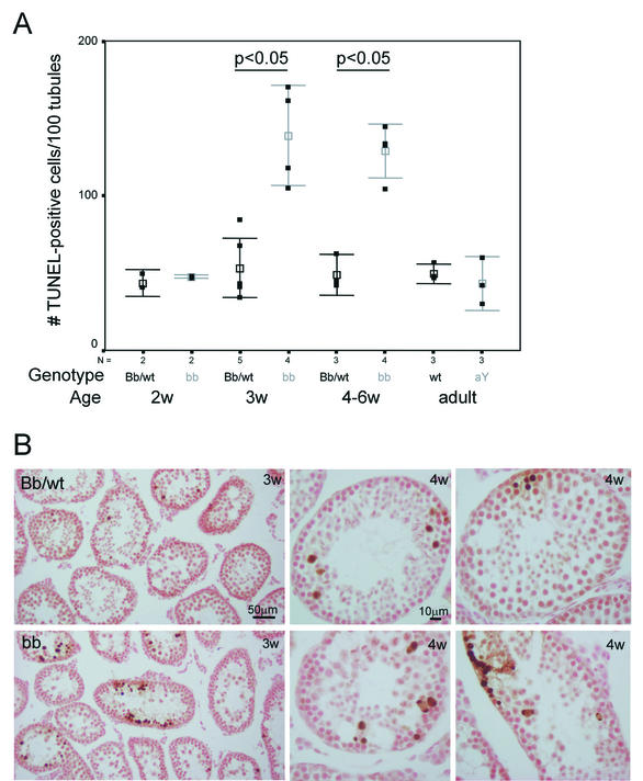
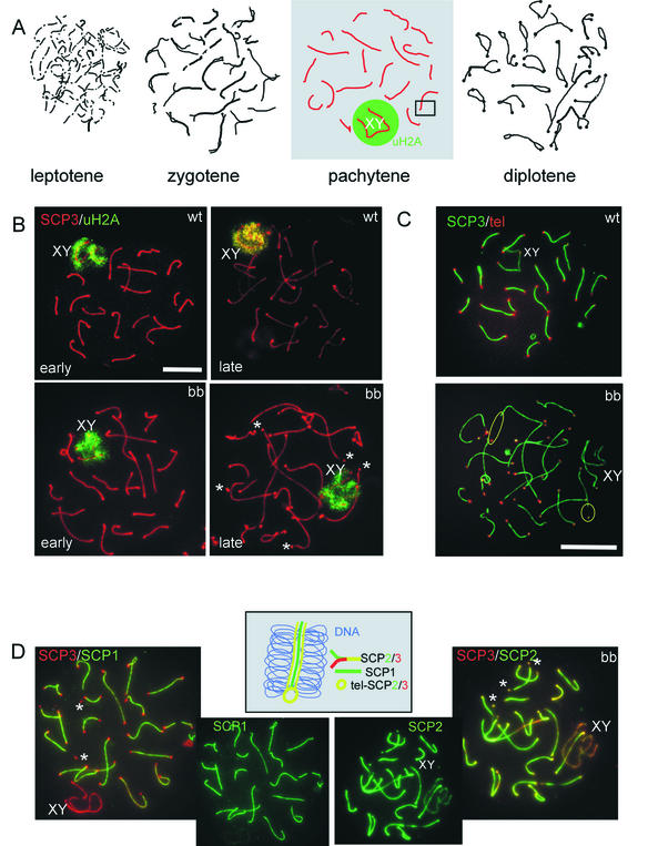
The ubiquitin-conjugating enzymes HR6A and HR6B are the two mammalian homologs of Saccharomyces cerevisiae RAD6. In yeast, RAD6 plays an important role in postreplication DNA repair and in sporulation. HR6B knockout mice are viable, but spermatogenesis is markedly affected during postmeiotic steps, leading to male infertility. In the present study, increased apoptosis of HR6B knockout primary spermatocytes was detected during the first wave of spermatogenesis, indicating that HR6B performs a primary role during the meiotic prophase. Detailed analysis of HR6B knockout pachytene nuclei showed major changes in the synaptonemal complexes. These complexes were found to be longer. In addition, we often found depletion of synaptonemal complex proteins from near telomeric regions in the HR6B knockout pachytene nuclei. Finally, we detected an increased number of foci containing the mismatch DNA repair protein MLH1 in these nuclei, reflecting a remarkable and consistent increase (20 to 25%) in crossing-over frequency. The present findings reveal a specific requirement for the ubiquitin-conjugating activity of HR6B in relation to dynamic aspects of the synaptonemal complex and meiotic recombination in spermatocytes.


Figures

FIG. 1.
FIG. 1.
Immunoblot analysis of HR6A and HR6B expression in germ cells from wild-type and HR6B knockout mouse testis. Highly purified spermatocytes and spermatids were isolated from wild-type (wt), HR6A knockout (aY), and HR6B knockout (bb) mouse testes. (A) Total protein extracts from wild-type cells were run on two-dimensional gels and subjected to isoelectric focusing in the first dimension and SDS-PAGE in the second dimension. HR6A and HR6B were detected with anti-HR6A/B. The positions of the two proteins are indicated; the pH increases from left to right in the gel. (B) Total protein extracts were run on SDS-PAGE gels and then analyzed by using anti-HR6A/B. The position of the 15K molecular weight marker is indicated. The lower panel in B shows part of the Ponceau red-stained blot, with several histone bands, as a control for equal protein loading of the gel. (C) Abundant presence of HR6A and/or HR6B proteins in highly purified spermatocytes (spc), round spermatids (rspt), and elongating spermatids (espt) isolated from wild-type mice.
FIG. 2.
FIG. 2.
Immunoblot of ubiquitinated proteins in germ cells from wild-type and HR6B knockout mouse testes. Preparations enriched in spermatocytes and spermatids were isolated in the presence of iodoacetamide by using a rapid procedure to try to minimize deubiquitination during isolation (see Materials and Methods). Immunoblots of total protein samples from isolated cell fractions of wild-type (wt) and HR6B knockout (bb) mice were analyzed by using an anti-ubiquitin antibody. For comparison, the total (tot) protein pattern is shown in the right lane, which is a Ponceau red-stained representative lane of the immunoblot. The asterisk indicates the position of ubiquitinated H2A/H2B, and the open arrow indicates the position of free ubiquitin. The positions of different molecular weight markers (in thousands) are also indicated.
FIG. 3.
FIG. 3.
Analysis of TUNEL-positive cells in HR6A and HR6B knockout mouse testes. (A) The numbers of TUNEL-positive cells per 100 tubules were determined in testis sections from wild-type (wt), HR6AY/− (aY), HR6B+/− (Bb), and HR6B−/− (bb) mice of different ages. The results for wild-type and HR6B+/− immature animals were pooled. The number of animals used in each group is indicated below the figure (N). The actual data are indicated as black squares, and the mean and standard error of the mean (SEM) values are indicated in black (wt and Bb) and gray (bb and aY). A significant increase (P < 0.05) in the amount of apoptotic spermatocytes per tubule cross section was found only for HR6B knockout mice that were 3 weeks old or older. (B) Representative photomicrographs of TUNEL staining in testis sections from wild-type or HR6B+/− (Bb/wt, upper panels) and HR6B−/− (bb, lower panels) mice that were 3 (3w) or 4 (4w) weeks old. Note the larger clusters of apoptotic cells in tubules of HR6B knockout mice.
FIG. 4.
FIG. 4.
SCs and the sex body in wild-type and HR6B knockout spermatocytes. (A) Schematic representation of the appearance of lateral elements of the SC during the different stages of the meiotic prophase. SC fragments are formed during leptotene. During zygotene, homologous chromosome pairing is initiated; this is visualized by the appearance of longer and thicker (in paired regions) axial and/or lateral elements of the SC. During pachytene, all autosomal chromosomes are fully paired, and each visible SC axis has a chromosome that consists of two chromatids on either side (the number of visible axes corresponds to the haploid number of chromosomes). The X and Y chromosomes pair only in the pseudoautosomal region. These chromosomes are heterochromatic and form the so-called sex body (indicated by XY), located in the nuclear periphery. During diplotene, the chromosomal axes start to separate but remain attached in the regions where crossing-over has occurred. Differences between wild-type (wt) and HR6B knockout (bb) spermatocytes were detected mainly during pachytene. Therefore, this stage is highlighted, and the photomicrographs in panels B, C, and D represent pachytene spermatocytes. XY indicates the sex body, and the green circleindicates that this area is enriched in ubiquitinated H2A (uH2A). (B) Spermatocyte spreads from wild-type and HR6B knockout mice stained with a polyclonal antibody against SCP3 (red) and a monoclonal antibody against ubiquitinated H2A (uH2A, green). Early and late pachytene spermatocytes are shown. The SC axes in many bb spermatocytes are longer and apparently thinner, and this effect is most pronounced during late pachytene. Also, loose-appearing SCP2/SCP3 beads at the ends of some SC axes can be observed. Sex body chromatin and uH2A staining appear normal. Upon longer exposure, uH2A also localizes to the telomeres of late pachytene and early diplotene cells in wild-type and in HR6B knockout spermatocytes (not shown). Asterisks indicate loose SCP2/SCP3 beads; XY indicates the sex body. Scale bar, 20 μm. (C) Meiotic spread preparations from wild-type (wt) and HR6B knockout (bb) mouse testis were hybridized to a mouse telomere probe (tel; red signal) and stained with anti-SCP3 (SCP3; green signal). In the HR6B knockout, a large number of the telomeres is not associated with the ends of the axes of the SCs but with loose bead-shaped SC fragments. For two of these telomeres, a yellow circle indicates the large distance between the telomere and the presumed beginning of the respective SC axis. XY indicates the X and Y chromosome pair. Scale bar, 20 μm. (D) HR6B knockout pachytene spermatocytes double stained with anti-SCP3 (red) and anti-SCP1 (green) (SCP3/SCP1) or with anti-SCP3 (red) and anti-SCP2 (green) (SCP3/SCP2). Single stains with anti-SCP1 and anti-SCP2 are also shown. Note that SCP2, but not SCP1, colocalizes to the loose SCP2/SCP3 beads stained with SCP3. In the middle, a schematic interpretation is shown of a fragment from the SC complex, enlarged from the box indicated in the pachytene nucleus in panel A. It visualizes localization of SCP3 and SCP2 on the lateral elements that have formed on the chromosomal axes, whereas SCP1 is a component of the transverse filaments that connect the lateral elements of the SC during chromosome pairing (no staining of SCP1 is observed on unpaired X and Y chromosomal axes). SCP1 is also absent from near telomeric DNA. In these regions SCP2 and SCP3 are both present (tel-SCP2/3).
FIG. 5.
FIG. 5.
Numbers of detached SCP2/SCP3 beads, and the length of the SC per cell in spermatocytes from wild-type and HR6B knockout mice. (A) Total length of the SC (in micrometers) per nucleus of wild-type (wt) pachytene and early diplotene spermatocytes and of HR6B knockout (bb) pachytene spermatocytes. Individual measurements are indicated in gray. Mean and SEM values (black squares and error bars, respectively) are indicated. P values are indicated above the lines connecting the two compared groups. N indicates the number of nuclei that were investigated. Nuclei were from three different animals in each group. (B) Frequency distribution of the number of loose SCP2/SCP3 beads per pachytene nucleus of wild-type (wt; black bars) and HR6B knockout (bb; hatched bars) mice. Thirty-eight nuclei from four different animals were analyzed for each group. For the wild type, all nuclei fell in the first category of zero to two loose SCP2/SCP3 beads; of these nuclei, 76% had no loose beads at all, and there was only 1 nucleus that showed 2 loose SCP2/SCP3 beads. For the HR6B knockout animals, only 24% of all analyzed nuclei had no loose SCP2/SCP3 beads.
FIG. 6.
FIG. 6.
Increased frequency of meiotic recombination during pachytene in HR6B−/− spermatocytes. (A) Meiotic spread preparations from wild-type (wt) and HR6B knockout (bb) mouse testes and from wild-type and HR6A knockout (aa) mouse fetal ovaries (E18) were stained with anti-SCP3 (SCP3, red signal) and anti-MLH1 (MLH1, green signal). In the HR6B knockout (bb), the number of MLH1 foci is increased (29 foci in this nucleus) compared to the wild type (23 foci). The average number of foci between wild-type and HR6A knockout oocytes does not differ (the number of foci for these two individual nuclei are 33 and 30, respectively). The SC structure of the knockout pachytene spermatocyte nucleus shown here has relatively few abnormalities. An increased number of MLH1 foci is found in bb nuclei that appear relatively normal and also in bb nuclei that show long and thin SC axes and/or loose SCP2/SCP3 beads (the latter is not shown). XY indicates the sex body. Scale bar, 20 μm. (B) The number of MLH1 foci per spermatocyte nucleus is indicated on the y axis. Please note that this axis starts at 21 foci. The gray squares indicate the average number of foci per nucleus per animal (N = the number of animals tested). For each animal at least 10 nuclei were counted. In black, the mean value and SEM are indicated (black square and error bars, respectively). MLH1 foci were counted in wild-type (wt), HR6A knockout (aY), and HR6B knockout (bb) spermatocytes. (C) Diakinesis/metaphase I spread preparations of wild-type (wt) and HR6B knockout (bb) spermatocyte nuclei. Bivalents showing two crossover events are indicated by and asterix. The bivalents containing the X and Y chromosome are indicated by XY. Note that the centromeric regions of the chromosomes are more heavily stained by DAPI. Scale bar, 10 μm.

Similar articles

Cited by

References

    1. Adams, M. D., S. E. Celniker, R. A. Holt, C. A. Evans, J. D. Gocayne, P. G. Amanatides, S. E. Scherer, P. W. Li, R. A. Hoskins, R. F. Galle, R. A. George, S. E. Lewis, S. Richards, M. Ashburner, S. N. Henderson, G. G. Sutton, J. R. Wortman, M. D. Yandell, Q. Zhang, L. X. Chen, R. C. Brandon, Y. H. Rogers, R. G. Blazej, M. Champe, B. D. Pfeiffer, K. H. Wan, C. Doyle, E. G. Baxter, G. Helt, C. R. Nelson, G. L. Gabor, J. F. Abril, A. Agbayani, H. J. An, C. Andrews-Pfannkoch, D. Baldwin, R. M. Ballew, A. Basu, J. Baxendale, L. Bayraktaroglu, E. M. Beasley, K. Y. Beeson, P. V. Benos, B. P. Berman, D. Bhandari, S. Bolshakov, D. Borkova, M. R. Botchan, J. Bouck, et al. 2000. The genome sequence of Drosophila melanogaster. Science 287:2185-2195. - PubMed
    1. Anderson, L. K., A. Reeves, L. M. Webb, and T. Ashley. 1999. Distribution of crossing over on mouse synaptonemal complexes using immunofluorescent localization of MLH1 protein. Genetics 151:1569-1579. - PMC - PubMed
    1. Baarends, W. M., J. W. Hoogerbrugge, H. P. Roest, M. Ooms, J. Vreeburg, J. H. J. Hoeijmakers, and J. A. Grootegoed. 1999. Histone ubiquitination and chromatin remodeling in mouse spermatogenesis. Dev. Biol. 207:322-333. - PubMed
    1. Baarends, W. M., R. van der Laan, and J. A. Grootegoed. 2000. Specific aspects of the ubiquitin system in spermatogenesis. J. Endocrinol. Investig. 23:597-604. - PubMed
    1. Baker, S. M., A. W. Plug, T. A. Prolla, C. E. Bronner, A. C. Harris, X. Yao, D. M. Christie, C. Monell, N. Arnheim, A. Bradley, T. Ashley, and R. M. Liskay. 1996. Involvement of mouse Mlh1 in DNA mismatch repair and meiotic crossing over. Nat. Genet. 13:336-342. - PubMed

Publication types

MeSH terms