. 2003 Feb;23(4):1181-95.
doi: 10.1128/MCB.23.4.1181-1195.2003.

batman Interacts with polycomb and trithorax group genes and encodes a BTB/POZ protein that is included in a complex containing GAGA factor

Affiliations

batman Interacts with polycomb and trithorax group genes and encodes a BTB/POZ protein that is included in a complex containing GAGA factor

M Faucheux et al. Mol Cell Biol. 2003 Feb.

Abstract

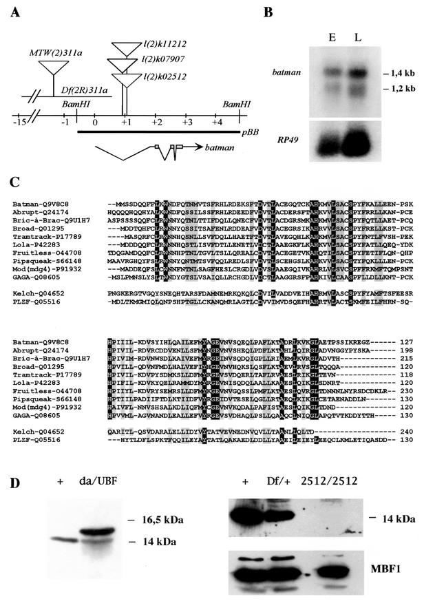
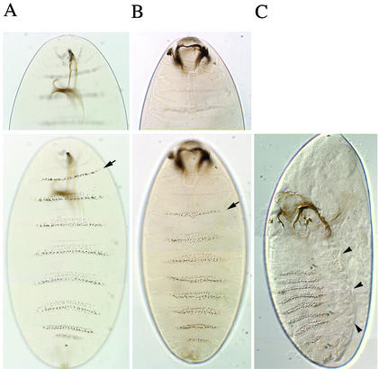
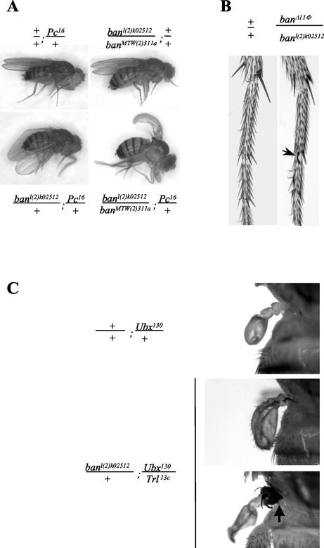
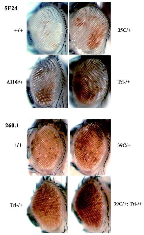
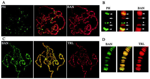
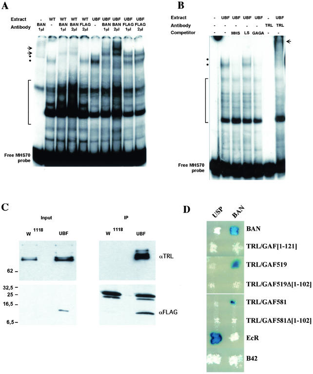
Polycomb and trithorax group genes maintain the appropriate repressed or activated state of homeotic gene expression throughout Drosophila melanogaster development. We have previously identified the batman gene as a Polycomb group candidate since its function is necessary for the repression of Sex combs reduced. However, our present genetic analysis indicates functions of batman in both activation and repression of homeotic genes. The 127-amino-acid Batman protein is almost reduced to a BTB/POZ domain, an evolutionary conserved protein-protein interaction domain found in a large protein family. We show that this domain is involved in the interaction between Batman and the DNA binding GAGA factor encoded by the Trithorax-like gene. The GAGA factor and Batman codistribute on polytene chromosomes, coimmunoprecipitate from nuclear embryonic and larval extracts, and interact in the yeast two-hybrid assay. Batman, together with the GAGA factor, binds to MHS-70, a 70-bp fragment of the bithoraxoid Polycomb response element. This binding, like that of the GAGA factor, requires the presence of d(GA)n sequences. Together, our results suggest that batman belongs to a subset of the Polycomb/trithorax group of genes that includes Trithorax-like, whose products are involved in both activation and repression of homeotic genes.


Figures

FIG. 1.
FIG. 1.
The batman transcription unit. (A) Molecular map of the ban locus. The ban gene is transcribed from distal to proximal relative to the centromere. The two BamHI sites delimit the ban transcription unit as inferred from rescue assays using the pBB fragment (5,405 bp). Three lethal noncomplementing P[LacW] insertions are positioned in the first intron of ban. Df(2R)311a (24) uncovers the 5′ region of ban up to the first intron. This deficiency is the only one available that affects ban without affecting the neighboring Pcl gene, located 18 kb downstream of ban. The sequence corresponding to ORF127 is boxed on the ban transcript. (B) Northern blot analysis of embryonic (E) and third-instar larva (L) RNA probed with ban cDNA (top) and RP49 cDNA (bottom) as a control. (C) Multiple sequence alignment of D. melanogaster BTB/POZ proteins ordered according to decreasing similarity to BAN as calculated with the BLASTP algorithm. The alignment was generated using CLUSTALW. The cytoplasmic D. melanogaster Kelch protein and the human PLZF protein, for which a crystal structure has been determined (1), are set apart. The N-terminal nuclear BTB signature, characteristic of the Tramtrack subfamily and not conserved in Kelch (4, 87), is boxed. Conserved amino acids are boxed in black, and similarities are boxed in gray. The coordinates on the right refer to the position of the last amino acid included in the alignment from the sequences listed in GenBank. (D) Western blot analysis of larval proteins using the batC11 antibody. Extracts from third-instar larvae of the appropriate genotype were separated on sodium dodecyl sulfate-18% polyacrylamide gels. +/, w1118; da/UBF:da:Gal4/UBF larvae were obtained by crossing da:Gal4 homozygous flies to UBF homozygotes; Df/+, Df(2R)PC4/+ larvae; 2512/2512, banl(2)k02512/banl(2)k02512 escaper third-instar larvae were selected among the progeny of banl(2)k02512/CyO-GFP heterozygous flies based on the absence of GFP expression. Anti-MBF1 antibody (bottom panel) was used as a control for gel loading.
FIG. 2.
FIG. 2.
Cuticular defects in banl(2)k02512/banl(2)k02512 embryos deprived of the maternal contribution of ban. (A and B) Focus is on the anterior part in the top panels and on the denticular belts in the bottom panels. The two mutant embryos (B and C) illustrate the various ranges of cuticular defects. When compared to wild-type embryos (A), the position of the first abdominal denticle belt (arrow) is moved posteriorly in ban mutant embryos (B), indicating defects in head involution associated with altered mouth hooks. Dorsal closure defects were always observed, however, with various degrees of severity, from a hole in the dorsal cuticle (not shown) to the absence of closure (C). (C) Arrowheads point to the edge of the dorsal cuticle.
FIG. 3.
FIG. 3.
ban homeotic phenotypes. (A) Cbx phenotype of heteroallelic ban combinations in a Pc16 heterozygous context. Heteroallelic banDf(2R)311a/banl(2)k02512 flies as well as Pc16/+ flies have wild-type wings. Double-heterozygous banl(2)k02512/+; Pc16/+ flies display a curving of the wings that is indicative of the Cbx phenotype. This phenotype is enhanced in heteroallelic banDf(2R)311a/banl(2)k02512; Pc16/+ flies. (B) Sex comb teeth normally absent in wild-type males (left) are observed on the mesothoracic tarsus in banΔ11Φ/banl(2)k02512 males (right). (C) Enhancement of the genetic interaction between Trl and Ubx. Ubx130/+ flies display slightly enlarged halters (top panel) compared to the wild type. The expressivity of this transformation is higher in Trl13c/Ubx130 flies, in which a clear transformation of halter to wing was present in 6.3% (n = 142) of the flies, whereas it was never found in Ubx130/+ flies. In banl(2)k02512/+; Trl13c/Ubx130 flies, this transformation was much more frequent (18% [n = 204]; middle and bottom panels) and was often accompanied by a notal transformation (arrow in bottom panel).
FIG. 4.
FIG. 4.
Similar effects of ban and Trl loss-of-function mutations on the PRE-dependent pairing-sensitive silencing of white reporters. Three white deletion mutant alleles generated from banDf(2R)311a (39C and 35C) and banl(2)k02512 (Δ11Φ) were assayed in a heterozygous context for the pairing-sensitive silencing of either the X-linked, 3.8-kb Fab-7 PRE line 5F24 25,2 (5F24) (86), or the 260-bp iab-7 PRE line 260.1 (47). In the latter case, ban alleles were first recombined with the 260.1 insertion on the second chromosome. In all cases (35C/+ and Δ11Φ/+ in 5F24 25,2 females and 39C 260.1 males and females), derepression of the white transgene was observed in ban heterozygotes, closely resembling what is observed in Trl13c (Trl-/+) heterozygotes. Derepression was increased in double heterozygotes 39C 260.1/260.1; Trl13c/+ (39C/+; Trl-/+). For each combination, effects were compared between siblings of the same gender and the appropriate genotype.
FIG. 5.
FIG. 5.
Comparison of the distributions of BAN, PH, and TRL/GAF proteins on larval salivary polytene chromosomes as detected by double immunostaining. Simultaneous immunostaining was performed for the following combinations: PH-FLAG (green) and BAN (red) (A and B) and BAN-FLAG (green) and TRL/GAF (red) (C and D). In each panel, a merge image is shown in the middle. (B) A representative chromosomal region displaying four adjacent BAN binding sites is enlarged. Among these BAN sites, two are shared with PH (arrowheads) and two are specific for BAN (arrows). In the same region, one PH binding site is not shared with BAN (asterisk). (D) Enlargement of a chromosomal region from panel C shows the colocalization of BAN and TRL/GAF binding sites.
FIG. 6.
FIG. 6.
(A and B) Gel shift analysis of MHS-70 binding activities. (A) Binding of proteins in nuclear extracts from w1118 embryos (WT) or da:Gal4, UBF/+ embryos (UBF) was analyzed in a gel shift assay using the MHS-70 fragment as a probe in the absence (−) or in the presence of BAN or FLAG antibodies as indicated. The positions of BAN-containing low-mobility complexes in the absence (dots) or the presence (arrows) of BAN antibody are indicated. The position of nonspecific complexes that are not displaced by the addition of unlabeled probe is indicated (bracket). (B) Binding of proteins in a nuclear extract from da:Gal4, UBF/+ embryos (UBF) was analyzed in a gel shift assay using the MHS-70 labeled probe in the absence (−) or in the presence of competitors DNAs or TRL/GAF antibody (TRL) as indicated. Positions of the BAN containing complexes in the absence (dots) or the presence (arrow) of TRL/GAF antibody are indicated. The position of unspecific complexes that are not competed against by a 100-fold molar excess of MHS-70 probe is indicated (bracket). (C) Coimmunoprecipitation of BAN-FLAG with theTRL/GAF. Nuclear protein extracts from w1118 or da:Gal4/UBF (UBF) larvae were immunoprecipitated (IP) with anti-FLAG beads and analyzed by Western blotting using anti-TRL/GAF (TRL) or anti-FLAG (FLAG) antibodies. Aliquots (1/60) of the input extracts (input) were analyzed in parallel using the indicated antibody. The positions of protein molecular mass markers (in kilodaltons) are shown on the left. (D) Interaction of BAN and GAF in a yeast two-hybrid assay. Each patch corresponds to diploid yeast colonies coexpressing either the BAN protein (right column) or the USP (left column) as a control; both fused to the DNA binding domain of LexA; and BAN, full-length or truncated TRL/GAF isoforms, or EcR, all fused to the B42 transactivation domain (rows). Interactions resulted in the activation of a lacZ reporter gene placed under the control of LexA binding sites and were revealed in a β-galactosidase assay. Interaction (84) between the EcR-B1 isoform (EcR) and USP provided a positive control, while expression of the B42 domain alone served as a negative control.

Similar articles

Cited by

References

    1. Ahmad, K. F., C. K. Engel, and G. G. Prive. 1998. Crystal structure of the BTB domain from PLZF. Proc. Natl. Acad. Sci. USA 95:12123-12128. - PMC - PubMed
    1. Albagli, O., P. Dhordain, C. Deweindt, G. Lecocq, and D. Leprince. 1995. The BTB/POZ domain: a new protein-protein interaction motif common to DNA- and actin-binding proteins. Cell Growth Differ. 6:1193-1198. - PubMed
    1. Antoniewski, C., M. Laval, A. Dahan, and J. A. Lepesant. 1994. The ecdysone response enhancer of the Fbp1 gene of Drosophila melanogaster is a direct target for the EcR/USP nuclear receptor. Mol. Cell. Biol. 14:4465-4474. - PMC - PubMed
    1. Aravind, L., and E. V. Koonin. 1999. Fold prediction and evolutionary analysis of the POZ domain: structural and evolutionary relationship with the potassium channel tetramerization domain. J. Mol. Biol. 285:1353-1361. - PubMed
    1. Bardwell, V. J., and R. Treisman. 1994. The POZ domain: a conserved protein-protein interaction motif. Genes Dev. 8:1664-1677. - PubMed

Publication types

MeSH terms