. 2003 Feb;23(4):1260-8.
doi: 10.1128/MCB.23.4.1260-1268.2003.

Inactivation of the nuclear receptor coactivator RAP250 in mice results in placental vascular dysfunction

Affiliations

Inactivation of the nuclear receptor coactivator RAP250 in mice results in placental vascular dysfunction

Per Antonson et al. Mol Cell Biol. 2003 Feb.

Abstract

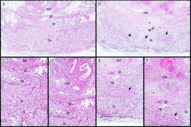
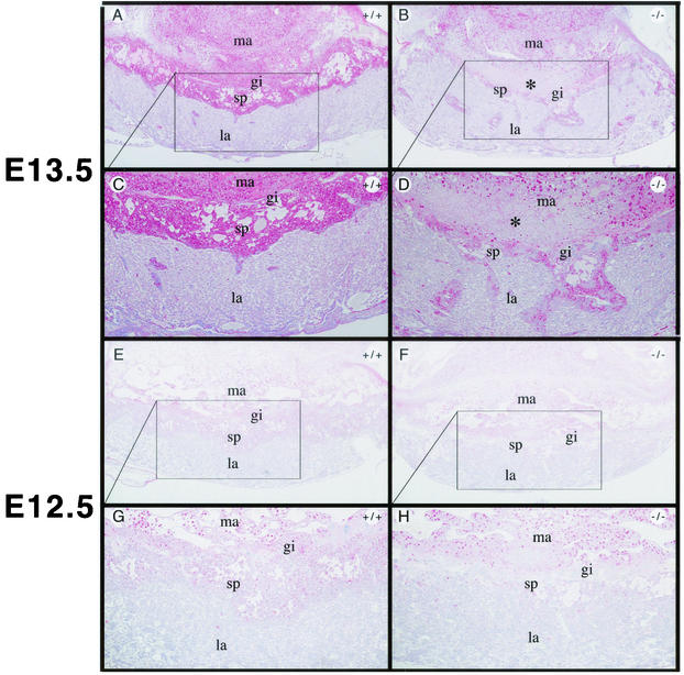
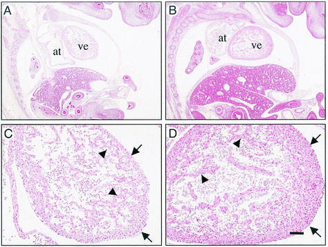
Coactivators constitute a diverse group of proteins that are essential for optimal transcriptional activity of nuclear receptors. In the past few years many coactivators have been identified but it is still unclear whether these proteins interact indiscriminately with all nuclear receptors and whether there is some redundancy in their functions. We have previously cloned and characterized RAP250 (ASC-2/PRIP/TRBP/NRC), an LXXLL-containing coactivator for nuclear receptors. In order to study its biological role, Rap250 null mice were generated by gene targeting. Here we show that genetic disruption of Rap250 results in embryonic lethality at embryonic day (E) 13.5. Histological examination of placentas revealed a dramatically reduced spongiotrophoblast layer, a collapse of blood vessels in the region bordering the spongiotrophoblast, and labyrinthine layers in placentas from Rap250(-/-) embryos. These findings suggest that the lethality of Rap250(-/-) embryos is the result of obstructed placental blood circulation. Moreover, the transcriptional activity of PPAR gamma is reduced in fibroblasts derived from Rap250(-/-) embryos, suggesting that RAP250 is an essential coactivator for this nuclear receptor in the placenta. Our results demonstrate that RAP250 is necessary for placental development and thus essential for embryonic development.


Figures

FIG. 1.
FIG. 1.
Targeted disruption of the mouse Rap250 gene. (A) Structures of the wild-type Rap250 allele, targeting vector, and targeted allele are shown with the BamHI (B), HindIII (H), KpnI (K), and XbaI (X) restriction sites and primers for PCR screening. The locations of probes used in Southern blot analysis are indicated. Black boxes indicate exons. The exon numbering is adapted from the work of Zhu et al. (42). (B) Homologous recombination in ES cells. Southern blot analysis of DNA from two positive ES clones, clone 1B8 and 2D2, digested with BamHI is shown. The 5-kb band for the wild-type allele (WT) and the 11-kb band for the targeted allele (KO) are indicated. The probes used are indicated in Fig. 1A. (C) Southern blot analysis of mouse yolk sac DNA digested with BamHI. (D) PCR genotyping of mouse embryo DNA. The bottom band, using the U6 and L8 primers, represents the wild-type allele and the top band, using the KOF1 and L3 primers, represents the mutated allele. M, molecular marker (1-kb ladder). (E) Photo of wild-type and Rap250−/− embryos at E13.5. No obvious abnormalities or significant size differences were detected between wild-type and Rap250−/− littermates.
FIG. 2.
FIG. 2.
Absence of Rap250 mRNA and protein in Rap250−/− embryonic fibroblasts. (A) RT-PCR analysis of total RNA from wild-type (+/+) and Rap250−/− (−/−) mouse embryonic fibroblasts. Expression of Rap250 is detected in wild-type but not null cells, and actin mRNA is detected in both cell types. M, molecular marker (1-kb ladder); 0, negative control without DNA. (B) Western blot analysis of nuclear extracts from wild-type (WT) and null (KO) mouse embryonic fibroblasts using RAP250 polyclonal antibodies. RAP250 protein, indicated by an arrow, is detected in wild-type but not null fibroblasts.
FIG. 3.
FIG. 3.
Developmental defects in Rap250−/− placentas, as shown by hematoxylin and eosin staining of Rap250+/+ and Rap250−/− placentas at E13.5. Overview pictures of wild-type (A) and Rap250−/− (B) placentas at E13.5 are shown. (C and D) Higher-power views of panel A. (E and F) Higher-power views of panel B. The spongiotrophoblast layer is markedly reduced in null placentas compared to the wild type, and the labyrinthine layer contains islands of spongiotrophoblast-like cells, indicated by arrows. The labyrinthine layer is less dense in null placentas than in wild-type placentas. Note an area of cell death in null placentas in the maternal decidua bordering the giant cell layer indicated by an asterisk. gi, giant cells; la, labyrinthine layer; ma, maternal decidua; sp, spongiotrophoblast layer.
FIG. 4.
FIG. 4.
Reduced spongiotrophoblast layer in Rap250−/− placentas, as shown by PAS staining, which stains glycogen-containing cells, of sections of wild-type and Rap250−/− placenta. Glycogen-containing cells appear in pink. (A and B) Sections of PAS staining of wild-type and Rap250−/− placentas, respectively, at E13.5. (C and D) Higher-power views of panels A and B, respectively. The number of stained cells in the spongiotrophoblast layer is markedly reduced in Rap250−/− placentas compared to wild-type placentas. While the border between the labyrinth and spongiotrophoblast layers is distinct in wild-type placentas, it is not as sharp in null placentas. The labyrinthine layer of Rap250−/− placentas contains several islands of glycogen-containing spongiotrophoblast-like cells that are not seen in wild-type placentas. An area with cell death in the maternal decidua of Rap250−/− placentas is indicated by an asterisk. (E and F) PAS staining of sections of wild-type and Rap250−/− placentas, respectively, at E12.5. (G and H) Higher-power views of panels E and F. The number of stained cells in the spongiotrophoblast layer is markedly reduced in Rap250−/− placentas compared to wild-type placentas. gi, giant cells; la, labyrinthine layer; ma, maternal decidua; sp, spongiotrophoblast layer.
FIG. 5.
FIG. 5.
Reduced expression of prolactin-like proteins in Rap250−/− placentas. Staining of wild-type and Rap250−/− placentas with antibodies against prolactin. Immunohistochemical analysis of wild-type (A and E) and Rap250−/− (B and F) placentas at E13.5 and E12.5 with antibodies against prolactin is shown. (C, D, G, and H) Higher-power views of panels A, B, E, and F, respectively. The number of stained cells in the spongiotrophoblast layer is markedly reduced in Rap250−/− placentas compared to wild-type placentas. gi, giant cells; la, labyrinthine layer; ma, maternal decidua; sp, spongiotrophoblast layer.
FIG. 6.
FIG. 6.
Vascular defects in Rap250−/− placentas. The collapse of vessels bordering the labyrinth and the spongiotrophoblast region of Rap250−/− placentas at E13.5 is shown. Immunohistochemical analysis was performed for Rap250−/− (A and C) and Rap250+/− (E and G) extra-embryonic tissues. Panels A, C, E, and G show staining with antibodies against von Willebrand's factor. Note the release of von Willebrand's factor in the collapsed vessel of the labyrinth and the spongiotrophoblast region of the Rap250−/− placenta. In contrast, the placental vessels in the labyrinth region were intact and positive staining was only detected in the endothelial cells. Similar vessels of Rap250+/− placentas were unaffected. (B, D, F, and H) TUNEL staining to detect DNA fragmentation. An area juxtaposed to the collapsed vessels shown in panels B and D stained positive with TUNEL staining (marked with an asterisk). Bars: A, B, E, and F, 125 μm; C, D, G, and H, 50 μm.
FIG. 7.
FIG. 7.
Histological abnormalities of hearts from Rap250−/− embryos at E13.5, as shown by hematoxylin and eosin staining of sagittal sections of Rap250−/− and wild-type embryos. (A and B) Hematoxylin and eosin staining of sagittal sections of Rap250−/− and wild-type embryos, respectively. Myocardial walls are thinner in Rap250−/− embryos than in wild-type littermates. at, atrium; ve, ventricle. Bar = 400 μm. (C and D) Hypoplasia in ventricular walls (arrows) and degeneration of trabecular zone (arrowheads) in Rap250−/− embryos and wild-type littermates, respectively. Bar = 100 μm.
FIG. 8.
FIG. 8.
Defective neural development in Rap250−/− mice. (A and B) Neopallial cortex of E13.5 embryos. Note the significantly thinner layer (arrows) of the Rap250−/− embryo (A) compared to that of the wild-type embryo (B). Bar = 50 μm. (C and D) Immunohistochemical analysis of neopallial cortex from Rap250−/− and wild-type embryos, respectively, at E13.5 with antibodies against MAP-2B. Less-differentiated neurons are seen in the neopallial cortex of Rap250−/− embryos than in the wild type. Bar = 25 μm. (E and F) Overview of E13.5 brains, as shown by hematoxylin and eosin staining of sagittal sections. Note the olfactory lobe in the wild type (arrows) (F) and the enlarged third ventricle (stars) in the Rap250−/− embryo (E). Bar = 400 μm.
FIG. 9.
FIG. 9.
Reduced PPARγ-mediated transcriptional activation in Rap250−/− MEFs. Fibroblasts isolated from wild-type and Rap250−/− embryos were cotransfected with 0.5 μg of Gal4-luciferase reporter plasmid, 0.5 μg of pGAL4-DBD, pGal4-PPARγ, pGal4-ERRβ, or pGal4-RXRα, and 0.2 μg of pCMVβ and cultured in the absence (−) or presence (+) of ligand (10 μM BRL49653 for PPARγ or 10 μM 9-cis-retinoic acid for RXRα). Cells were harvested 24 h after transfection and assayed for luciferase and β-galactosidase activities. The luciferase activity was adjusted with β-galactosidase activity for each cellular extract to correct for transfection efficiency. The results represent means ± standard deviations of a representative experiment performed in duplicate and are represented as fold increases compared to the activity of pGAL4-DBD.

Similar articles

Cited by

References

    1. Anzick, S. L., J. Kononen, R. L. Walker, D. O. Azorsa, M. M. Tanner, X. Y. Guan, G. Sauter, O. P. Kallioniemi, J. M. Trent, and P. S. Meltzer. 1997. AIB1, a steroid receptor coactivator amplified in breast and ovarian cancer. Science 277:965-968. - PubMed
    1. Barak, Y., M. C. Nelson, E. S. Ong, Y. Z. Jones, P. Ruiz-Lozano, K. R. Chien, A. Koder, and R. M. Evans. 1999. PPAR gamma is required for placental, cardiac, and adipose tissue development. Mol. Cell 4:585-595. - PubMed
    1. Caira, F., P. Antonson, M. Pelto-Huikko, E. Treuter, and J.-Å. Gustafsson. 2000. Cloning and characterization of RAP250, a novel nuclear receptor coactivator. J. Biol. Chem. 275:5308-5317. - PubMed
    1. Chen, H. W., R. J. Lin, R. L. Schiltz, D. Chakravarti, A. Nash, L. Nagy, M. L. Privalsky, Y. Nakatani, and R. M. Evans. 1997. Nuclear receptor coactivator ACTR is a novel histone acetyltransferase and forms a multimeric activation complex with P/CAF and CBP/p300. Cell 90:569-580. - PubMed
    1. Fondell, J. D., H. Ge, and R. G. Roeder. 1996. Ligand induction of a transcriptionally active thyroid hormone receptor coactivator complex. Proc. Natl. Acad. Sci. USA 93:8329-8333. - PMC - PubMed

Publication types

MeSH terms

Substances