. 2003 Feb;23(4):1292-303.
doi: 10.1128/MCB.23.4.1292-1303.2003.

Growth arrest and DNA damage-inducible protein GADD34 targets protein phosphatase 1 alpha to the endoplasmic reticulum and promotes dephosphorylation of the alpha subunit of eukaryotic translation initiation factor 2

Affiliations

Growth arrest and DNA damage-inducible protein GADD34 targets protein phosphatase 1 alpha to the endoplasmic reticulum and promotes dephosphorylation of the alpha subunit of eukaryotic translation initiation factor 2

Matthew H Brush et al. Mol Cell Biol. 2003 Feb.

Abstract

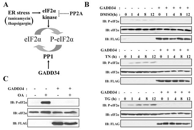
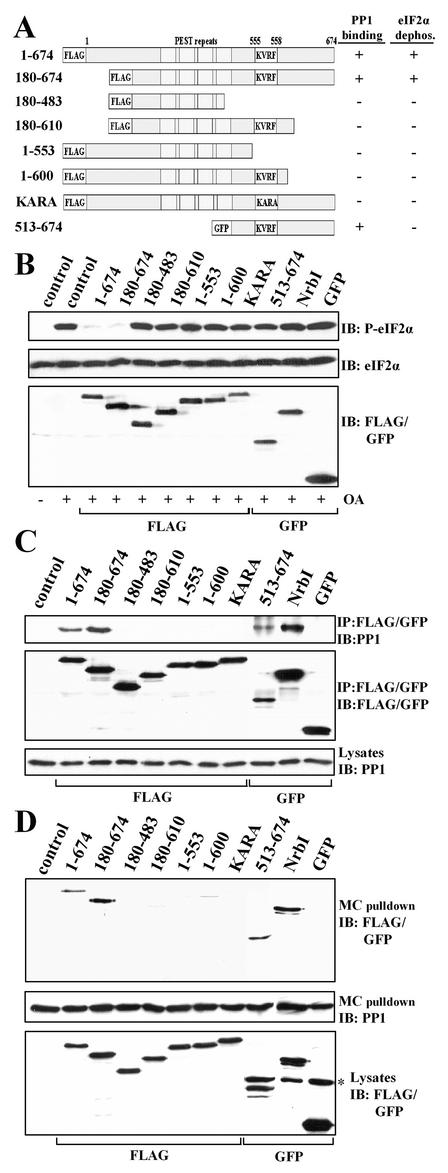
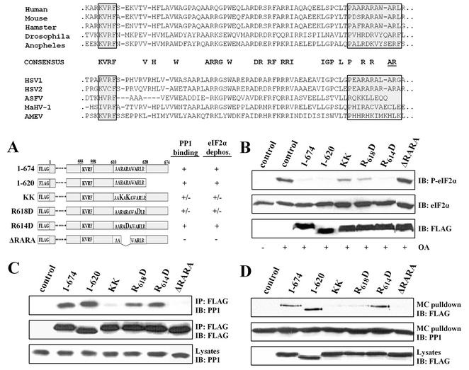
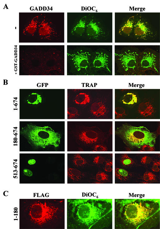
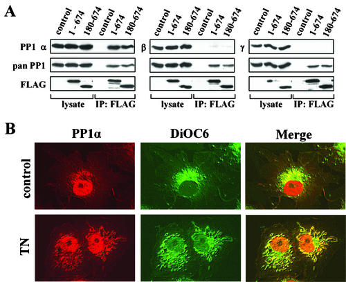
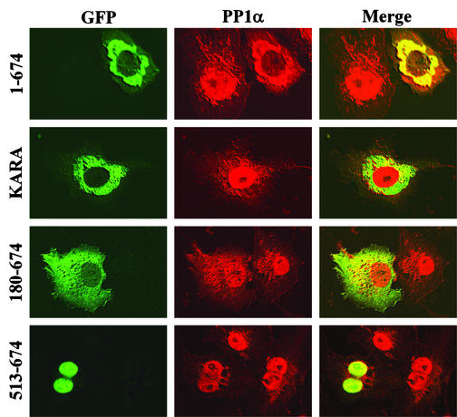
The growth arrest and DNA damage-inducible protein, GADD34, associates with protein phosphatase 1 (PP1) and promotes in vitro dephosphorylation of the alpha subunit of eukaryotic translation initiation factor 2, (eIF-2 alpha). In this report, we show that the expression of human GADD34 in cultured cells reversed eIF-2 alpha phosphorylation induced by thapsigargin and tunicamycin, agents that promote protein unfolding in the endoplasmic reticulum (ER). GADD34 expression also reversed eIF-2 alpha phosphorylation induced by okadaic acid but not that induced by another phosphatase inhibitor, calyculin A (CA), which is a result consistent with PP1 being a component of the GADD34-assembled eIF-2 alpha phosphatase. Structure-function studies identified a bipartite C-terminal domain in GADD34 that encompassed a canonical PP1-binding motif, KVRF, and a novel RARA sequence, both of which were required for PP1 binding. N-terminal deletions of GADD34 established that while PP1 binding was necessary, it was not sufficient to promote eIF-2 alpha dephosphorylation in cells. Imaging of green fluorescent protein (GFP)-GADD34 proteins showed that the N-terminal 180 residues directed the localization of GADD34 at the ER and that GADD34 targeted the alpha isoform of PP1 to the ER. These data provide new insights into the mode of action of GADD34 in assembling an ER-associated eIF-2 alpha phosphatase that regulates protein translation in mammalian cells.


Figures

FIG. 1.
FIG. 1.
Regulation of eIF-2α phosphorylation in HEK293T cells. (A) A model for ER stress signaling induced by tunicamycin and thapsigargin. These stimuli activate PERK (23) and other eIF-2α kinases, such as PKR (45), which phosphorylate eIF-2α at serine-51 and inhibit protein translation. GADD34 expression in cells enhances PP1 activity, which dephosphorylates eIF-2α. The inactivation of eIF-2α kinase(s) is most likely mediated by non-GADD34-associated PP2A-like phosphatases. (B) Treatment of HEK293T cells with DMSO (0.1% vol/vol) had little effect on eIF-2α phosphorylation, as monitored by immunoblotting (IB) with an anti-phosphoserine-51-eIF-2α antibody. In contrast, treatment with 5 μg of tunicamycin (TN)/ml or 500 nM thapsigargin (TG) resulted in a time-dependent (measured in hours) increase in eIF-2α phosphorylation. This increase was severely attenuated by the expression of FLAG-GADD34. (C) Treatment of cells with 1 μM OA (OA) for 1 h resulted in a significant increase in eIF-2α phosphorylation that was also blocked by expression of FLAG-GADD34. Total eIF-2α and FLAG-GADD34 levels were monitored by immunoblotting (IB) with anti-eIF-2α and anti-FLAG antibodies.
FIG. 2.
FIG. 2.
Cell-permeable phosphatase inhibitors increase eIF-2α phosphorylation. (A) eIF-2α phosphorylation was increased by treatment of HEK293T cells for 30 min with increasing concentrations of OA. FLAG-GADD34 expression attenuated the eIF-2α phosphorylation elicited by all concentrations of OA. (B) Treatment of HEK293T cells for 30 min with increasing concentrations of CA also promoted eIF-2α phosphorylation. However, FLAG-GADD34 expression failed to reverse eIF-2α phosphorylation induced by the presence of CA at concentrations above 100 nM. (C) Representative dose-response curves for OA (circles)- and CA (squares)-induced eIF-2α phosphorylation in the presence (open shapes) and absence (filled shapes) of GADD34 expression are shown. eIF-2α phosphorylation, monitored by laser densitometry analysis of anti-phosphoserine-51-eIF-2α antibody immunoblots, is expressed as a percentage of maximum response.
FIG. 3.
FIG. 3.
Structure-function analysis of GADD34 as a component of a cellular eIF-2α phosphatase. (A) A schematic representation of theGADD34 mutants analyzed in this study, with blocks highlighting the N-terminal tag, the central PEST repeats, and the consensus KVRF PP1-binding motif. The presence and absence of eIF-2α phosphatase activity (panel B) and PP1 binding (panels C and D) displayed by GADD34 proteins are summarized at the right side of panel A: + indicates a positive function and − indicates an absence of function. All proteins were FLAG tagged with the exception of GFP-tagged GADD34(513-674). (B) HEK293T cells expressing the GADD34 proteins were treated with 1 μM OA for 1 h, and eIF-2α phosphorylation was assayed by immunoblotting with an anti-phosphoserine-51-eIF-2α antibody. NrbI represents a fragment of the neuronal actin-binding protein (amino acids 286 to 552) known to bind PP1. Immunoblotting for total eIF-2α demonstrated equal protein loading, and anti-FLAG/GFP immunoblots (IB) showed equivalent expression of GADD34 proteins. Control, lysates of untransfected cells. (C) The GADD34 proteins were immunoprecipitated (IP) using anti-FLAG M2 beads or an anti-GFP polyclonal antibody. The immunoprecipitates were subjected to SDS-PAGE, transferred to a PVDF membrane, and immunoblotted (IB) with anti-FLAG, anti-GFP, and anti-PP1 antibodies. Control, lysates of untransfected cells. (D) Cellular PP1 complexes were affinity isolated on microcystin-LR-Sepharose as described in Materials and Methods. The microcystin-bound proteins were subjected to SDS-PAGE and subjected to immunoblotting (IB) with anti-FLAG, anti-GFP, and anti-PP1 antibodies. The asterisk indicates a nonspecific band recognized by anti-GFP antibody. Control, lysates of untransfected cells.
FIG. 4.
FIG. 4.
Identification of a novel PP1-binding domain in GADD34. (Top panel) Alignment of C-terminal sequences of GADD34 proteins from human, hamster, mouse, fruit fly (Drosophila), and mosquito (Anapholes) sources is shown along with alignment of GADD34-related proteins from HSV-1 and HSV-2, African swine fever virus (ASFV), macropodid herpesvirus (MaHV-1), and amsacta moorei entomopoxvirus (AMEV) sources. Highly conserved residues are highlighted in bold letters. The consensus KVRF PP1-binding motif and an arginine- and alanine-rich (AlaArg) region are highlighted by boxes shaded in gray. Conserved arginine and alanine residues specifically implicated in ICP34.5-mediated assembly of an eIF-2α phosphatase complex (11) are underlined. (A) A schematic of FLAG-GADD34 proteins with the amino acids (shown in bold letters) mutated within the full-length GADD34(1-674). The eIF-2α phosphatase and PP1-binding activities associated with individual GADD34 proteins are summarized on the right side of the panel: +, positive function; −, absence of function; +/−, reduced function. ΔRARA represents an internal deletion of residues 612-615 from FLAG-GADD34. (B) HEK293T cells expressing individual GADD34 proteins were treated with OA, and eIF-2α phosphorylation, total eIF-2α, and FLAG-GADD34 were analyzed by immunoblotting (IB) as described above. Control, lysates of untransfected cells. (C) Immunoprecipitation of GADD34 proteins expressed in HEK293T cells was undertaken using anti-FLAG M2 beads, and the immunoprecipitates (IP) were subjected to immunoblotting (IB) with anti-FLAG and anti-PP1 antibodies. Control, lysates of untransfected cells. (D) Cellular PP1 complexes were affinity isolated using microcystin-LR-Sepharose, and the bound proteins were subjected to immunoblotting (IB) with anti-FLAG and anti-PP1 antibodies. Control, lysates of untransfected cells.
FIG. 5.
FIG. 5.
Subcellular localization of GADD34 in COS-7 cells. (A) COS-7 cells were immunostained using an anti-GADD34 antibody (red) as described in Materials and Methods. The cells were also stained with the ER dye, DiOC6 (green), and the two images were merged (yellow) to allow examination of their colocalization. In the bottom row (+GST-GADD34), the anti-GADD34 antibody was blocked by the presence of excess recombinant GST-GADD34(513-674) and the cells were incubated with the antibody in the continued presence of antigen. (B) WT GFP-GADD34(1-674), GFP-GADD34(180-674), and GFP-GADD34(513-674) (shown in green) expressing COS-7 cells were immunostained using an anti-TRAP antibody (red) to identify the ER. The cells were analyzed using scanning laser confocal microscopy, and images were merged (yellow) as described in Materials and Methods. (C) Cells expressing FLAG-GADD34(1-180) were stained using the anti-FLAG antibody (red) and DiOC6 (green).
FIG. 6.
FIG. 6.
GADD34 binds the α isoform of PP1 and dictates its subcellular distribution. (A) Immunoprecipitation (IP) of FLAG-GADD34(1-674) and FLAG-GADD34(180-674) from HEK293T cells was undertaken using anti-FLAG M2 beads. The lysates and immunoprecipitates were subjected to immunoblotting with antibodies specific for the α, β, and γ1 isoforms of PP1, as well as with anti-FLAG and anti-PP1 (pan) antibodies, to assess levels of FLAG-GADD34 and total PP1. (B) Immunostaining untreated COS-7 cells (control) and cells treated with 5 μg of tunicamycin (TN)/ml for 8 h with anti-PP1α (red). The ER was costained using DiOC6 (green), and the panels were merged as described in Materials and Methods.
FIG. 7.
FIG. 7.
GADD34-mediated targeting of PP1α in mammalian cells. COS-7 cells expressing either WT GFP-GADD34(1-674), the non-PP1-binding mutant GFP-KARA, GFP-GADD34(180-674), or GFP-GADD34(513-674) (shown in green) were immunostained with an anti-PP1α antibody (red). The cells were analyzed using scanning laser confocal microscopy, and the images were merged (yellow) as described in Materials and Methods.

Similar articles

Cited by

References

    1. Adler, H. T., R. Chinery, D. Y. Wu, S. J. Kussick, J. M. Payne, A. J. Fornace, Jr., and D. C. Tkachuk. 1999. Leukemic HRX fusion proteins inhibit GADD34-induced apoptosis and associate with the GADD34 and hSNF5/INI1 proteins. Mol. Cell. Biol. 19:7050-7060. - PMC - PubMed
    1. Aggen, J. B., A. C. Nairn, and R. Chamberlin. 2000. Regulation of protein phosphatase-1. Chem. Biol. 7:R13-R23. - PubMed
    1. Andreassen, P. R., F. B. Lacroix, E. Villa-Moruzzi, and R. L. Margolis. 1998. Differential subcellular localization of protein phosphatase-1 α, γ1, and δ isoforms during both interphase and mitosis in mammalian cells. J. Cell Biol. 141:1207-1215. - PMC - PubMed
    1. Babu, S. V., and K. V. Ramaiah. 1996. Type 1 phosphatase inhibitors reduce the restoration of guanine nucleotide exchange activity of eukaryotic initiation factor 2B inhibited reticulocyte lysates rescued by hemin. Arch. Biochem. Biophys. 327:201-208. - PubMed
    1. Bollen, M. 2001. Combinatorial control of protein phosphatase-1. Trends Biochem. Sci. 26:426-431. - PubMed

Publication types

MeSH terms