. 2003 Feb;23(4):1316-33.
doi: 10.1128/MCB.23.4.1316-1333.2003.

Rho family GTPases are required for activation of Jak/STAT signaling by G protein-coupled receptors

Affiliations

Rho family GTPases are required for activation of Jak/STAT signaling by G protein-coupled receptors

Stéphane Pelletier et al. Mol Cell Biol. 2003 Feb.

Abstract

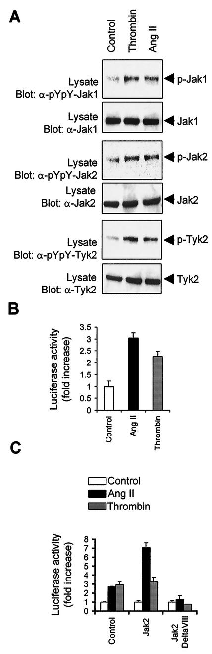
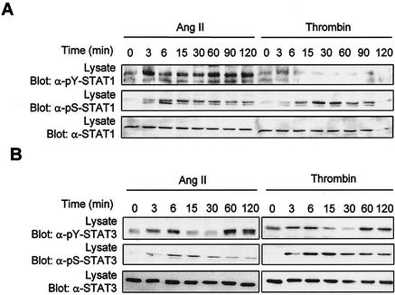
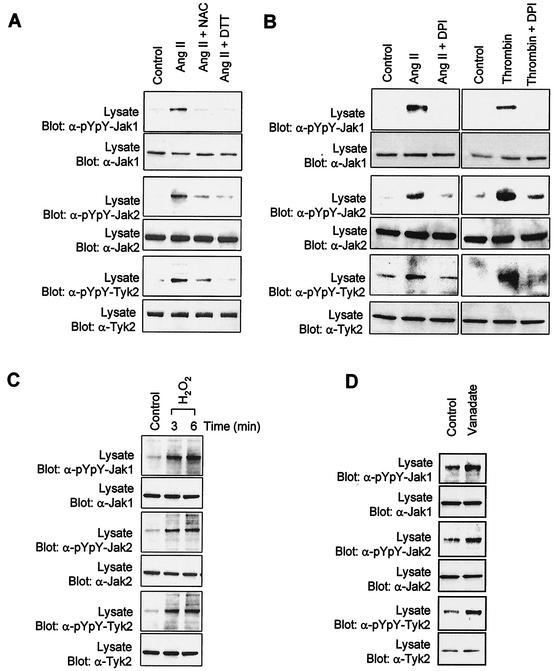
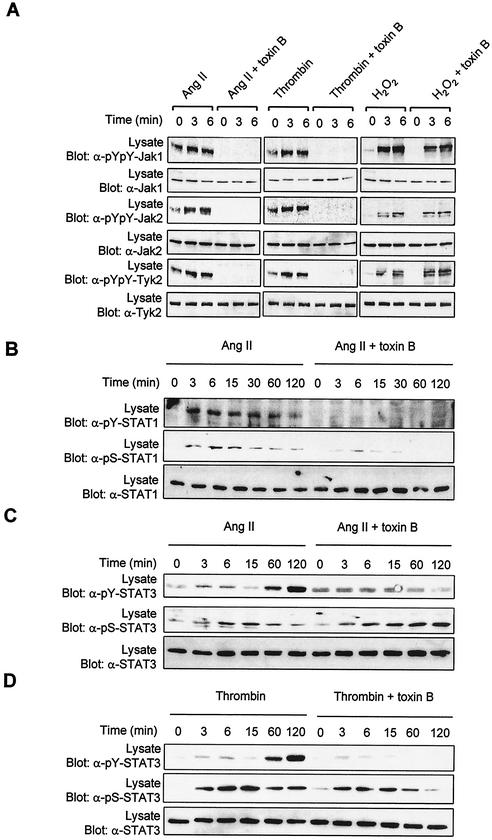
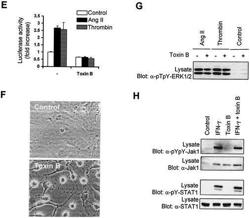
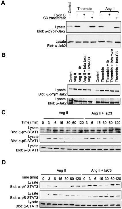
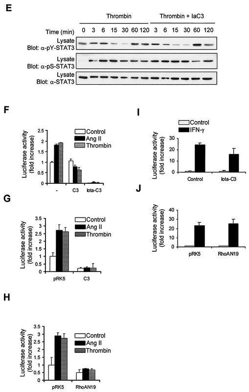
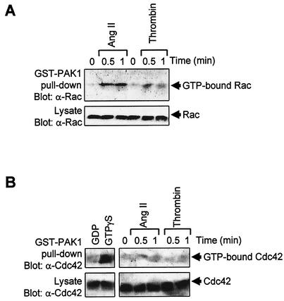
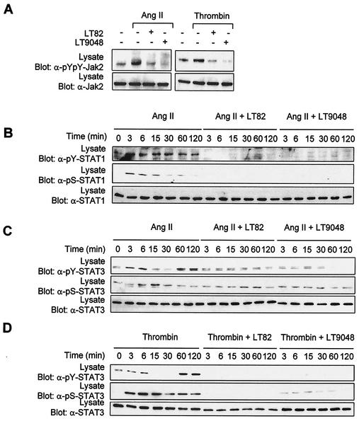
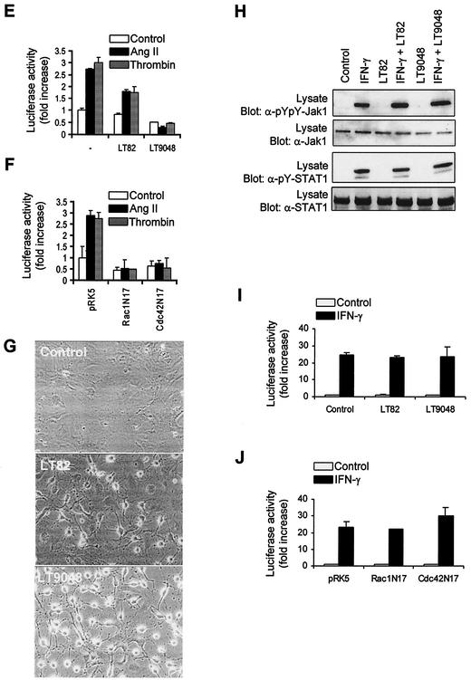
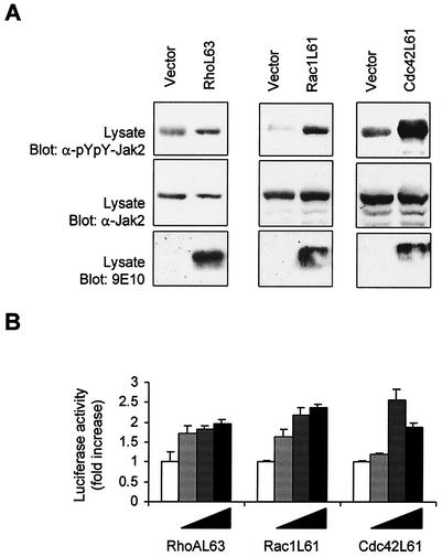
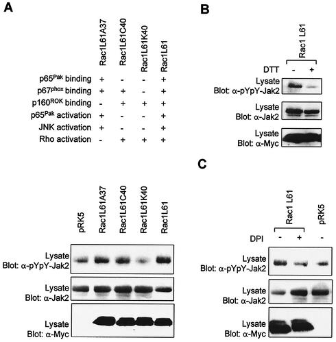
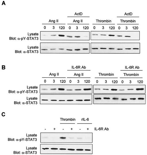
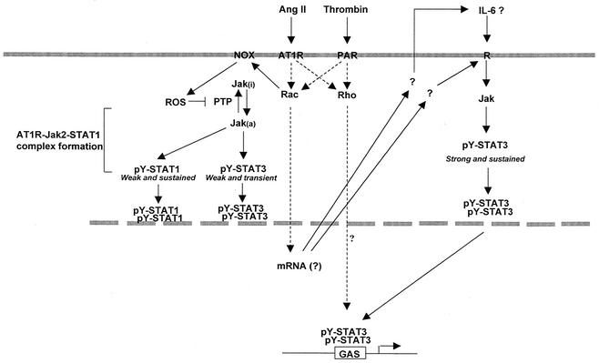
As do cytokine receptors and receptor tyrosine kinases, G protein-coupled receptors (GPCRs) signal to Janus kinases (Jaks) and signal transducers and activators of transcription (STATs). However, the early biochemical events linking GPCRs to this signaling pathway have been unclear. Here we show that GPCR-stimulated Rac activity and the subsequent generation of reactive oxygen species are necessary for activating tyrosine phosphorylation of Jaks and STAT-dependent transcription. The requirement for Rac activity can be overcome by addition of hydrogen peroxide. Expression of activated mutants of Rac1 is sufficient to activate Jak2 and STAT-dependent transcription, and the activation of Jak2 correlates with the ability of Rac1 to bind to NADPH oxidase subunit p67(phox). We further show that GPCR agonists stimulate tyrosine phosphorylation of STAT1 and STAT3 proteins in a Rac-dependent manner. The tyrosine phosphorylation of STAT3 is biphasic; the first peak of phosphorylation is weak and correlates with rapid activation of Jaks by GPCRs, whereas the second peak is stronger and requires the synthesis of an autocrine factor. Rho also plays an essential role in the induction of STAT transcriptional activity. Our results highlight a novel role for Rho GTPases in mediating the regulatory effects of GPCRs on STAT-dependent gene expression.


Figures

FIG. 1.
FIG. 1.
GPCR agonists stimulate Jak activity and STAT-dependent transcription in vascular SMC. (A) Quiescent rat vascular SMC were stimulated or not (Control) with thrombin (1 U/ml) or Ang II (100 nM) for 3 min. The activation of Jaks was monitored by immunoblotting of total lysate proteins with phospho-specific antibodies to activation loop tyrosine residues (pYpY). Expression levels of Jak1, Jak2, and Tyk2 were analyzed by reprobing the membrane with isoform-specific antibodies. Results are representative of five experiments. (B) Vascular SMC were transfected with pGL-2xIFP53GAS-luc reporter plasmid. After 24 h, the cells were serum starved for 48 h and stimulated with Ang II or thrombin for 24 h. The activity of luciferase was measured and normalized to that of β-galactosidase. (C) Vascular SMC were transfected with the pGL-2xIFP53GAS-luc reporter together with 500 ng of pcDNA3, pEF-BOS-Jak2, or pEF-BOS-JAk2ΔVIII. Serum-starved cells were stimulated with Ang II or thrombin, and luciferase activity was measured. The luciferase data are presented as increase over unstimulated control and represent the means of triplicate determinations (error bars, standard errors). (B and C) Results are representative of three independent experiments.
FIG. 2.
FIG. 2.
GPCRs stimulate tyrosine and serine phosphorylation of STAT1 and STAT3 in vascular SMC. Quiescent vascular SMC were stimulated with thrombin or Ang II for the indicated times. The activation of STAT1 (A) and STAT3 (B) was monitored by immunoblotting of total lysate proteins with phospho-specific antibodies to the C-terminal tyrosine of STAT1 (Tyr701) and STAT3 (Tyr705). Serine phosphorylation of STAT1 and STAT3 was monitored by immunoblotting with phospho-specific antibodies to Ser727. The expression levels of STAT1 and -3 were analyzed by reprobing the membrane with isoform-specific antibodies. Results presented are representative of three experiments.
FIG. 3.
FIG. 3.
Activation of Jaks by GPCRs is dependent on the production of ROS. (A) Quiescent vascular SMC were pretreated or not for 30 min with the antioxidant N-acetyl-l-cysteine (NAC) (30 mM) or DTT (30 mM) and then stimulated with Ang II for 3 min. (B) Quiescent cells were pretreated or not for 1 h with the NADPH oxidase inhibitor DPI (10 μM) and stimulated with Ang II or thrombin for 3 min. (C) Quiescent cells were incubated with 250 μM H2O2 for the indicated times. (D) Quiescent cells were incubated with 500 μM vanadate for the indicated times. The activation of Jaks was monitored by immunoblotting of total lysate proteins with phospho-specific antibodies to activating tyrosine residues. Results are representative of at least three independent experiments.
FIG. 4.
FIG. 4.
Rho GTPases are required for activation of the Jak/STAT pathway by GPCRs. (A to D) Quiescent vascular SMC were pretreated or not for 3 h with C. difficile toxin B (5 ng/ml) and then stimulated with Ang II, thrombin, or H2O2 for the indicated times. The activation of Jaks(A), STAT1 (B), and STAT3 (C and D) was monitored by immunoblotting of total lysate proteins with phospho-specific antibodies to activating tyrosine residues. (B to D) Ser727 phosphorylation of STAT1/3 was assessed by immunoblotting with phospho-specific antibodies. (E) Vascular SMC were transfected with pGL-2xIFP53GAS-luc reporter plasmid. Serum-starved cells were pretreated or not for 6 h with toxin B (0.5 ng/ml) and then stimulated with Ang II or thrombin for 24 h. The activity of luciferase was measured and normalized to that of β-galactosidase. Data are expressed as increase over unstimulated control and represent the means of triplicate determinations (error bars, standard errors). (A to E) Results are representative of three independent experiments. (F) Phase-contrast micrographs showing the effect of toxin B on the morphology of vascular SMC. (G) Quiescent cells were pretreated or not with toxin B and then stimulated with Ang II or thrombin for 5 min. The activating phosphorylation of Erk1/Erk2 was monitored by immunoblotting of lysate proteins with a phospho-specific antibody. (H) Quiescent vascular SMC were pretreated or not with toxin B and then stimulated with IFN-γ for 15 min. The activation of Jak1 and STAT1 was monitored as described above.
FIG. 4.
FIG. 4.
Rho GTPases are required for activation of the Jak/STAT pathway by GPCRs. (A to D) Quiescent vascular SMC were pretreated or not for 3 h with C. difficile toxin B (5 ng/ml) and then stimulated with Ang II, thrombin, or H2O2 for the indicated times. The activation of Jaks(A), STAT1 (B), and STAT3 (C and D) was monitored by immunoblotting of total lysate proteins with phospho-specific antibodies to activating tyrosine residues. (B to D) Ser727 phosphorylation of STAT1/3 was assessed by immunoblotting with phospho-specific antibodies. (E) Vascular SMC were transfected with pGL-2xIFP53GAS-luc reporter plasmid. Serum-starved cells were pretreated or not for 6 h with toxin B (0.5 ng/ml) and then stimulated with Ang II or thrombin for 24 h. The activity of luciferase was measured and normalized to that of β-galactosidase. Data are expressed as increase over unstimulated control and represent the means of triplicate determinations (error bars, standard errors). (A to E) Results are representative of three independent experiments. (F) Phase-contrast micrographs showing the effect of toxin B on the morphology of vascular SMC. (G) Quiescent cells were pretreated or not with toxin B and then stimulated with Ang II or thrombin for 5 min. The activating phosphorylation of Erk1/Erk2 was monitored by immunoblotting of lysate proteins with a phospho-specific antibody. (H) Quiescent vascular SMC were pretreated or not with toxin B and then stimulated with IFN-γ for 15 min. The activation of Jak1 and STAT1 was monitored as described above.
FIG. 5.
FIG. 5.
Rho is necessary for the transcriptional activation of STATs by GPCRs. (A) Quiescent vascular SMC were pretreated or not for 3 h with C. difficile toxin B (5 ng/ml) or for 48 h with C. botulinum C3 transferase (20 μg/ml). (B) Quiescent cells were pretreated or not for 24 h with Iota toxin subunit Ib (7 μg/ml), Iota toxin (Ia and Ib) (each subunit, 7 μg/ml), or Iota-C3 fusion protein (7 μg of Ia-C3 and Ib/ml). The cells were then stimulated with Ang II or thrombin for 3 min. The activation of Jak2 was monitored by immunoblotting of lysate proteins with a phospho-specific antibody to activating tyrosine residues. (C to E) Quiescent cells were pretreated or not for 24 h with Iota-C3 fusion protein, and then stimulated with Ang II or thrombin for the indicated times. The activation and serine phosphorylation of STAT1 (C) and STAT3 (D and E) were monitored by immunoblotting with phospho-specific antibodies. (F) Vascular SMC were transfected with pGL-2xIFP53GAS-luc reporter plasmid. Serum-starved cells were pretreated or not for 48 h with C3 transferase (10 μg/ml) or for 6 h with Iota-C3 fusion toxin, and then stimulated with Ang II or thrombin for 24 h. The activity of luciferase was measured and normalized to that of β-galactosidase. (G and H) Vascular SMC were transfected with the pGL-2xIFP53GAS-luc reporter together with 500 ng of pEF-Myc-C3 transferase (G) or pRK5-MycRhoAN19 (H). Serum-starved cells were stimulated with Ang II or thrombin for 24 h, and luciferase activity was measured. Luciferase data are presented as increase over unstimulated control and represent the means of triplicate determinations (error bars, standard errors). (A to H) Results arerepresentative of three independent experiments. (i) Vascular SMC were transfected with pGL-2xIFP53GAS-luc reporter plasmid. Serum-starved cells were pretreated or not with Iota-C3 fusion toxin, and then stimulated with IFN-γ for 6 h. The activity of luciferase was measured and normalized to that of β-galactosidase. (J) Vascular SMC were transfected with the GAS reporter together with 500 ng of pRK5-MycRhoAN19. Serum-starved cells were stimulated with IFN-γ for 6 h, and luciferase activity was measured. Results are representative of three experiments.
FIG. 5.
FIG. 5.
Rho is necessary for the transcriptional activation of STATs by GPCRs. (A) Quiescent vascular SMC were pretreated or not for 3 h with C. difficile toxin B (5 ng/ml) or for 48 h with C. botulinum C3 transferase (20 μg/ml). (B) Quiescent cells were pretreated or not for 24 h with Iota toxin subunit Ib (7 μg/ml), Iota toxin (Ia and Ib) (each subunit, 7 μg/ml), or Iota-C3 fusion protein (7 μg of Ia-C3 and Ib/ml). The cells were then stimulated with Ang II or thrombin for 3 min. The activation of Jak2 was monitored by immunoblotting of lysate proteins with a phospho-specific antibody to activating tyrosine residues. (C to E) Quiescent cells were pretreated or not for 24 h with Iota-C3 fusion protein, and then stimulated with Ang II or thrombin for the indicated times. The activation and serine phosphorylation of STAT1 (C) and STAT3 (D and E) were monitored by immunoblotting with phospho-specific antibodies. (F) Vascular SMC were transfected with pGL-2xIFP53GAS-luc reporter plasmid. Serum-starved cells were pretreated or not for 48 h with C3 transferase (10 μg/ml) or for 6 h with Iota-C3 fusion toxin, and then stimulated with Ang II or thrombin for 24 h. The activity of luciferase was measured and normalized to that of β-galactosidase. (G and H) Vascular SMC were transfected with the pGL-2xIFP53GAS-luc reporter together with 500 ng of pEF-Myc-C3 transferase (G) or pRK5-MycRhoAN19 (H). Serum-starved cells were stimulated with Ang II or thrombin for 24 h, and luciferase activity was measured. Luciferase data are presented as increase over unstimulated control and represent the means of triplicate determinations (error bars, standard errors). (A to H) Results arerepresentative of three independent experiments. (i) Vascular SMC were transfected with pGL-2xIFP53GAS-luc reporter plasmid. Serum-starved cells were pretreated or not with Iota-C3 fusion toxin, and then stimulated with IFN-γ for 6 h. The activity of luciferase was measured and normalized to that of β-galactosidase. (J) Vascular SMC were transfected with the GAS reporter together with 500 ng of pRK5-MycRhoAN19. Serum-starved cells were stimulated with IFN-γ for 6 h, and luciferase activity was measured. Results are representative of three experiments.
FIG. 6.
FIG. 6.
GPCR agonists activate Rac but not Cdc42 in vascular SMC. (A and B) Quiescent vascular SMC were stimulated with Ang II or thrombin for the times indicated. The activity of Rac1 (A) and Cdc42 (B) was determined by measuring the amount of GTP-loaded protein bound to GST-Pak as described in Materials and Methods. Control assays were carried out by incubating lysates of unstimulated cells with either GTPγS (positive control) or GDP (negative control) for 15 min at 25°C prior to incubation with GST-Pak1 beads. Results are representative of three experiments.
FIG. 7.
FIG. 7.
Role of Rac and Cdc42 in Jak2 activation and induction of STAT-dependent transcription by GPCRs. (A) Quiescent vascular SMC were pretreated or not for 4 h with C. sordellii LT82 (5 μg/ml) or LT9048 (5 μg/ml), and then stimulated with thrombin or Ang II for 3 min. The activation of Jak2 was monitored by immunoblotting of lysate proteins with a phospho-specific antibody. (B to D) Quiescent cells were pretreated or not for 4 h with LT82 or LT9048 and then stimulated with Ang II or thrombin for the indicated times. The activation and serine phosphorylation of STAT1 (B) and STAT3 (C and D) were monitored by immunoblotting with phospho-specific antibodies. (E) Vascular SMC were transfected with pGL-2xIFP53GAS-luc reporter plasmid. Serum-starved cells were pretreated or not for 4 h with LT82 or LT9048, and then stimulated with Ang II or thrombin for 24 h. The activity of luciferase was measured and normalized to β-galactosidase. (F) Vascular SMC were transfected with the pGL-2xIFP53GAS-luc reporter together with 500 ng of pRK5-MycRac1N17 or pRK5-MycCdc42N17. Serum-starved cells were stimulated with Ang II or thrombin, and luciferase activity was measured. Luciferase data are presented as increase over unstimulated control and represent the means of triplicate determinations (error bars, standard error). (A to F) Results are representative of three independent experiments. (G) Phase-contrast micrographs showing the effects of LT82 and LT9048 on the morphology of vascular SMC. (H) Quiescent cells were pretreated or not with LT82 or LT9048 and then stimulated with IFN-γ for 15 min. The activation of Jak1 and STAT1 was monitored as described above. (I) Vascular SMC were transfected with the GAS reporter, and treated or not with LT82 or LT9048. Serum-starved cells were then stimulated with IFN-γ for 6 h, and luciferase activity was measured. (J) Vascular SMC were transfected with the GAS reporter together with 500 ng of pRK5-MycRac1N17 or pRK5-Cdc42N17. Serum-starved cells were stimulated with IFN-γ for 6 h, and luciferase activity was measured. Results are representative of three experiments.
FIG. 7.
FIG. 7.
Role of Rac and Cdc42 in Jak2 activation and induction of STAT-dependent transcription by GPCRs. (A) Quiescent vascular SMC were pretreated or not for 4 h with C. sordellii LT82 (5 μg/ml) or LT9048 (5 μg/ml), and then stimulated with thrombin or Ang II for 3 min. The activation of Jak2 was monitored by immunoblotting of lysate proteins with a phospho-specific antibody. (B to D) Quiescent cells were pretreated or not for 4 h with LT82 or LT9048 and then stimulated with Ang II or thrombin for the indicated times. The activation and serine phosphorylation of STAT1 (B) and STAT3 (C and D) were monitored by immunoblotting with phospho-specific antibodies. (E) Vascular SMC were transfected with pGL-2xIFP53GAS-luc reporter plasmid. Serum-starved cells were pretreated or not for 4 h with LT82 or LT9048, and then stimulated with Ang II or thrombin for 24 h. The activity of luciferase was measured and normalized to β-galactosidase. (F) Vascular SMC were transfected with the pGL-2xIFP53GAS-luc reporter together with 500 ng of pRK5-MycRac1N17 or pRK5-MycCdc42N17. Serum-starved cells were stimulated with Ang II or thrombin, and luciferase activity was measured. Luciferase data are presented as increase over unstimulated control and represent the means of triplicate determinations (error bars, standard error). (A to F) Results are representative of three independent experiments. (G) Phase-contrast micrographs showing the effects of LT82 and LT9048 on the morphology of vascular SMC. (H) Quiescent cells were pretreated or not with LT82 or LT9048 and then stimulated with IFN-γ for 15 min. The activation of Jak1 and STAT1 was monitored as described above. (I) Vascular SMC were transfected with the GAS reporter, and treated or not with LT82 or LT9048. Serum-starved cells were then stimulated with IFN-γ for 6 h, and luciferase activity was measured. (J) Vascular SMC were transfected with the GAS reporter together with 500 ng of pRK5-MycRac1N17 or pRK5-Cdc42N17. Serum-starved cells were stimulated with IFN-γ for 6 h, and luciferase activity was measured. Results are representative of three experiments.
FIG. 8.
FIG. 8.
Activation of Jak2 and STAT-dependent transcription by Rho GTPases. (A) COS-7 cells were transfected with pEF-BOS-Jak2 (250 ng) together with 250 ng of pRK5-MycRhoAL63 (left panel), pRK5-MycRac1L61 (middle panel), or pRK5-MycCdc42L61 (right panel). The cells were serum starved for 24 h and the activation of Jak2 was monitored by immunoblotting of total lysate proteins with a phospho-specific antibody to activating tyrosine residues. Expression level of Jak2 and Rho GTPases was analyzed by immunoblotting with anti-Jak2 and anti-Myc antibodies, respectively. (B) Vascular SMC were transfected with the pGL-2xIFP53GAS-luc reporter together with increasing amounts (from 50 to 400 ng) of pRK5-MycRhoAL63, pRK5-MycRac1L61 or pRK5-MycCdc42L61. The cells were serum starved for 18 h and luciferase activity was measured. Results are representative of three independent experiments.
FIG. 9.
FIG. 9.
Effect of Rac1 effector loop mutants on Jak2 activity. (A) COS-7 cells were transfected with pEF-BOS-Jak2 (100 ng) together with 250 ng of the indicated mutants of Rac1. The cells were serum starved for 24 h, and the activation of Jak2 was monitored by immunoblotting with a phospho-specific antibody. Expression levels of Jak2 and Rac were analyzed by immunoblotting with anti-Jak2 and anti-Myc antibodies, respectively. (B and C) Same as panel A, except that COS-7 cells were treated for 1 h with DTT (30 mM) or DPI (10 μM) prior to harvesting. Results are representative of three independent experiments.
FIG. 10.
FIG. 10.
Evidence that late-phase activation of STAT3 is mediated by an autocrine factor. (A) Vascular SMC were treated or not with ActD (5 μg/ml) for 60 min and then stimulated with Ang II or thrombin for the times indicated. Activation of STAT3 was monitored by immunoblotting of lysate proteins with a phospho-specific antibody. (B) Quiescent vascular SMC were incubated for 60 min in the absence or presence of a neutralizing antibody to IL-6 receptor, prior to stimulation with Ang II or thrombin. Activation of STAT3 was monitored as described above. (C) Growth-arrested HeLa cells were incubated or not with the IL-6 receptor antibody and then stimulated with thrombin for 2 h. Activation of STAT3 was monitored as above. Results are representative of three experiments.
FIG. 11.
FIG. 11.
Proposed model for the role of Rho GTPases in the regulation of Jak activation and STAT-dependent transcription in response to GPCR engagement.

Similar articles

Cited by

References

    1. Aktories, K., G. Schmidt, and I. Just. 2000. Rho GTPases as targets of bacterial protein toxins. Biol. Chem. 381:421-426. - PubMed
    1. Ali, M. S., P. P. Sayeski, and K. E. Bernstein. 2000. Jak2 acts as both a STAT1 kinase and as a molecular bridge linking STAT1 to the angiotensin II AT1 receptor. J. Biol. Chem. 275:15586-15593. - PubMed
    1. Ali, M. S., P. P. Sayeski, L. B. Dirksen, D. J. Hayzer, M. B. Marrero, and K. E. Bernstein. 1997. Dependence on the motif YIPP for the physical association of Jak2 kinase with the intracellular carboxyl tail of the angiotensin II AT1 receptor. J. Biol. Chem. 272:23382-23388. - PubMed
    1. Ali, M. S., P. P. Sayeski, A. Safavi, M. Lyles, and K. E. Bernstein. 1998. Janus kinase 2 (Jak2) must be catalytically active to associate with the AT1 receptor in response to angiotensin II. Biochem. Biophys. Res. Commun. 249:672-677. - PubMed
    1. Babior, B. M. 1999. NADPH oxidase: an update. Blood 93:1464-1476. - PubMed

Publication types

MeSH terms

Substances