. 2003 Feb;23(4):1390-402.
doi: 10.1128/MCB.23.4.1390-1402.2003.

Functional cross-antagonism between transcription factors FLI-1 and EKLF

Affiliations

Functional cross-antagonism between transcription factors FLI-1 and EKLF

Joëlle Starck et al. Mol Cell Biol. 2003 Feb.

Abstract

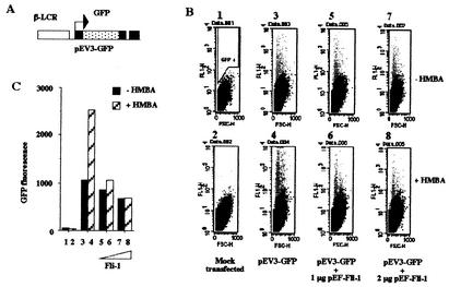
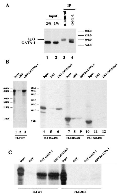
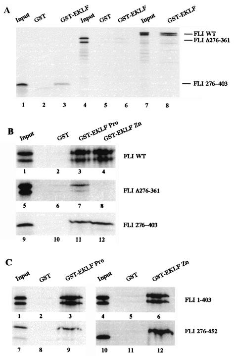
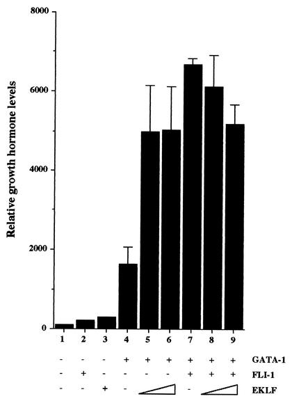
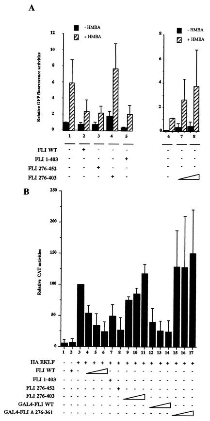
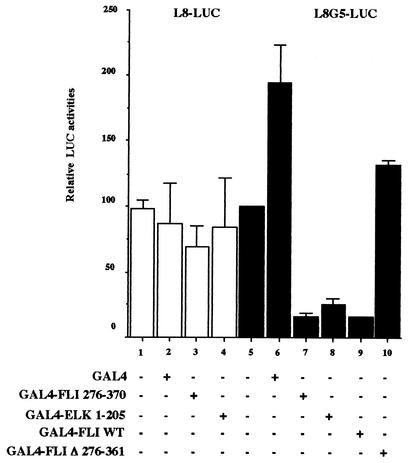
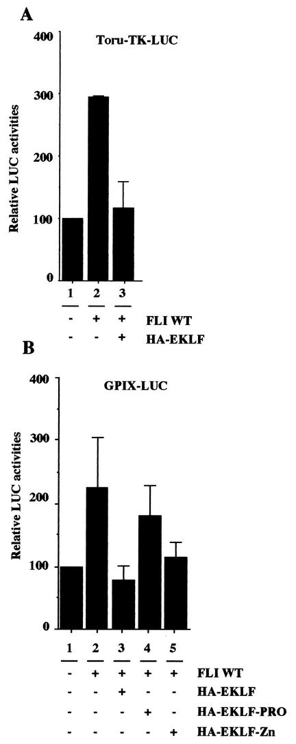
FLI-1 is an ETS family transcription factor which is overexpressed in Friend erythroleukemia and contributes to the blockage of differentiation of erythroleukemic cells. We show here that FLI-1 represses the transcriptional activity of the beta-globin gene promoter in MEL cells and interacts with two of its critical transactivators, GATA-1 and EKLF. Unexpectedly, FLI-1 enhances the stimulating activity of GATA-1 on a GATA-1-responsive promoter but represses that of EKLF on beta-globin and an EKLF-responsive artificial promoters. This repressive effect of FLI-1 requires the ETS DNA binding domain and its association with either the N- or C-terminal domain, which themselves interact with EKLF but not with GATA-1. Furthermore, the FLI-1 ETS domain alone behaves as an autonomous repression domain when linked to the Gal4 DNA binding domain. Taken together, these data indicate that FLI-1 represses EKLF-dependent transcription due to the repression activity of its ETS domain and its indirect recruitment to erythroid promoters by protein-protein interaction with EKLF. Reciprocally, we also show that EKLF itself represses the FLI-1-dependent megakaryocytic GPIX gene promoter, thus further suggesting that functional cross-antagonism between FLI-1 and EKLF might be involved in the control of the erythrocytic versus megakaryocytic differentiation of bipotential progenitors.


Figures

FIG. 1.
FIG. 1.
Schematic structure of the FLI-1 and EKLF proteins and deletion or fusion mutants derived thereof. Proteins encoded by the expression plasmids used in this study are depicted. The ETS DNA binding domain of FLI-1 (aa 276 to 360) and the three-zinc-fingers DNA binding domain of EKLF (aa 293 to 376) are shown as black boxes. GAL4 (aa 1 to 147) and HA (21 aa) encoded sequences are shown as hatched boxes, and ER (aa 281 to 599) encoded sequences are shown as a grey box. Other domains are shown as white boxes. Details of the constructions are given in Material and Methods.
FIG. 2.
FIG. 2.
Enforced expression of FLI-1 inhibits β-globin gene promoter activity in MEL cells. (A) Schematic structure of the pEV3-GFP reporter construct. (B) MEL cells were transfected with a fixed amount of the pEV3-GFP plasmid in the presence or absence of increasing doses of the FLI-1 expression vector pEF-FLI-1 as indicated. Immediately following transfection, each set of transfected cells was cultured for 72 h in the absence (upper panels) or in the presence (lower panels) of 5 mM HMBA. The mean fluorescence and the number of GFP-positive cells were then determined by FACS analysis on forward-scattering (FSC) versus fluorescence (FL1) records. The gate used to identify positive cells (GFP+: grey spots) is shown on panel 1. (C) Quantitative analysis of the data presented in panel B. Total fluorescence activities were quantified by determining the product of the percentage of GFP-positive cells and their mean fluorescence value. Each lane corresponds, respectively, to the different panels presented in panel B. Panels B and C show typical results obtained from at least three different transfection experiments.
FIG. 3.
FIG. 3.
Transactivation activity of FLI-1 is not required to inhibit β-globin gene transcription. MEL cells were transiently cotransfected with the pEV3-GFP and FLI-responsive pTORU-TK-LUC reporter plasmids in the presence of the same amount of expression vectors encoding wild-type FLI-1 or FLI-ER proteins. Immediately following transfection, transfected cells were cultured for 72 h in the presence or absence of HMBA and in the presence of increasing doses of hydroxy-tamoxifen (OHT) (150 or 750 nM). The figure shows typical results of GFP (A) and luciferase (B) activities obtained from two different experiments.
FIG. 4.
FIG. 4.
Interaction between FLI-1 and GATA-1 proteins. (A) MEL cell nuclear extracts were immunoprecipitated with an irrelevant antibody (lane 3) or a FLI-1 specific monoclonal antibody (lane 4). Immunoprecipitated proteins were then analyzed by Western blotting using a GATA-1-specific antibody. Lanes 1 and 2 correspond, respectively, to 2 and 1% of the total cell extract used for immunoprecipitation. The positions of immunoglobulin G (Ig G) and GATA-1 proteins are indicated to the left. (B and C) Interaction of 35S-labeled deletion mutants (B), point mutant (C), or full-length FLI-1 protein (B and C) with GST, GST-GATA-1, and GST-Zn-GATA-1 was investigated by GST pull-down assays. Equal amounts of 35S-labeled proteins were used in each reaction, and 5% of the input proteins are shown in the input lanes.
FIG. 5.
FIG. 5.
Interaction between FLI-1 and EKLF proteins. Interaction between 35S-labeled deletion mutants or full-length FLI-1 proteins and GST, GST-EKLF, GST-EKLF PRO, or GST-EKLF-Zn was investigated by GST-pull down assays. Equal amounts of 35S-labeled proteins were used in each reaction, and 5% of the input proteins are shown in the input lanes. The nature of the 35S-labeled FLI-1 protein and that of the GST or GST fusion protein used in the reactions are indicated to the right and on the top of each panel, respectively. Note that due to the presence of an alternative initiation codon at position 33 (56), all the in vitro-translated FLI-1 proteins including the N-terminal domain appear as doublets.
FIG. 6.
FIG. 6.
FLI-1 and EKLF stimulate the transcriptional activity of GATA-1 in nonerythroid cells. 3T3 cells were cotransfected with a fixed amount (0.25 μg) of the GATA-1-responsive pM1Gα-GH reporter plasmid and with expression vectors encoding GATA-1 (0.5 μg in lanes 4 to 9), FLI-1 (0.75 μg in lanes 2 and 7 to 9), or EKLF (0.25 or 0.5 μg in lanes 5 and 8 or 6 and 9, respectively) as indicated. Growth hormone levels accumulated in the supernatants of transfected cells 24 h posttransfection were determined by ELISA. Results are expressed as percentages of the level obtained in lane 1 (mean and standard deviation from three different experiments).
FIG. 7.
FIG. 7.
FLI-1 represses the transcriptional activity of EKLF. (A) MEL cells were cotransfected with a fixed amount of the pEV3-GFP reporter plasmid and various amounts of expression plasmids encoding EKLF or FLI-1 as indicated. Immediately following transfection, each set of transfected cells was cultured for 72 h in the absence (black bars) or in the presence (hatched bars) of 5 mM HMBA. Quantification of GFP fluorescence was performed by FACS analysis as described in Fig. 2. (B) 3T3 cells were cotransfected with fixed amounts of the EKLF-responsive pC1G3-CAT and the CMV-β-Gal reporter plasmids and various amounts of expression plasmids encoding either FLI-1 or a HA-tagged version of EKLF as indicated. CAT and β-Gal activities were determined 24 h posttransfection. Results are expressed as relative values of CAT activities after standardization on β-Gal activities.
FIG. 8.
FIG. 8.
Mapping of FLI-1 domains involved in repression of EKLF activity. (A) MEL cells were cotransfected in the same conditions as for Fig. 7A with a fixed amount of the pEV3-GFP reporter plasmid and expression vectors encoding the indicated deletion mutants of FLI-1. Results are expressed as relative GFP fluorescence activities (mean and standard deviation from three different experiments). (B) 3T3 cells were cotransfected in the same conditions as for Fig. 7B with a fixed amount of pC1G3-CAT and the CMV-β-Gal reporters and HA-EKLF expression plasmid with or without expression plasmids encoding the indicated FLI-1 proteins. CAT activities were standardized on β-Gal activities, and results are expressed as percentages of the standardized CAT activity obtained for the pC1G3-CAT reporter transfected with HA-EKLF alone (lane 3) (mean and standard deviation from three different experiments).
FIG. 9.
FIG. 9.
Autonomous repression activity of the FLI-1 ETS domain. COS cells were cotransfected with a fixed amount of the CMV-β-Gal and either L8-LUC or L8G5-LUC reporter plasmids as well as a fixed amount of expression plasmid encoding the LexA-VP16 fusion protein and a fixed amount of expression plasmids encoding the indicated Gal4 fusion proteins. Luciferase and β-Gal activities in transfected cells were determined 24 h posttransfection. Luciferase activities were standardized on β-Gal activities, and final results are expressed as percentages of the standardized luciferase activity obtained for L8G5-LUC reporter transfected in the presence of LexA-VP16 alone (lane 5) (mean and standard deviation from three different experiments).
FIG. 10.
FIG. 10.
EKLF represses the transcriptional activity of FLI-1. COS cells were cotransfected with a fixed amount of the TORU-TK-LUC (A) or GPIX-LUC (B) and CMV-β-Gal reporter plasmids with or without expression vectors encoding wild-type FLI-1, wild-type HA-EKLF, or deletion mutants of HA-EKLF as indicated. Luciferase activities were standardized on β-Gal activities, and final results are expressed as percentages of the standardized luciferase activity obtained for the luciferase reporter alone (lane 1) (mean and standard deviation from two different experiments).
FIG. 11.
FIG. 11.
GST-EKLF and GST-FLI-1 do not cross-inhibit their DNA binding activities. (A) GST-FLI-1 DNA binding activity was analyzed by gel shift assay using the GGAA probe either alone (lane 3) or in the presence of increasing excess of GST-EKLF (1-, 5-, or 10-fold excess, respectively, in lanes 4, 5, and 6). Control lanes correspond, respectively, to the probe incubated without protein (lane 1), with GST alone (lane 2), or with GST-FLI-1 and a 10-fold excess of GST (lane 7). The free probe was allowed to run out of the gel in order to facilitate the detection of putative tripartite complexes between GST-EKLF, GST-FLI-1, and DNA and for that reason is not visible on the autoradiograph. (B) Reciprocal experiment performed as for panel A but using the CAC probe.
FIG. 12.
FIG. 12.
Cross-antagonistic model of EKLF and FLI-1 activities in erythroid versus megakaryocytic differentiation. Schematic representation of the cell fate determination of erythrocytic and megakaryocytic cell types from the common bipotential progenitor. EKLF and FLI-1 cooperate with GATA-1 and contribute in a positive manner to produce erythroid or megakaryocytic cells, respectively. Simultaneously, EKLF and FLI-1 contribute in a negative manner through the cross-inhibition of their positive activities.

Similar articles

Cited by

References

    1. Afrikanova, I., E. Yeh, D. Bartos, S. S. Watowich, and G. D. Longmore. 2002. Oncogene cooperativity in Friend erythroleukemia: erythropoietin receptor activation by the env gene of SFFV leads to transcriptional upregulation of PU.1, independent of SFFV proviral insertion. Oncogene 21:1272-1284. - PMC - PubMed
    1. Athanasiou, M., P. A. Clausen, G. J. Mavrothalassitis, X. K. Zhang, D. K. Watson, and D. G. Blair. 1996. Increased expression of the ETS-related transcription factor FLI-1/ERGB correlates with and can induce the megakaryocytic phenotype. Cell Growth Differ. 7:1525-1534. - PubMed
    1. Athanasiou, M., G. Mavrothalassitis, L. Sun-Hoffman, and D. G. Blair. 2000. FLI-1 is a suppressor of erythroid differentiation in human hematopoietic cells. Leukemia 14:439-445. - PubMed
    1. Barnache, S., F. Wendling, C. Lacombe, N. Denis, M. Titeux, W. Vainchenker, and F. Moreau-Gachelin. 1998. Spi-1 transgenic mice develop a clonal erythroleukemia which does not depend on p53 mutation. Oncogene 16:2989-2995. - PubMed
    1. Barnache, S., P. Mayeux, B. Payrastre, and F. Moreau-Gachelin. 2001. Alterations of the phosphoinositide 3-kinase and mitogen-activated protein kinase signaling pathways in the erythropoietin-independent Spi-1/PU.1 transgenic proerythroblasts. Blood 98:2372-2381. - PubMed

Publication types

MeSH terms

LinkOut - more resources